Unlocking Nature's Pharmacy: A Comprehensive Guide to ADMET Profiling of Natural Products in Modern Drug Discovery
This article provides a systematic review and practical guide for researchers and drug development professionals on the critical role of Absorption, Distribution, Metabolism, Excretion, and Toxicity (ADMET) profiling in natural...
Unlocking Nature's Pharmacy: A Comprehensive Guide to ADMET Profiling of Natural Products in Modern Drug Discovery
Abstract
This article provides a systematic review and practical guide for researchers and drug development professionals on the critical role of Absorption, Distribution, Metabolism, Excretion, and Toxicity (ADMET) profiling in natural product-based drug discovery. It explores the unique ADMET challenges posed by natural product scaffolds, details contemporary in silico, in vitro, and in vivo methodologies for their evaluation, addresses common pitfalls in optimization, and compares their properties with synthetic libraries. The content synthesizes current strategies to transform promising natural leads into viable drug candidates with favorable pharmacokinetic and safety profiles, bridging the gap between traditional medicine and contemporary pharmaceutical development.
Why Natural Products Pose Unique ADMET Challenges: From Complex Scaffolds to Bioavailability
Within the landscape of drug discovery, the assessment of Absorption, Distribution, Metabolism, Excretion, and Toxicity (ADMET) is the critical filter that determines the translational success of a candidate molecule. This principle is magnified in the context of natural products (NPs), which offer unparalleled structural diversity but are often hampered by complex and suboptimal ADMET profiles. A core thesis in contemporary NP research posits that early, parallel, and quantitative evaluation of ADMET properties is non-negotiable for de-risking the development of natural product-derived therapeutics. This whitepaper serves as a technical guide to the core methodologies defining this gatekeeper role.
Quantitative Landscape of ADMET Failure
A significant majority of clinical-stage failures are attributed to poor pharmacokinetics or unacceptable toxicity, underscoring the necessity of robust preclinical ADMET screening. Recent analyses of drug development pipelines provide the following quantitative context:
Table 1: Primary Causes of Clinical Attrition (Recent Analysis)
| Attrition Phase | Primary Cause | Estimated Percentage |
|---|---|---|
| Preclinical to Phase I | Poor PK/ADMET & Toxicity | ~40% |
| Phase II | Lack of Efficacy | ~52% |
| Phase III | Lack of Efficacy / Safety | ~50% |
| Overall (All Phases) | Poor PK/ADMET & Toxicity | ~30% |
Table 2: Key ADMET Property Benchmarks for Oral Drugs
| Property | Ideal Range / Outcome | High-Risk Indicator |
|---|---|---|
| Aqueous Solubility | > 100 µM | < 10 µM |
| Caco-2 Permeability (Papp) | > 1 x 10⁻⁶ cm/s | < 1 x 10⁻⁷ cm/s |
| Microsomal Half-life (Human) | > 30 minutes | < 15 minutes |
| Plasma Protein Binding | < 95% bound | > 99% bound |
| hERG Inhibition (IC50) | > 10 µM | < 1 µM |
| CYP450 Inhibition (3A4, 2D6) | IC50 > 10 µM | IC50 < 1 µM |
Core Experimental Methodologies
In Vitro Absorption & Permeability: Caco-2 Assay Protocol
This assay models intestinal epithelial transport.
- Cell Culture: Maintain Caco-2 cells in DMEM with 20% FBS, 1% NEAA. Seed on collagen-coated transwell inserts (1.12 cm², 0.4 µm pore) at high density (e.g., 100,000 cells/insert). Culture for 21-28 days to allow full differentiation and tight junction formation, monitoring transepithelial electrical resistance (TEER > 500 Ω·cm²).
- Experiment Setup: Pre-warm transport buffer (HBSS-HEPES, pH 7.4). Test compound (typically 10-100 µM) is added to the donor compartment (apical for A→B, basolateral for B→A). Receiver compartment contains blank buffer.
- Sampling & Analysis: Collect samples from the receiver side at 30, 60, 90, and 120 minutes. Replenish with fresh buffer. Analyze samples via LC-MS/MS to quantify compound concentration.
- Data Calculation: Calculate apparent permeability (Papp) = (dQ/dt) / (A * C₀), where dQ/dt is the transport rate, A is the membrane area, and C₀ is the initial donor concentration. Calculate efflux ratio = Papp(B→A) / Papp(A→B). An efflux ratio > 2 suggests active efflux (e.g., by P-glycoprotein).
In Vitro Metabolic Stability: Liver Microsomal Incubation
- Incubation Preparation: Prepare incubation mix (final volume 100 µL): 0.1 M phosphate buffer (pH 7.4), human or species-specific liver microsomes (0.5 mg protein/mL), test compound (1 µM), and MgCl₂ (5 mM). Pre-incubate at 37°C for 5 minutes.
- Reaction Initiation & Quenching: Start the reaction by adding NADPH (1 mM final concentration). At predetermined time points (0, 5, 10, 20, 30, 45 minutes), remove 50 µL aliquot and quench in 100 µL of ice-cold acetonitrile containing an internal standard.
- Sample Analysis: Vortex, centrifuge (15,000 x g, 10 min), and analyze the supernatant via LC-MS/MS. Monitor the peak area of the parent compound relative to the internal standard.
- Data Calculation: Plot Ln(% parent remaining) vs. time. The slope (k) is the elimination rate constant. Calculate in vitro half-life: t₁/₂ = 0.693 / k. Intrinsic clearance (CLint) is calculated as CLint = (0.693 / t₁/₂) * (Incubation Volume / Microsomal Protein).
High-Throughput Toxicity Screening: hERG Inhibition Patch Clamp
The gold standard for assessing cardiac risk via potassium channel blockade.
- Cell Preparation: Stable hERG-expressing HEK293 or CHO cells are cultured. For assay, cells are harvested and placed in the recording chamber perfused with extracellular solution.
- Electrophysiology Setup: Use a patch clamp amplifier in whole-cell configuration. A borosilicate glass pipette (3-5 MΩ) is filled with intracellular solution. After achieving a Giga-ohm seal and whole-cell access, initiate the voltage protocol.
- Voltage Protocol & Drug Application: Hold at -80 mV, step to +20 mV for 2 sec (to activate channels), then step to -50 mV for 2 sec (to elicit hERG tail current). Repeat every 10-15 seconds. After stable baseline recording, perfuse the test compound at increasing concentrations (e.g., 0.1, 1, 10 µM).
- Data Analysis: Measure the peak tail current amplitude after each depolarizing step. Plot normalized current amplitude vs. compound concentration and fit the data with a Hill equation to determine the IC₅₀ value.
Visualizing ADMET Pathways & Workflows
Title: Iterative ADMET Optimization Cycle in NP Discovery
Title: Metabolic Activation & Idiosyncratic Toxicity Pathway
The Scientist's Toolkit: Key Research Reagent Solutions
Table 3: Essential Materials for Core ADMET Assays
| Reagent / Material | Function / Application | Key Consideration |
|---|---|---|
| Caco-2 Cell Line | Model for intestinal permeability and active transport. | Passage number and culture duration are critical for differentiation. |
| Pooled Human Liver Microsomes (HLM) | Contains major CYP450 enzymes for metabolic stability and metabolite ID studies. | Use gender/ethnicity-pooled or individual donors for variability assessment. |
| Recombinant CYP450 Isozymes | Individual enzymes (CYP3A4, 2D6, etc.) for reaction phenotyping. | Determines which specific enzyme is responsible for metabolism. |
| hERG-Expressing Cell Line (e.g., HEK293-hERG) | In vitro model for cardiac liability screening. | Requires functional validation via reference inhibitors (e.g., E-4031). |
| LC-MS/MS System (Triple Quadrupole) | Quantitative bioanalysis for parent compound and metabolites. | High sensitivity and specificity required for low-concentration PK samples. |
| High-Throughput Automated Patch Clamp System | Higher-throughput functional screening for ion channel effects. | Bridges gap between fluorescence assays and manual patch clamp. |
| Phospholipid Vesicle Preparation (PAMPA) | Artificial membrane for passive permeability screening. | Useful for early, low-cost ranking of compound libraries. |
Natural products (NPs) remain a cornerstone of drug discovery, providing privileged scaffolds with unmatched structural diversity and potent bioactivity against a wide array of therapeutic targets. However, their integration into modern pharmaceutical pipelines is consistently hampered by suboptimal Absorption, Distribution, Metabolism, Excretion, and Toxicity (ADMET) profiles. This whitepaper, framed within a broader thesis on the ADMET properties of NPs, explores this central paradox and provides a technical guide for researchers to navigate these challenges through advanced experimental and computational strategies.
The Core of the Paradox: Data Comparison
Table 1: Bioactivity vs. Pharmacokinetic Properties of Representative Natural Product Classes
| Natural Product Class | Example Compound | Typical IC50/EC50 (nM) | Oral Bioavailability (%) | Plasma Protein Binding (%) | CYP450 Inhibition (Major Isoform) | Clinical Status |
|---|---|---|---|---|---|---|
| Polyketides | Rapamycin | 0.1 - 10 | ~15 | ~92 | 3A4 (Moderate) | Approved |
| Alkaloids | Berberine | 100 - 1000 | < 5 | ~80 | 2D6, 3A4 (Strong) | Research/Herbal |
| Terpenoids | Paclitaxel | 1 - 10 | N/A (IV only) | 89-98 | 2C8, 3A4 (Substrate) | Approved |
| Polyphenols | Curcumin | 1000 - 10000 | < 1 | High | 3A4 (Weak) | Preclinical |
| Glycosides | Digoxin | 0.5 - 2 | 60-80 | ~25 | 3A4 (P-gp Substrate) | Approved |
Table 2: Common ADMET Liabilities of Natural Product Scaffolds
| ADMET Liability | Structural Correlate in NPs | Consequence | Mitigation Strategy |
|---|---|---|---|
| Poor Solubility | High molecular weight, lipophilicity, crystal lattice | Low oral absorption, erratic IV formulation | Prodrug, nano-formulation, salt formation |
| Low Permeability | Multiple H-bond donors/acceptors, glycosylation | Poor intestinal absorption, low BBB penetration | Structural simplification, glycoside removal |
| Rapid Metabolism | Susceptible ester/phenol groups, specific motifs | High clearance, short half-life | Blocking metabolically labile sites, deuteration |
| Efflux by P-gp | Overlapping substrate pharmacophore | Reduced intestinal uptake, brain exposure | Co-administration of P-gp inhibitors, analog design |
| Toxicity | Reactive functional groups (e.g., epoxides) | Off-target effects, organ toxicity | Structural modification, targeted delivery |
Experimental Protocols for Assessing NP Pharmacokinetics
Protocol: Parallel Artificial Membrane Permeability Assay (PAMPA)
Objective: To predict passive transcellular permeability, a key factor for oral absorption. Methodology:
- Plate Preparation: A 96-well filter plate (acceptor plate) is coated with 5 µL of a 1% (w/v) lecithin solution in dodecane to form the artificial membrane.
- Assay Buffer: Use a pH 7.4 phosphate buffer for both donor and acceptor compartments to simulate intestinal conditions.
- Compound Dosing: Add 150 µL of NP test solution (50 µM in buffer) to the donor plate (lower compartment).
- Incubation: Carefully place the acceptor plate (filter plate) on top. Seal the system and incubate for 4-16 hours at 25°C without agitation.
- Quantification: Analyze compound concentration in both donor and acceptor wells using HPLC-MS/MS.
- Calculation: Calculate effective permeability (Pe) using the formula: (Pe = -\ln(1 - \frac{[Acceptor]}{[Equilibrium]}) / (A \times (1/VD + 1/V_A) \times t)), where A is membrane area, V is volume, and t is time.
Protocol: Metabolic Stability in Human Liver Microsomes (HLM)
Objective: To determine the intrinsic clearance of an NP via Phase I hepatic metabolism. Methodology:
- Incubation Mix: Prepare a 100 µL reaction containing 0.5 mg/mL HLM protein, 1 µM test NP, and 1 mM NADPH in 100 mM potassium phosphate buffer (pH 7.4). Pre-incubate for 5 minutes at 37°C.
- Reaction Initiation: Start the reaction by adding NADPH. Run in parallel with a negative control (no NADPH).
- Time Course Sampling: Aliquot 50 µL of the reaction mixture at time points 0, 5, 15, 30, and 60 minutes into a stop solution (200 µL acetonitrile with internal standard).
- Protein Precipitation: Vortex, then centrifuge at 14,000 rpm for 10 minutes to pellet protein.
- Analysis: Inject supernatant onto LC-MS/MS to determine parent compound concentration remaining.
- Data Analysis: Plot Ln(% remaining) vs. time. The slope (k) is used to calculate intrinsic clearance: (Cl_{int} = k \times (\text{incubation volume}) / (\text{microsomal protein amount})).
Visualization of Pathways and Workflows
Diagram Title: NP Drug Discovery PK Attrition Pathway
Diagram Title: Key Metabolic & Efflux Pathways for NPs
The Scientist's Toolkit: Key Research Reagent Solutions
Table 3: Essential Reagents and Kits for NP ADMET Profiling
| Reagent/Kits | Vendor Examples (Current) | Primary Function in NP Research |
|---|---|---|
| PAMPA Plate System | Corning Gentest, MilliporeSigma (MSS) | High-throughput assessment of passive membrane permeability for early absorption prediction. |
| Pooled Human Liver Microsomes (HLM) | XenoTech, Corning Life Sciences, BioIVT | In vitro evaluation of Phase I metabolic stability and metabolite identification. |
| Cryopreserved Hepatocytes | Lonza, BioIVT, CellzDirect | More physiologically relevant model for integrated Phase I & II metabolism and transporter studies. |
| Transporter-Expressing Cell Lines (e.g., MDCK-MDR1, Caco-2) | ATCC, Sigma-Aldrich | Functional assays for P-glycoprotein and other efflux transporter substrate identification. |
| Recombinant Human CYP450 Enzymes | BD Biosciences, Thermo Fisher | Isoform-specific metabolism studies to identify key enzymes involved in NP clearance. |
| LC-MS/MS System with High-Resolution MS | Sciex, Thermo Fisher, Waters | Essential for quantitative bioanalysis and structural elucidation of NPs and their metabolites. |
| Physicochemical Property Assay Kits (Solubility, LogD) | Sirius Analytical, Cyprotex | Determination of critical parameters like thermodynamic solubility and lipophilicity (LogD7.4). |
Natural products (NPs) are a prolific source of novel pharmacophores, yet their inherent structural complexity often presents significant ADMET (Absorption, Distribution, Metabolism, Excretion, and Toxicity) challenges. To successfully translate NPs into viable drugs, a profound understanding of how key molecular features govern pharmacokinetic and toxicological profiles is essential. This guide dissects the primary structural features—Molecular Weight (MW), LogP, Hydrogen Bond Donor/Acceptor count (HBD/HBA), and Structural Complexity—that must be optimized to navigate the delicate balance between efficacy and desirable ADMET properties in NP-based drug discovery.
Core Features: Definitions, Rules, and Impact on ADMET
Molecular Weight (MW)
Definition: The sum of atomic weights of all atoms in a molecule. ADMET Impact: MW is a primary determinant of passive diffusion. Increasing MW generally correlates with decreased oral bioavailability and membrane permeability due to reduced transcellular passive diffusion. It also influences distribution volume and clearance mechanisms.
LogP (Partition Coefficient)
Definition: The logarithm of the partition coefficient of a compound between n-octanol and water, measuring lipophilicity. ADMET Impact: LogP critically influences absorption, plasma protein binding, volume of distribution, and penetration of blood-brain barrier. Excessive lipophilicity (high LogP) is linked to poor aqueous solubility, increased metabolic clearance, and higher risk of promiscuous binding and toxicity.
Hydrogen Bond Donors (HBD) & Acceptors (HBA)
Definitions:
- HBD: Count of -OH and -NH groups.
- HBA: Count of oxygen and nitrogen atoms with lone pairs. ADMET Impact: HBD count strongly influences permeability (e.g., via the Rule of 5). High counts can hinder transcellular passive diffusion due to desolvation energy penalties. Both HBD and HBA affect solubility, binding to transporters, and metabolic susceptibility.
Structural Complexity
Definition: A multifaceted descriptor often quantified via metrics like fraction of sp³ hybridized carbons (Fsp³), chiral center count, and bond connectivity indices. ADMET Impact: Complexity influences molecular shape, solubility, and specific interactions with metabolic enzymes and off-target proteins. Higher Fsp³ often correlates with improved solubility and success in development. Complexity can be both a liability (e.g., metabolic hotspots) and an asset (e.g., target selectivity).
Table 1: Established Rules and Quantitative Guidelines for Key Molecular Features
| Feature | Optimal Range (for Oral Drugs) | "Rule of 5" Violation Threshold | Impact Beyond Threshold on ADMET |
|---|---|---|---|
| Molecular Weight | ≤ 500 Da | > 500 Da | Reduced permeability, potential for decreased oral absorption. |
| LogP | 1 – 3 (often compound-dependent) | > 5 | Poor solubility, increased metabolic clearance, higher toxicity risk. |
| HBD Count | ≤ 5 | > 5 | Significantly reduced membrane permeability. |
| HBA Count | ≤ 10 | > 10 | Reduced permeability, increased polarity. |
| Structural Complexity | Fsp³ > 0.42; Chiral centers < 5* | N/A | Higher Fsp³ often improves solubility & developability. Excessive chirality complicates synthesis & PK. |
Note: These are general trends, not absolute rules. *Based on recent drug approval analyses.
Table 2: ADMET Consequences of Feature Deviation in Natural Product Optimization
| Structural Feature | If Too Low | If Too High |
|---|---|---|
| MW | Limited target engagement, rapid clearance. | Poor permeability, low oral bioavailability. |
| LogP | Poor membrane permeation, high renal clearance. | Low solubility, high metabolic turnover, plasma protein binding, toxicity. |
| HBD Count | May reduce target affinity for certain targets. | Severely limits passive diffusion, reduces absorption. |
| Structural Complexity (Low Fsp³) | "Flat" molecules prone to promiscuity, poor solubility. | Excessive complexity may hinder synthesis and introduce metabolic instability. |
Experimental Protocols for Key Assessments
Protocol for Determining LogP (Shake-Flask Method)
Objective: To experimentally determine the partition coefficient (LogP) of a compound. Materials: See Scientist's Toolkit. Method:
- Saturate n-octanol and buffer (e.g., phosphate buffer pH 7.4) with each other overnight.
- Dissolve the test compound at a low concentration (e.g., 0.5 mg/mL) in the pre-saturated octanol phase.
- Combine 1 mL of the drug-octanol solution with 1 mL of pre-saturated buffer in a vial.
- Shake vigorously for 1 hour at constant temperature (e.g., 25°C) to reach equilibrium.
- Centrifuge the mixture (3000 rpm, 10 min) to achieve complete phase separation.
- Carefully separate the two phases.
- Quantify the drug concentration in each phase using a validated analytical method (e.g., HPLC-UV).
- Calculate LogP = log₁₀(Concentration in octanol / Concentration in buffer).
Protocol for Parallel Artificial Membrane Permeability Assay (PAMPA)
Objective: To predict passive transcellular permeability. Method:
- Prepare a lipid solution (e.g., 2% w/v phosphatidylcholine in dodecane).
- Add this lipid solution to a hydrophobic filter membrane in a donor plate to form the artificial membrane.
- Fill donor wells with test compound solution in buffer (pH 7.4).
- Place acceptor plate (containing buffer only) underneath the donor plate.
- Incubate the sandwich plate for a set period (e.g., 4-16 hours) under controlled conditions.
- Quantify compound concentration in both donor and acceptor wells post-incubation via HPLC-MS.
- Calculate effective permeability (Pₑ): (Pe = - \frac{2.303 VD}{A(t-\tau{lag})} \frac{CA(t)}{CD(0)}), where VD is donor volume, A is filter area, t is time, τₗₐ is lag time, CA is acceptor concentration, and CD(0) is initial donor concentration.
Visualization of Key Concepts and Workflows
Diagram 1: NP Lead Optimization for ADMET
Diagram 2: PAMPA Experimental Workflow
The Scientist's Toolkit: Key Research Reagents and Materials
Table 3: Essential Materials for Key ADMET Profiling Experiments
| Item | Function/Application | Key Consideration |
|---|---|---|
| n-Octanol (water-saturated) | Organic phase for LogP determination. | Must be pre-saturated with aqueous buffer to prevent phase volume shift. |
| Phosphate Buffer Saline (PBS), pH 7.4 | Aqueous phase for LogP & PAMPA; mimics physiological pH. | Ionic strength impacts partitioning; must be pre-saturated with octanol. |
| Phosphatidylcholine (e.g., Egg PC) | Lipid for forming the artificial membrane in PAMPA. | Source and purity can affect permeability values. |
| Dodecane | Inert solvent for dissolving lipids in PAMPA. | Provides stable, reproducible membrane formation. |
| Multi-well PAMPA Plate | Specialized plate with donor/acceptor compartments and filter. | Plate design (filter type, well volume) is assay-critical. |
| HPLC-MS System | Quantification of analyte concentrations in permeability/LogP assays. | Requires high sensitivity for low compound concentrations post-assay. |
| 96-well Filter Plates | For phase separation in high-throughput LogP shake-flask methods. | Plate material must be compatible with organic solvents. |
Within the broader thesis on the ADMET (Absorption, Distribution, Metabolism, Excretion, and Toxicity) properties of natural products in drug discovery research, three core hurdles consistently impede the successful development of bioactive candidates: poor aqueous solubility, low intestinal permeability, and metabolic instability. These pharmacokinetic deficiencies, despite promising in vitro pharmacodynamic activity, are primary causes of preclinical and clinical attrition. This technical guide provides an in-depth analysis of these hurdles, detailing contemporary assessment methodologies and mitigation strategies relevant to natural product-derived compounds, which are often complex and challenging from a physicochemical standpoint.
Poor Solubility
Solubility is the fundamental first step for oral absorption. Poor aqueous solubility (<100 µg/mL) leads to low and variable bioavailability.
Quantitative Solubility Data for Natural Product Scaffolds
Table 1: Representative Solubility Profiles of Key Natural Product Chemotypes
| Natural Product Class | Representative Compound | Aqueous Solubility (µg/mL) | Log P (Predicted) |
|---|---|---|---|
| Flavonoids | Quercetin | 2.1 | 2.82 |
| Terpenoids | Paclitaxel | ~0.3 | 3.96 |
| Alkaloids | Berberine | >1000 (as salt) | 2.76 |
| Polyphenols | Curcumin | 0.6 | 3.29 |
| Saponins | Ginsenoside Rb1 | 154 | -0.34 |
Key Experimental Protocol: Thermodynamic Solubility Measurement (Shake-Flask Method)
Objective: To determine the equilibrium solubility of a solid compound in a specific buffer at a given temperature and pH. Materials: Compound in pure, crystalline form; buffer (e.g., phosphate-buffered saline, pH 7.4); orbital shaker incubator; HPLC system with UV detection. Procedure:
- Prepare a saturated solution by adding an excess of solid compound to 1-5 mL of buffer in a sealed vial.
- Agitate the suspension at a constant temperature (e.g., 25°C or 37°C) for 24-72 hours to reach equilibrium.
- After equilibration, separate the undissolved solid by centrifugation (e.g., 10,000 x g, 10 minutes) and filtration (0.45 µm membrane filter).
- Dilute the clear supernatant appropriately and quantify the dissolved compound concentration using a validated HPLC-UV method against a standard curve.
- Confirm equilibrium by measuring concentration from samples taken at two time points (e.g., 24h and 48h).
Low Permeability
For oral drugs, permeability across the intestinal epithelium is critical. It is governed by passive transcellular/paracellular diffusion and active transport processes.
In Vitro Permeability Models: Data Comparison
Table 2: Comparative Permeability of Model Compounds Across Standard Assays
| Assay Model | Caco-2 Apparent Permeability (Papp x10^-6 cm/s) | PAMPA Papp (x10^-6 cm/s) | Key Utility |
|---|---|---|---|
| High Permeability Std (Metoprolol) | 20.5 ± 3.2 | 15.8 ± 2.1 | Passive transcellular marker |
| Low Permeability Std (Atenolol) | 0.8 ± 0.3 | 1.2 ± 0.4 | Paracellular marker |
| Efflux Substrate (Digoxin) | 1.5 (A-B), 25.1 (B-A) | N/A | P-gp efflux identification |
| Sample Nat. Product (Resveratrol) | 18.3 ± 4.1 | 12.7 ± 3.5 | Moderate passive permeability |
Key Experimental Protocol: Caco-2 Cell Monolayer Permeability Assay
Objective: To assess intestinal permeability and identify efflux transporter involvement for a test compound. Materials: Caco-2 cells (passage 40-55); Transwell inserts (polycarbonate membrane, 0.4 µm pore, 12 mm diameter); DMEM culture medium; HBSS transport buffer; LC-MS/MS system. Procedure:
- Seed Caco-2 cells on Transwell inserts at high density (~100,000 cells/cm²). Culture for 21-28 days, changing medium every 2-3 days, until transepithelial electrical resistance (TEER) exceeds 300 Ω·cm².
- On the assay day, wash monolayers twice with pre-warmed HBSS buffer.
- Add test compound (typically 10-100 µM) to the donor compartment (Apical, A, for A-B transport; Basolateral, B, for B-A transport). Add fresh buffer to the receiver compartment.
- Incubate plates on an orbital shaker (37°C, 5% CO₂). Sample from the receiver compartment at designated times (e.g., 30, 60, 90, 120 min) and replace with fresh buffer.
- Analyze sample concentrations using LC-MS/MS. Calculate apparent permeability (Papp) using the formula: Papp = (dQ/dt) / (A * C₀), where dQ/dt is the flux rate, A is the membrane area, and C₀ is the initial donor concentration.
- Calculate the efflux ratio: ER = Papp(B-A) / Papp(A-B). An ER > 2 suggests active efflux.
Metabolic Instability
Hepatic metabolism, primarily by Cytochrome P450 (CYP) enzymes, often leads to rapid clearance and short half-lives.
Metabolic Stability Parameters in Liver Microsomes
Table 3: In Vitro Intrinsic Clearance (CLint) of Reference Compounds
| Compound | Species Liver Microsomes | In vitro t1/2 (min) | CLint (µL/min/mg protein) | Predicted Hepatic Extraction |
|---|---|---|---|---|
| Verapamil (High CL) | Human | 8.2 | 169.1 | High |
| Diazepam (Low CL) | Human | 132.5 | 10.5 | Low |
| Sample Nat. Product (Capsaicin) | Human | 25.7 | 54.0 | Moderate |
Key Experimental Protocol: Metabolic Stability in Liver Microsomes
Objective: To determine the in vitro intrinsic clearance (CLint) of a compound via phase I oxidative metabolism. Materials: Pooled human or species-specific liver microsomes; NADPH regeneration system (Solution A: NADP⁺, glucose-6-phosphate; Solution B: glucose-6-phosphate dehydrogenase); potassium phosphate buffer (100 mM, pH 7.4); test compound; LC-MS/MS system. Procedure:
- Prepare incubation mixture containing liver microsomes (0.5 mg protein/mL), test compound (1 µM), and phosphate buffer in a water bath at 37°C.
- Pre-incubate for 5 minutes. Initiate the reaction by adding the NADPH regeneration system.
- At predetermined time points (e.g., 0, 5, 15, 30, 45, 60 minutes), remove an aliquot (e.g., 50 µL) and quench it with an equal volume of ice-cold acetonitrile containing an internal standard.
- Vortex, centrifuge (≥10,000 x g, 10 min) to precipitate proteins, and analyze the supernatant by LC-MS/MS to determine the parent compound remaining.
- Plot the natural logarithm of the percentage remaining versus time. The slope (k) is the elimination rate constant.
- Calculate in vitro half-life: t1/2 = 0.693 / k.
- Calculate in vitro intrinsic clearance: CLint = (0.693 / t1/2) * (Incubation Volume / Microsomal Protein).
The Scientist's Toolkit: Key Research Reagent Solutions
Table 4: Essential Materials for ADMET Profiling Experiments
| Reagent/Material | Primary Function | Example Vendor/Product |
|---|---|---|
| Caco-2 Cell Line | In vitro model of human intestinal permeability and efflux transport. | ATCC HTB-37 |
| Pooled Human Liver Microsomes | Source of CYP enzymes for metabolic stability and metabolite identification studies. | Corning Gentest, XenoTech |
| PAMPA Plate | High-throughput, non-cell-based model for predicting passive transcellular permeability. | pION PAMPA Evolution System |
| Biorelevant Dissolution Media | Simulates gastric and intestinal fluids for solubility and dissolution testing. | Biorelevant.com FaSSIF/FeSSIF |
| Recombinant CYP Enzymes | Isoform-specific reaction phenotyping to identify metabolizing enzymes. | BD Supersomes |
| LC-MS/MS System | Sensitive and specific quantification of drugs and metabolites in complex matrices. | SCIEX Triple Quad, Agilent QQQ |
| Transwell Permeable Supports | Cell culture inserts for establishing polarized cell monolayers for transport studies. | Corning Costar |
Visualizations
Title: The Sequential ADMET Hurdles Limiting Oral Bioavailability
Title: Integrated ADMET Screening Workflow for Lead Optimization
Thesis Context: Within the broader thesis on the ADMET (Absorption, Distribution, Metabolism, Excretion, and Toxicity) properties of natural products in drug discovery, this analysis presents a critical examination of success and failure. The inherent structural complexity of natural products offers potent biological activity but poses significant ADMET challenges, making early-stage profiling paramount to differentiate promising leads from costly failures.
Natural products (NPs) and their derivatives constitute a substantial portion of approved small-molecule drugs. However, their high attrition rate in clinical development is frequently linked to suboptimal ADMET profiles. This guide contrasts specific NPs that succeeded due to favorable pharmacokinetics with those that failed due to ADMET liabilities, providing a technical framework for their evaluation.
Case Studies: A Comparative Analysis
Excellent ADMET Profile: Metformin (Galega officinalis-derived)
Originally derived from the French lilac (Galega officinalis), metformin's prodrug success is rooted in its exemplary ADMET characteristics.
Key ADMET Data: Table 1: Quantitative ADMET Profile of Metformin
| Parameter | Value / Profile | Implication |
|---|---|---|
| Oral Bioavailability | 50-60% | High and consistent systemic exposure. |
| Permeability (Caco-2) | Low (P-gp substrate) | Absorption via organic cation transporters (OCTs), not passive diffusion. |
| Protein Binding | Negligible (<5%) | High fraction of free, pharmacologically active drug. |
| Volume of Distribution | ~63-276 L | Extensive tissue distribution, primarily to intestinal wall and liver. |
| Metabolism | Not metabolized by CYP450s | Low risk of drug-drug interactions (DDIs). |
| Excretion | Renal excretion (>90% unchanged); t₁/₂ ~6.5 hours | Predictable clearance, requires renal function monitoring. |
| Major Toxicity | Lactic acidosis (rare) | Risk mitigated by contraindication in severe renal impairment. |
Experimental Protocol for Key Assay: Transporter-Mediated Uptake (Caco-2)
- Objective: To demonstrate OCT-dependent apical-to-basal transport of metformin.
- Materials: Caco-2 cell monolayers (21-day culture on transwell inserts), HBSS buffer, Metformin (radiolabeled or LC-MS/MS quantifiable), OCT inhibitor (e.g., Cimetidine).
- Procedure:
- Wash monolayers twice with pre-warmed HBSS.
- Add metformin (10 µM) to the apical chamber. Include inhibitor control (e.g., 1 mM cimetidine).
- Incubate at 37°C with gentle shaking. Aliquot samples from the basal chamber at 15, 30, 60, and 90 minutes.
- Analyze metformin concentration via LC-MS/MS.
- Calculate apparent permeability (Papp): Papp = (dQ/dt) / (A * C₀), where dQ/dt is the transport rate, A is the membrane area, and C₀ is the initial donor concentration.
- Interpretation: A significant reduction in Papp in the presence of cimetidine confirms transporter-mediated uptake, explaining its high bioavailability despite low passive permeability.
Failed ADMET Profile: Pyrrolizidine Alkaloids (e.g., Retrorsine)
These plant toxins exemplify how metabolic activation leads to irreversible toxicity, rendering them unusable as drugs.
Key ADMET Data: Table 2: Quantitative ADMET Liabilities of Retrorsine (Pyrrolizidine Alkaloid)
| Parameter | Value / Profile | Implication & Liability |
|---|---|---|
| Oral Bioavailability | High | Efficient systemic absorption of the protoxin. |
| Metabolism (Activation) | Hepatic CYP3A4/2B6 to dehydropyrrolizidine (DHP) | Bioactivation generates highly reactive electrophiles. |
| Protein Binding | Reactive metabolites bind covalently | Mechanism-based inactivation of enzymes/proteins; DNA adduct formation. |
| Distribution | Widespread; reactive metabolites are short-lived | Toxicity is organ-specific (hepatotoxic, pneumotoxic). |
| Excretion | Renal and biliary | Reactive intermediates cause damage before excretion. |
| Major Toxicity | Hepatotoxicity (SOS), pneumotoxicity, genotoxicity | Unacceptable safety margin. Irreversible, dose-dependent toxicity. |
Experimental Protocol for Key Assay: CYP450-Mediated Metabolic Activation & GSH Trapping
- Objective: To detect reactive metabolite formation from retrorsine using a glutathione (GSH) trapping assay.
- Materials: Human liver microsomes (HLM), NADPH regenerating system, Retrorsine, Glutathione (GSH), LC-MS/MS system.
- Procedure:
- Incubation: In a final volume of 200 µL, combine HLM (1 mg/mL), retrorsine (50 µM), GSH (5 mM), and MgCl₂ in phosphate buffer (pH 7.4).
- Pre-incubate for 5 min at 37°C. Initiate reaction by adding NADPH regenerating system.
- Incubate for 60 min at 37°C. Terminate with 200 µL of ice-cold acetonitrile.
- Vortex, centrifuge (14,000g, 10 min), and analyze supernatant by LC-MS/MS.
- MS Method: Use precursor ion scanning for m/z 272 (pyridinium ion characteristic of GSH conjugates of dehydropyrrolizidines) or neutral loss scanning for 129 Da (loss of pyroglutamic acid from GSH conjugates).
- Interpretation: Identification of a GSH-retrorsine adduct confirms the formation of reactive DHP intermediates, pinpointing the metabolic toxicity liability.
Visualization of Key Pathways and Workflows
Decision Flow for ADMET Profiling
Pyrrolizidine Alkaloid Metabolic Activation Pathway
The Scientist's Toolkit: Key Research Reagent Solutions
Table 3: Essential Reagents for Natural Product ADMET Profiling
| Reagent / Material | Function in ADMET Assessment |
|---|---|
| Caco-2 Cell Line | Gold-standard in vitro model for predicting intestinal absorption and permeability. |
| Human Liver Microsomes (HLM) | Contains major CYP450 enzymes for studying Phase I metabolism, intrinsic clearance, and metabolite identification. |
| Recombinant CYP450 Isozymes | Used to identify specific cytochrome P450 enzymes responsible for metabolite formation. |
| Cryopreserved Hepatocytes | Intact cell system for assessing both Phase I/II metabolism, transporter effects, and cytotoxicity. |
| LC-MS/MS System | Essential for quantitative bioanalysis (e.g., permeability, stability) and qualitative metabolite profiling. |
| Glutathione (GSH) | Nucleophilic trapping agent used in assays to detect short-lived, reactive electrophilic metabolites. |
| ATPase Assay Kit (P-gp) | To determine if a natural product is a substrate or inhibitor of the P-glycoprotein efflux transporter. |
| Plasma Protein Binding Kit (e.g., Rapid Equilibrium Dialysis) | To determine the fraction of drug bound to plasma proteins, impacting free concentration and volume of distribution. |
Modern ADMET Evaluation Toolbox: From In Silico Predictions to Advanced Assays for Natural Products
Within the broader thesis on the ADMET (Absorption, Distribution, Metabolism, Excretion, and Toxicity) properties of natural products in drug discovery research, in silico prediction has become an indispensable first-pass filter. The immense structural diversity of natural product libraries presents both a unique opportunity and a significant challenge. Traditional experimental ADMET profiling is resource-intensive and low-throughput, creating a bottleneck. This technical guide details the contemporary integration of Quantitative Structure-Activity Relationship (QSAR) models and advanced Artificial Intelligence (AI) algorithms to efficiently triage and prioritize natural product candidates with favorable pharmacokinetic and safety profiles.
Core Methodologies and Protocols
Data Curation and Preparation Protocol
The foundation of any reliable predictive model is high-quality, curated data.
Data Sourcing: Gather experimental ADMET data from public repositories (e.g., ChEMBL, PubChem, DrugBank) and proprietary assays. Key endpoints include:
- Absorption: Caco-2 permeability, Human Intestinal Absorption (HIA).
- Distribution: Plasma Protein Binding (PPB), Volume of Distribution (Vd).
- Metabolism: Cytochrome P450 (CYP) inhibition/induction data.
- Excretion: Clearance (CL).
- Toxicity: hERG inhibition, Ames mutagenicity, hepatotoxicity.
Standardization: Apply chemical standardization rules (e.g., using RDKit or OpenBabel) to normalize molecular structures: removal of salts, neutralization of charges, tautomer standardization, and representation in a canonical form (e.g., SMILES).
Descriptor Calculation: Generate numerical representations (descriptors) for each molecule. Common types include:
- 1D/2D Descriptors: Molecular weight, logP (octanol-water partition coefficient), topological polar surface area (TPSA), hydrogen bond donors/acceptors.
- 3D Descriptors: Pharmacophoric features, molecular shape.
- Fingerprints: Extended Connectivity Fingerprints (ECFP), Molecular ACCess System (MACCS) keys.
Dataset Splitting: Partition data into training (~70-80%), validation (~10-15%), and hold-out test sets (~10-15%) using techniques like stratified splitting to maintain endpoint distribution.
QSAR Model Development Workflow
The classic QSAR approach establishes a mathematical relationship between molecular descriptors and a biological endpoint.
Feature Selection: Reduce dimensionality and mitigate overfitting using methods like Recursive Feature Elimination (RFE), genetic algorithms, or LASSO regression.
Model Building: Apply machine learning algorithms:
- Regression (for continuous endpoints like logD, CL): Partial Least Squares (PLS), Support Vector Regression (SVR), Random Forest Regression.
- Classification (for binary endpoints like hERG inhibition): Random Forest, Gradient Boosting Machines (XGBoost, LightGBM), Support Vector Machines (SVM).
Validation & Qualification: Rigorously assess model performance.
- Internal Validation: Use k-fold cross-validation on the training set.
- External Validation: Evaluate predictive power on the independent hold-out test set.
- Metrics: Report R², RMSE for regression; AUC-ROC, accuracy, sensitivity, specificity for classification.
Advanced AI/Deep Learning Architectures
Deep learning models automatically learn feature representations from raw molecular input, capturing complex, non-linear relationships.
Input Representation:
- Graph Neural Networks (GNNs): Atoms as nodes, bonds as edges. The molecular graph is the direct input.
- Sequence-based Models: SMILES strings are tokenized and treated as sequences for Recurrent Neural Networks (RNNs) or Transformers.
Model Training: Train models (e.g., Graph Convolutional Networks (GCNs), Message Passing Neural Networks (MPNNs), or SMILES-based Transformers) using optimized loss functions (MSE, Cross-Entropy) and adaptive optimizers (Adam).
Multi-task Learning: A single model is trained simultaneously on multiple ADMET endpoints, leveraging shared knowledge to improve generalizability and data efficiency—a key advantage for data-poor natural products.
Table 1: Performance Benchmark of Different Model Types on Key ADMET Endpoints
| ADMET Endpoint | Model Type | Algorithm | Test Set AUC-ROC / R² | Key Descriptors/Features |
|---|---|---|---|---|
| hERG Inhibition | Conventional ML | Random Forest | 0.88 | logP, TPSA, pKa, presence of basic nitrogen |
| hERG Inhibition | Deep Learning | Graph Neural Network | 0.92 | Learned topological & charge patterns |
| Human Hepatic Clearance | Conventional ML | XGBoost Regression | R² = 0.63 | logD, #Rotatable bonds, CYP2D6 substrate likelihood |
| Caco-2 Permeability | Deep Learning | SMILES Transformer | 0.91 | Learned sequence patterns related to permeability |
| AMES Mutagenicity | Multi-task AI | Multi-task DNN | 0.89 | Shared molecular representations across toxicity endpoints |
Table 2: Publicly Available ADMET Datasets for Natural Product-Like Compounds
| Database Name | # Compounds | ADMET Endpoints Covered | Link (as of 2024) |
|---|---|---|---|
| ChEMBL | >2M | Extensive (CYP, hERG, PPB, Solubility, etc.) | https://www.ebi.ac.uk/chembl/ |
| PK-DB | ~40k | Concentration-time profiles, CL, Vd | https://pk-db.com/ |
| Tox21 | ~10k | Nuclear receptor signaling, stress response | https://tripod.nih.gov/tox21/ |
| NCATS Inxight Drugs | ~4k | Approved drugs with PK data | https://drugs.ncats.io/ |
Visualized Workflows and Pathways
Diagram: In Silico ADMET Screening Workflow
Diagram: Multi-task Deep Learning Model Architecture
The Scientist's Toolkit: Research Reagent Solutions
| Tool/Resource Category | Specific Item/Software | Primary Function in In Silico ADMET |
|---|---|---|
| Cheminformatics Suites | RDKit, OpenBabel, ChemAxon | Chemical standardization, descriptor calculation, fingerprint generation, and basic property calculation. |
| Machine Learning Platforms | Scikit-learn, XGBoost, LightGBM | Building, training, and validating conventional QSAR models (RF, SVM, GBM). |
| Deep Learning Frameworks | PyTorch, TensorFlow, DeepChem | Developing and deploying graph neural networks and other deep learning architectures for molecules. |
| Molecular Modeling Suites | Schrödinger Suite, MOE, OpenEye | Advanced 3D descriptor calculation, pharmacophore modeling, and structure-based ADMET insights. |
| ADMET Prediction Servers | SwissADME, pkCSM, ProTox-III | Quick, web-based preliminary profiling using published models; useful for benchmarking. |
| Curated Databases | ChEMBL, PubChem BioAssay | Essential sources of high-quality experimental ADMET data for model training and validation. |
| Programming Languages | Python (Primary), R | Glue language for integrating pipelines, data analysis, and visualization. |
| High-Performance Computing | GPU Clusters (NVIDIA), Cloud (AWS, GCP) | Accelerates training of deep learning models on large molecular datasets. |
Within the pursuit of novel therapeutics from natural products, the profiling of Absorption, Distribution, Metabolism, Excretion, and Toxicity (ADMET) properties is a critical gatekeeper. Early-stage, high-throughput in vitro assays provide essential data to triage compounds with poor pharmacokinetic profiles. This technical guide details core methodologies—solubility, permeability, and metabolic stability assays—framed within the context of evaluating the drug-like potential of complex natural product libraries in modern drug discovery pipelines.
Kinetic Solubility Assay (Nephelometry)
This assay determines the solubility of a compound under physiologically relevant conditions (e.g., pH 7.4 phosphate buffer), predicting its likelihood of dissolving in the gastrointestinal tract.
Experimental Protocol:
- Stock Solution Preparation: Prepare a 10 mM stock of the natural product in DMSO.
- Dilution: Dilute the stock into pre-warmed (37°C) PBS (pH 7.4) in a 96-well plate to a final typical test concentration of 50-200 µM. Final DMSO concentration should be ≤1%.
- Incubation: Shake the plate at 37°C for 1-4 hours.
- Measurement: Measure turbidity (nephelometry) by recording light scattering at 620-650 nm using a plate reader. A parallel UV-vis quantification of supernatant (after filtration or centrifugation) can be used for confirmation.
- Data Analysis: Solubility is calculated from a standard curve of the compound. Results are categorized as soluble (<10 µg/mL), moderately soluble (10-60 µg/mL), or highly soluble (>60 µg/mL).
Table 1: Kinetic Solubility Classification for Natural Products
| Solubility (µg/mL) | Classification | Interpretation for Natural Products |
|---|---|---|
| < 10 | Poor | High risk for inadequate absorption; may require formulation. |
| 10 – 60 | Moderate | May be acceptable; requires monitoring in later assays. |
| > 60 | High | Favorable for oral absorption. |
Permeability Assays
Permeability assays predict a compound's ability to cross biological membranes, such as the intestinal epithelium.
PAMPA (Parallel Artificial Membrane Permeability Assay)
A non-cell-based, high-throughput model of passive transcellular permeability.
Experimental Protocol:
- Plate Preparation: Use a donor-acceptor plate system. Coat a hydrophobic filter membrane in the donor plate with a lipid solution (e.g., 2% lecithin in dodecane) to form the artificial membrane.
- Buffer Addition: Add pH 7.4 buffer (acceptor) and pH 6.5 or 7.4 buffer containing test compound (donor) to respective compartments.
- Incubation: Sandwich the plates and incubate at 25°C or 37°C for 4-16 hours without agitation.
- Quantification: Analyze compound concentration in both donor and acceptor wells post-incubation using UV spectrometry or LC-MS/MS.
- Calculation: Permeability (Pe) is calculated: (Pe = \frac{-ln(1 - \frac{[Acceptor]}{[Equilibrium]})}{A \times (\frac{1}{VD} + \frac{1}{V_A}) \times t}) where A=membrane area, V=volume, t=time.
Caco-2 Cell Monolayer Assay
A gold-standard cell-based model that predicts active and passive transport, including efflux by transporters like P-glycoprotein.
Experimental Protocol:
- Cell Culture: Seed Caco-2 cells at high density on semi-permeable filter inserts. Culture for 21-28 days to allow differentiation into enterocyte-like monolayers. Confirm integrity via Transepithelial Electrical Resistance (TEER > 300 Ω·cm²).
- Dosing: Add test compound to the donor compartment (apical for A→B transport, basolateral for B→A transport). Include controls (e.g., high-permeability metoprolol, low-permeability atenolol, P-gp substrate digoxin).
- Incubation: Incubate at 37°C, 5% CO₂ for 1-2 hours. Sample from both compartments.
- Bioanalysis: Quantify compound concentrations using LC-MS/MS.
- Calculation: Determine Apparent Permeability ((P{app})): (P{app} = (\frac{dQ}{dt}) / (A \times C0)), where dQ/dt is transport rate, A is membrane area, C₀ is initial donor concentration. Calculate Efflux Ratio (ER) = (P{app(B→A)} / P_{app(A→B)}).
Table 2: Permeability Classifications from Caco-2 and PAMPA Assays
| Assay | Papp (x10⁻⁶ cm/s) | Pe (x10⁻⁶ cm/s) | Classification | Expected Human Absorption |
|---|---|---|---|---|
| Caco-2 | > 10 | — | High | Well absorbed (>90%) |
| 1 - 10 | — | Moderate | Variable (50-90%) | |
| < 1 | — | Low | Poorly absorbed (<20%) | |
| PAMPA | — | > 4.0 | High (Passive) | Likely well absorbed |
| — | 0.4 - 4.0 | Moderate | Possibly absorbed | |
| — | < 0.4 | Low | Poor passive absorption | |
| Efflux Ratio (Caco-2) | > 2 | — | Potential Efflux Substrate | Risk of reduced absorption/efflux |
Metabolic Stability Assay (Microsomal Incubation)
This assay measures the intrinsic clearance of a compound using liver microsomes (human or rodent), predicting its in vivo hepatic metabolism rate.
Experimental Protocol:
- Incubation Setup: Prepare incubation mixture (final volume 100-200 µL) containing: 0.1-1 mg/mL liver microsomes, 1 µM test compound, 1 mM NADPH regenerating system (or 1 mM NADPH) in phosphate buffer (pH 7.4). Include controls (no NADPH, no microsomes).
- Reaction Initiation: Start reaction by adding NADPH/regenerating system. Incubate at 37°C with shaking.
- Time Course Sampling: Aliquot reaction mixture at multiple time points (e.g., 0, 5, 15, 30, 45, 60 min) into a plate containing cold acetonitrile with internal standard to stop the reaction.
- Sample Processing: Centrifuge to precipitate proteins. Analyze supernatant via LC-MS/MS to determine parent compound remaining.
- Data Analysis: Plot Ln(% parent remaining) vs. time. The slope (k) is the elimination rate constant. Calculate in vitro half-life (t{1/2} = \frac{0.693}{k}) and intrinsic clearance (CL{int} = \frac{0.693}{t_{1/2}} \times \frac{incubation\ volume}{microsomal\ protein}).
Table 3: Interpretation of Metabolic Stability Data
| Microsomal t₁/₂ (min) | CLint (µL/min/mg) | Stability Classification | Prognosis for Natural Products |
|---|---|---|---|
| > 60 | < 10 | Low Clearance | Favorable metabolic stability. |
| 15 - 60 | 10 - 40 | Moderate Clearance | May require further optimization. |
| < 15 | > 40 | High Clearance | High risk of rapid first-pass metabolism. |
Visualizations
High-Throughput Kinetic Solubility Assay Workflow
Sequential ADMET Screening for Natural Product Triaging
Decision Flow for Permeability Assay Selection
The Scientist's Toolkit: Key Research Reagent Solutions
| Reagent/Material | Function & Application | Key Considerations |
|---|---|---|
| Caco-2 Cell Line (HTB-37) | Differentiates into enterocyte-like monolayers for predictive permeability/efflux studies. | Use passages 20-40; rigorous QC of monolayer integrity via TEER and marker compounds. |
| PAMPA Plate Systems | Multi-well plates with artificial lipid membranes for high-throughput passive permeability screening. | Select lipid composition (e.g., Brain, Intestinal) to best mimic the target barrier. |
| Pooled Human Liver Microsomes | Contains cytochrome P450s and UGTs for standardized metabolic stability and reaction phenotyping. | Use gender/ethnicity-pooled lots; verify activity with probe substrates. |
| NADPH Regenerating System | Sustains NADPH supply for Phase I oxidative reactions in microsomal incubations. | Superior to single-dose NADPH for longer incubations, maintaining linear kinetics. |
| LC-MS/MS System | Gold-standard for quantitative bioanalysis of parent compound in complex in vitro matrices. | Enables multiplexed analysis from multiple assay types; requires stable isotope internal standards. |
| HTS-Compatible 96/384-Well Plates | Standardized plate format for automation in solubility, PAMPA, and stability assays. | Ensure material compatibility (e.g., low binding for hydrophobic natural products). |
| Transepithelial Electrical Resistance (TEER) Meter | Validates the integrity and confluence of Caco-2 monolayers prior to permeability experiments. | Critical QC step; TEER > 300 Ω·cm² is a standard acceptance criterion. |
Within the critical assessment of ADMET (Absorption, Distribution, Metabolism, Excretion, and Toxicity) properties in drug discovery, metabolism evaluation is a cornerstone for predicting drug-drug interactions (DDIs) and safety profiles. Natural products (NPs) present a unique challenge due to their inherent chemical complexity, potential for promiscuous enzyme interactions, and the presence of minor constituents that may act as potent modulators of metabolic enzymes. A systematic assessment of cytochrome P450 (CYP450) enzyme inhibition/induction, comprehensive metabolite identification, and definitive reaction phenotyping is essential to de-risk NP-based lead compounds and guide their structural optimization for clinical success.
CYP450 Enzyme Inhibition and Induction Assessment
CYP450s are responsible for the metabolism of ~70-80% of clinically used drugs. NPs or their metabolites can inhibit or induce these enzymes, leading to potentially severe DDIs.
2.1 Quantitative Data: Key CYP450 Isoforms and Probe Substrates
| CYP Isoform | Proportion of Drug Metabolism (%) | Typical Probe Substrate | Reaction Measured |
|---|---|---|---|
| 1A2 | ~10% | Phenacetin | O-deethylation to acetaminophen |
| 2B6 | ~7% | Bupropion | Hydroxylation |
| 2C8 | ~5% | Amodiaquine | N-deethylation |
| 2C9 | ~15% | Diclofenac | 4'-hydroxylation |
| 2C19 | ~10% | S-Mephenytoin | 4'-hydroxylation |
| 2D6 | ~20-25% | Dextromethorphan | O-demethylation |
| 3A4/5 | ~30-50% | Midazolam / Testosterone | 1'-hydroxylation / 6β-hydroxylation |
2.2 Experimental Protocols
CYP450 Inhibition (IC₅₀ Determination)
- Incubation: Combine human liver microsomes (HLM, 0.1 mg/mL), NADPH-regenerating system, probe substrate (at ~Km concentration), and varying concentrations of the NP/test compound in phosphate buffer (pH 7.4).
- Control: Include positive control inhibitors (e.g., Ketoconazole for CYP3A4) and solvent controls.
- Reaction: Initiate with NADPH, incubate at 37°C for linear time (e.g., 5-10 min), and terminate with an organic solvent (e.g., acetonitrile).
- Analysis: Quantify metabolite formation of the probe substrate using LC-MS/MS.
- Data Analysis: Plot % remaining activity vs. log[inhibitor] to calculate IC₅₀ values. Follow-up experiments (time-dependency, pre-incubation) determine mechanism (reversible vs. time-dependent inhibition).
CYP450 Induction (mRNA Expression)
- Cell Culture: Use human hepatocytes (e.g., primary cryopreserved or HepaRG cells) cultured in induction-specified medium.
- Treatment: Expose cells to the NP/test compound, a vehicle control, and a positive control (e.g., Rifampin for CYP3A4/PXR) for 48-72 hours, with daily replenishment.
- RNA Isolation & qRT-PCR: Isolve total RNA and perform quantitative reverse transcription PCR to measure mRNA levels of target CYP isoforms (e.g., CYP3A4, CYP1A2).
- Data Analysis: Fold induction is calculated relative to vehicle control. Activity induction can be confirmed via CYP-specific activity assays in cell lysates.
2.3 Diagram: CYP450 Inhibition & Induction Assessment Workflow
Diagram Title: Workflow for CYP450 Inhibition and Induction Studies
Metabolite Identification (Met ID)
Structural elucidation of metabolites is vital for understanding biotransformation pathways and identifying potentially toxic or active species.
3.1 Experimental Protocol: High-Resolution Metabolite Profiling
- In Vitro Incubation: Incubate the NP (10-50 µM) with HLM, S9 fractions, or hepatocytes in the presence of NADPH. Include negative controls without cofactor.
- Sample Preparation: At designated time points, quench with cold acetonitrile, centrifuge, and analyze supernatant. Use pooled samples from multiple time points.
- LC-HRMS Analysis: Employ ultra-high-performance liquid chromatography (UHPLC) coupled to high-resolution mass spectrometry (HRMS) e.g., Q-TOF or Orbitrap).
- Chromatography: Use a C18 column with a water/acetonitrile gradient.
- MS Settings: Acquire data in both positive and negative electrospray ionization (ESI) modes. Use data-dependent acquisition (DDA) to trigger MS/MS scans on precursor ions.
- Data Processing: Use software (e.g., Compound Discoverer, MassHunter) to detect potential metabolites by comparing treated vs. control samples. Key filters: mass defect, isotopic pattern, and expected biotransformations (e.g., +15.9949 Da for oxidation, -0.9840 Da for deethylation).
- Metabolite Characterization: Interpret MS/MS fragmentation patterns to propose structures for major and pharmacologically relevant metabolites.
3.2 Diagram: Metabolite Identification & Characterization Workflow
Diagram Title: Metabolite Identification and Characterization Workflow
Reaction Phenotyping
This identifies the specific CYP450 isoform(s) responsible for the primary metabolic clearance of a compound.
4.1 Quantitative Data: Phenotyping System Contributions
| Experimental System | Measurement | Key Outcome |
|---|---|---|
| Individual cDNA-Expressed CYPs | Reaction rate (pmol/min/pmol P450) | Intrinsic activity of each isoform |
| Chemical Inhibition in HLM | % Inhibition by isoform-selective inhibitors | Relative contribution in a mixed system |
| Correlation Analysis | Correlation rate vs. marker activity in a bank of HLM (n≥10) | Statistical link to specific isoforms |
4.2 Experimental Protocol: Integrated Phenotyping
- Reaction with Recombinant Enzymes: Incubate the NP with individual cDNA-expressed human CYP isoforms (1A2, 2C9, 2C19, 2D6, 3A4). Determine the rate of metabolite formation for each.
- Chemical Inhibition in HLM: In HLM incubations, add selective chemical inhibitors (e.g., Furafylline for 1A2, Sulfaphenazole for 2C9, Ticlopidine for 2C19, Quinidine for 2D6, Ketoconazole for 3A4). Measure the % reduction in metabolite formation.
- Correlation Analysis: Use a characterized bank of individual human liver microsomal preparations. Measure the metabolic rate of the NP and the marker activity for each CYP isoform across all HLMs. Calculate correlation coefficients (r).
- Data Integration: The major metabolizing enzyme is confirmed when there is: a) high activity in a specific rCYP, b) >80% inhibition by its selective inhibitor in HLM, and c) a strong correlation (r > 0.85) with the marker activity in the HLM bank.
4.3 Diagram: Integrated Reaction Phenotyping Strategy
Diagram Title: Integrated Strategy for Reaction Phenotyping
The Scientist's Toolkit: Key Research Reagents & Materials
| Item | Function/Application | Example (for educational purposes) |
|---|---|---|
| Human Liver Microsomes (HLM) | Pooled, isoform-characterized preparation for in vitro inhibition and metabolite formation studies. | Xenobiotics HLM Pool (150-donor) |
| cDNA-Expressed Recombinant CYP Enzymes | Individual human CYP isoforms expressed in a standardized system (e.g., baculovirus) for reaction phenotyping. | Supersomes (CYP1A2, 2C9, 2D6, 3A4) |
| Cryopreserved Human Hepatocytes | Gold-standard cellular system for assessing CYP induction and integrated metabolism. | BioIVT Hepatocytes, Lot Hu4194 |
| NADPH Regenerating System | Provides constant supply of NADPH, the essential cofactor for CYP450 reactions. | Solution A (NADP+, Glucose-6-Phosphate) & B (G6P Dehydrogenase) |
| Isoform-Selective Chemical Inhibitors | Used in HLM to pharmacologically inhibit specific CYPs for phenotyping. | Ketoconazole (CYP3A4), Quinidine (CYP2D6), Furafylline (CYP1A2) |
| Probe Substrate Cocktails | Sets of isoform-specific substrates used simultaneously to assess multiple CYP activities. | LC-MS/MS Certified P450 Cocktail (e.g., Vivid) |
| LC-HRMS System | Essential instrument for high-resolution metabolite profiling and identification. | Thermo Q-Exactive Orbitrap coupled to Vanquish UHPLC |
A rigorous, multi-faceted assessment of metabolism is non-negotiable for advancing natural products in modern drug discovery. By systematically evaluating CYP450 inhibition/induction potential, identifying major and minor metabolites, and definitively phenotyping the enzymes responsible for clearance, researchers can accurately forecast clinical DDIs and metabolic stability. This integrated data informs structural modification to mitigate metabolic liabilities while preserving efficacy, thereby enhancing the success rate of NP-derived clinical candidates through the critical lens of ADMET optimization.
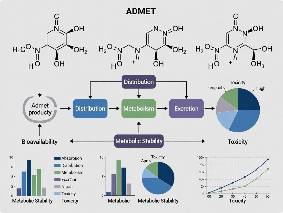
Within the context of drug discovery research, the investigation of ADMET (Absorption, Distribution, Metabolism, Excretion, and Toxicity) properties is critical for de-risking candidate compounds. Natural products, with their immense structural diversity and bioactivity, present unique challenges due to their complex chemistries and potential for unforeseen toxicities. This technical guide focuses on three pivotal early-stage toxicity screens—hepatotoxicity, cardiotoxicity (specifically hERG channel blockade), and genotoxicity—that are essential for advancing viable natural product-derived leads.
Hepatotoxicity Screening
Hepatotoxicity remains a leading cause of drug attrition and post-market withdrawal. Early screening employs both in vitro and computational methods.
Key Experimental Protocols:
- Primary Hepatocyte Assay: Isolated human or rat hepatocytes are cultured in sandwich configuration to maintain polarity and cytochrome P450 (CYP) activity. Test compounds are incubated for 24-72 hours. Viability is measured via ATP content (CellTiter-Glo) and membrane integrity via lactate dehydrogenase (LDH) release. Concurrent measurement of albumin and urea production assesses functional integrity.
- HepG2/C3A Spheroid Assay: HepG2/C3A cells are cultured to form 3D spheroids using low-adherence plates. Spheroids, which better mimic in vivo liver architecture, are treated with compounds for up to 14 days. High-content imaging analyzes nuclei count (Hoechst 33342), neutral lipid accumulation (Nile Red), and reactive oxygen species (ROS; CellROX Green).
- Mechanistic High-Content Screening (HCS): HepG2 cells are treated in 96-well plates for 24-48 hours. Cells are stained with multiparameter fluorescent probes: Hoechst 33342 (nuclei), MitoTracker Deep Red (mitochondrial mass/potential), BODIPY 493/503 (lipid droplets), and anti-γH2AX antibody (DNA damage). Automated microscopy captures images analyzed for over 20 phenotypic endpoints.
Table 1: Quantitative Endpoints in Hepatotoxicity Screening
| Endpoint | Assay/Method | Typical Threshold for Concern | Biological Significance |
|---|---|---|---|
| Cell Viability (IC50) | ATP content (CellTiter-Glo) | < 30 μM in primary hepatocytes | General cytotoxicity |
| Membrane Integrity | LDH Release | > 2-fold over vehicle control | Necrotic cell death |
| Mitochondrial Dysfunction | JC-1 Aggregate/Monomer Ratio | > 25% decrease in membrane potential | Apoptosis, energy crisis |
| Steatosis | Nile Red Intensity (HCS) | > 3-fold increase in lipid droplets | Fatty liver, metabolic disruption |
| Cholestasis | BSEP Inhibition IC50 | < 25 μM | Bile acid accumulation, intrinsic DILI risk |
| Reactive Metabolites | Glutathione (GSH) Depletion | > 50% depletion in 2h | Electrophile formation, oxidative stress |
Diagram 1: Key Pathways in Drug-Induced Liver Injury (DILI).
Cardiotoxicity: hERG Channel Blockade
Inhibition of the human Ether-à-go-go-Related Gene (hERG) potassium channel is a primary marker for drug-induced Long QT Syndrome (LQTS) and Torsades de Pointes (TdP) arrhythmia.
Key Experimental Protocols:
- Patch-Clamp Electrophysiology (Gold Standard): hERG-encoded Kv11.1 channels are expressed in mammalian cell lines (e.g., HEK293, CHO). Using whole-cell patch-clamp configuration, cells are voltage-clamped. A standard protocol depolarizes the cell to +20 mV, then repolarizes to -50 mV to elicit the characteristic hERG tail current. Concentration-dependent inhibition by the test compound is measured, generating an IC50.
- FluxOR Thallium Flux Assay: Cells stably expressing hERG are loaded with a thallium-sensitive dye. Upon addition of a test compound, a thallium-containing stimulus solution is added. Thallium influx through open hERG channels quenches dye fluorescence. Inhibitors reduce the fluorescence quenching rate, allowing medium-throughput IC50 determination.
- hERG Binding Assay (Radioligand Displacement): Cell membranes expressing hERG are incubated with a known radio-labeled hERG channel blocker (e.g., [³H]dofetilide) and increasing concentrations of the test compound. After incubation, bound and free radio-ligand are separated via filtration. Ki is calculated from the concentration causing 50% displacement (IC50) using the Cheng-Prusoff equation.
Table 2: hERG Screening Data Interpretation
| Assay Platform | Throughput | Key Measured Parameter | Safety Margin Threshold | Pros & Cons |
|---|---|---|---|---|
| Manual Patch-Clamp | Low | IC50 (current inhibition) | hERG IC50 / Cmax (free) > 30-50x | Gold standard, low throughput. |
| Automated Patch-Clamp | Medium-High | IC50 | hERG IC50 / Cmax (free) > 30-50x | Higher throughput, good fidelity. |
| Thallium Flux | High | IC50 (flux inhibition) | Used for early hazard ID, less quantitative. | High throughput, indirect measure. |
| Radioligand Binding | High | Ki (binding affinity) | Interpret with caution; functional confirm needed. | High throughput, measures direct binding. |
Diagram 2: hERG Block Link to Cardiac Arrhythmia.
Genotoxicity Screening
Genotoxicity assays identify compounds that cause genetic damage via DNA damage, mutation, or chromosomal aberrations, posing carcinogenic risk.
Key Experimental Protocols:
- Ames Test (Bacterial Reverse Mutation Assay): Salmonella typhimurium strains (e.g., TA98, TA100, TA1535, TA1537) with specific his- mutations are exposed to the test compound with and without S9 metabolic activation (rat liver homogenate). After incubation, revertant colonies (his+) are counted. A positive result is a concentration-dependent, reproducible 2-fold or greater increase in revertants over vehicle control.
- In Vitro Micronucleus (MNvit) Assay: Human lymphoblastoid TK6 cells or Chinese Hamster Lung (CHL) cells are treated with the test compound for 1.3–1.5 cell cycles (~24-28h) in the presence of cytochalasin B (which blocks cytokinesis, creating binucleated cells). Cells are harvested, fixed, and stained with DNA-specific dye (e.g., DAPI). Micronuclei (small, nuclear membrane-bound extranuclear bodies) in binucleated cells are scored manually or via flow cytometry.
- Comet Assay (Single Cell Gel Electrophoresis): Cells (e.g., TK6, HepG2) are treated, embedded in agarose on a slide, and lysed. DNA is unwalked under alkaline conditions (pH>13) and electrophoresed. Damaged DNA migrates from the nucleus, forming a "comet tail." Slides are stained with SYBR Gold and analyzed for % tail DNA or Olive Tail Moment using image analysis software.
Table 3: Core Genotoxicity Assay Battery (ICH S2(R1) Guideline)
| Assay | Endpoint | Test System | Metabolic Activation (S9) | Key Outcome |
|---|---|---|---|---|
| Ames Test | Gene Mutation | S. typhimurium & E. coli | +/- | Identifies point mutations & frameshifts. |
| In Vitro Micronucleus | Chromosomal Damage | Mammalian cells (TK6, CHL) | +/- | Identifies clastogens & aneugens (chromosome breakage/loss). |
| In Vitro Mouse Lymphoma TK | Gene Mutation & Clastogenicity | L5178Y Mouse Lymphoma cells | +/- | Detects mutations at tk locus & chromosomal events. |
Diagram 3: Genotoxicity Screening Workflow & Endpoints.
The Scientist's Toolkit: Research Reagent Solutions
Table 4: Essential Materials for Early Toxicity Screening
| Reagent / Kit / Material | Provider Examples | Primary Function in Tox Screening | |
|---|---|---|---|
| Cryopreserved Primary Human Hepatocytes | BioIVT, Lonza, Thermo Fisher | Gold-standard metabolically competent cells for hepatotoxicity & metabolic stability studies. | |
| HepG2/C3A Cell Line | ATCC | Common in vitro liver model for 2D and 3D (spheroid) hepatotoxicity assessment. | |
| CellTiter-Glo 2.0/3D | Promega | Luminescent assay for quantifying ATP as a marker of cell viability and cytotoxicity. | |
| MitoTox Complex I OXPHOS Profile Kit | Agilent Seahorse | Measures mitochondrial respiration & glycolysis in real-time to identify metabolic toxicity. | |
| hERG-HEK Cell Line | ATCC, Revvity | Stably expresses hERG channel for patch-clamp and flux-based assays. | |
| Patchliner or SyncroPatch Consumables | Nanion, Sophion | Nanion, Sophion | Consumables for automated planar patch-clamp recording of hERG and other ion channels. |
| FluxOR Thallium Influx Assay Kit | Thermo Fisher | Fluorescence-based, medium-throughput functional assay for hERG/K+ channel activity. | |
| Ames MPF 98/100 Kit | Moltox, Revvity | Miniaturized, pre-packaged Ames test using liquid micro-format, reducing test compound requirement. | |
| In Vitro MicroFlow Kit | Litron Laboratories | Flow cytometry-based in vitro micronucleus assay enabling high-speed, objective scoring. | |
| CometAssay Kit | Revvity, Trevigen | Optimized reagents and slides for performing the alkaline or neutral comet assay. | |
| Rat Liver S9 Fraction | Moltox, Thermo Fisher | Metabolic activation system containing CYPs and phase I/II enzymes for genotoxicity assays. | |
| Matrigel Matrix | Corning | Basement membrane extract for 3D cell culture, enabling hepatocyte spheroid formation. | |
| Multiparameter HCS Tox Kits | Thermo Fisher | Pre-configured dye sets for high-content imaging of mitochondrial health, oxidative stress, etc. |
Within the broader thesis on the ADMET (Absorption, Distribution, Metabolism, Excretion, and Toxicity) properties of natural products in drug discovery, a systematic approach to their early integration is paramount. Natural products (NPs) present unique challenges, including complex chemistry, limited availability, and unpredictable pharmacokinetics. This whitepaper details a stage-gate technical framework designed to incorporate ADMET evaluation at critical decision points, de-risking NP-based lead development.
The Stage-Gate Framework for Natural Products
The stage-gate process divides discovery into discrete stages separated by decision gates. ADMET data acts as a key gatekeeper.
Diagram Title: Stage-Gate Workflow for NP Discovery
Key ADMET Assays & Protocols by Stage
Stage 2: In Vitro Profiling
This stage employs medium-throughput assays to filter hits.
Table 1: Stage 2 Core ADMET Assays
| ADMET Property | Assay Name | Key Parameter Measured | Typical NP Acceptable Range | Throughput |
|---|---|---|---|---|
| Absorption | Parallel Artificial Membrane Permeability Assay (PAMPA) | Effective Permeability (Pe) | Pe (×10⁻⁶ cm/s) > 1.5 (High) | Medium-High |
| Metabolism | Microsomal Stability (Human/Rat) | Half-life (t₁/₂), % Parent Remaining | t₁/₂ > 30 min; % Remaining > 50% @ 1h | Medium |
| Toxicity | hERG Inhibition (Patch Clamp) | IC₅₀ (hERG channel) | IC₅₀ > 10 µM (Low risk) | Low |
| Toxicity | HepG2 Cell Viability (MTT) | CC₅₀ (Cytotoxicity) | CC₅₀ > 30 µM (or >10x efficacy conc.) | Medium |
| Solubility | Kinetic Solubility (Phosphate Buffer) | Solubility (µg/mL) | > 100 µg/mL in pH 7.4 buffer | High |
Protocol 3.1.1: Microsomal Stability Assay
- Objective: Determine metabolic half-life (t₁/₂) and intrinsic clearance (CLᵢₙₜ).
- Reagents: Test compound (10 mM stock in DMSO), human liver microsomes (0.5 mg/mL protein), NADPH regenerating system, phosphate buffer (pH 7.4), stop solution (acetonitrile with internal standard).
- Procedure:
- Pre-incubate microsomes and compound (1 µM final) in buffer at 37°C for 5 min.
- Initiate reaction by adding NADPH regenerating system. Final incubation volume: 100 µL.
- Aliquot 50 µL at time points: 0, 5, 15, 30, 45, 60 minutes into pre-chilled stop solution.
- Centrifuge (4000xg, 15 min, 4°C) to pellet proteins.
- Analyze supernatant via LC-MS/MS to quantify remaining parent compound.
- Plot % parent remaining vs. time. Calculate t₁/₂ using: t₁/₂ = 0.693 / k, where k is the elimination rate constant from linear regression of ln(% remaining) vs. time.
Stage 3: In Vivo Pharmacokinetics (PK)
Lead candidates undergo definitive PK studies.
Protocol 3.2.1: Rat Pharmacokinetic Study (IV/PO)
- Objective: Determine bioavailability, clearance, volume of distribution, and half-life.
- Animal Model: Male Sprague-Dawley rats (n=3 per route, ~250-300g), cannulated (jugular vein).
- Dosing: IV bolus (1 mg/kg via tail vein), PO gavage (5 mg/kg). Formulation: 5% DMSO, 10% Solutol HS-15, 85% saline (IV); 0.5% MC, 0.1% Tween 80 (PO).
- Sampling: Serial blood samples (~150 µL) pre-dose and at 2, 5, 15, 30 min, 1, 2, 4, 8, 12, 24h post-dose. Plasma separated by centrifugation (3000xg, 10 min, 4°C).
- Bioanalysis: Plasma proteins precipitated with acetonitrile. Analyze using a validated LC-MS/MS method. Generate concentration-time profiles.
- PK Analysis: Non-compartmental analysis (NCA) using WinNonlin or similar to calculate: AUC₀‑∞, Cₘₐₓ, Tₘₐₓ, CL, Vd, F (% bioavailability).
Table 2: Stage 3 Key In Vivo PK Parameters
| PK Parameter | Definition | Ideal Profile for an Oral NP Lead |
|---|---|---|
| Bioavailability (F%) | Fraction of dose reaching systemic circulation | > 20% (oral) |
| Clearance (CL) | Volume of plasma cleared per unit time | < 30% of liver blood flow |
| Volume of Distribution (Vd) | Apparent volume into which drug distributes | > 0.6 L/kg (good tissue penetration) |
| Half-life (t₁/₂) | Time for plasma concentration to halve | > 3 hours for QD/BID dosing |
| AUC₀‑∞ | Total drug exposure over time | Sufficient to cover efficacy target |
Pathways in Natural Product Metabolism & Toxicity
Understanding major metabolic pathways is critical for interpreting ADMET data.
Diagram Title: NP Metabolic Activation and Detox Pathways
The Scientist's Toolkit: Key Research Reagents & Solutions
Table 3: Essential Reagents for NP ADMET Studies
| Reagent/Solution | Supplier Examples | Primary Function in ADMET Studies |
|---|---|---|
| Human Liver Microsomes (HLM) | Corning, XenoTech, Thermo Fisher | In vitro metabolism studies (stability, metabolite ID) using CYP450 enzymes. |
| Caco-2 Cell Line | ATCC, Sigma-Aldrich | Model for predicting intestinal permeability and absorption potential. |
| Recombinant CYP Enzymes | Sigma-Aldrich, BD Biosciences | Isozyme-specific metabolism studies to identify major metabolizing enzymes. |
| hERG-Expressed Cell Line | ChanTest (Eurofins), Thermo Fisher | Screening for cardiac toxicity risk via inhibition of the hERG potassium channel. |
| NADPH Regenerating System | Promega, Cyprotex | Provides essential cofactor (NADPH) for oxidative in vitro metabolism assays. |
| Bio-Renewable Deep Well Solvents (DMSO, ACN) | Sigma-Aldrich (Milli-Q sourced) | High-purity solvents for compound storage, dilution, and LC-MS sample prep. |
| Phosphate Buffered Saline (PBS), pH 7.4 | Gibco (Thermo Fisher) | Physiological buffer for solubility, permeability, and cell-based assays. |
| Stable Isotope Labeled Internal Standards | Cambridge Isotope Labs, Clearsynth | Critical for accurate and precise quantification in LC-MS/MS bioanalysis. |
| Matrigel Basement Membrane Matrix | Corning | Used in advanced 3D cell culture models (e.g., spheroids) for hepatotoxicity screening. |
| PAMPA Plate System | pION, Corning | High-throughput tool for predicting passive transcellular permeability. |
Overcoming ADMET Liabilities: Strategic Optimization of Natural Product Leads
Within the context of modern drug discovery research focused on natural products, overcoming poor Absorption, Distribution, Metabolism, Excretion, and Toxicity (ADMET) properties is paramount. Many bioactive natural compounds, such as flavonoids, terpenoids, and alkaloids, exhibit promising therapeutic potential but are hampered by low aqueous solubility, poor membrane permeability, and/or extensive first-pass metabolism. These factors collectively result in suboptimal oral bioavailability, severely limiting their clinical translation. This technical guide details two synergistic, industrially relevant strategies to mitigate these challenges: rational prodrug design and advanced formulation engineering.
Prodrug Design: A Chemical Strategy
Prodrugs are bioreversible derivatives of active pharmaceutical ingredients (APIs), designed to transiently modify physicochemical properties. They are converted in vivo, enzymatically or chemically, to release the parent drug.
Key Functional Groups and Targeting Mechanisms
Common modifications target ionizable, hydroxyl, or carboxyl groups to alter solubility and permeability.
Table 1: Common Prodrug Linkages and Their Attributes
| Target Group | Prodrug Linkage/Strategy | Primary Goal | Typical Cleavage Mechanism |
|---|---|---|---|
| -OH / -COOH | Ester (e.g., acetate, phosphate) | Increase Lipophilicity (Permeability) or Aqueous Solubility | Esterases, Phosphatases |
| -COOH | Amino acid conjugates | Target peptide transporters | Enzymatic hydrolysis |
| Carbonyl | Schiff bases, Oximes | Improve crystalline stability | pH-dependent hydrolysis |
| General | Polymer conjugation (PEGylation) | Enhance solubility, prolong circulation | Enzymatic or hydrolytic cleavage |
| Phosphate/OH | Lipid conjugates (e.g., glycerides) | Enhance lymphatic uptake | Lipases in gut |
Experimental Protocol: In Vitro Hydrolysis Kinetics of Ester Prodrugs
Objective: To assess the chemical and enzymatic stability of a synthesized ester prodrug.
Materials:
- Prodrug and parent drug standards.
- Phosphate-buffered saline (PBS, pH 7.4) and simulated gastric fluid (SGF, pH 1.2).
- Liver microsomes or S9 fractions from relevant species (e.g., human, rat).
- Analytical method (HPLC or LC-MS) with validated calibration curves.
Procedure:
- Chemical Stability: Prepare prodrug solution (e.g., 10 µM) in PBS and SGF. Incubate at 37°C.
- Enzymatic Stability: Prepare incubation mixture containing liver microsomes (0.5 mg protein/mL) in PBS with NADPH regenerating system (for phase I) or without (for esterase activity). Add prodrug.
- Sampling: At predetermined time points (0, 5, 15, 30, 60, 120 min), withdraw aliquots and immediately quench with an equal volume of acetonitrile containing internal standard to precipitate proteins.
- Analysis: Centrifuge, analyze supernatant by HPLC/LC-MS to quantify remaining prodrug and appearance of parent drug.
- Data Analysis: Calculate half-life (t₁/₂) of prodrug disappearance and rate constant (k).
Title: Prodrug Candidate Screening and Evaluation Workflow
Formulation Approaches: A Physical and Physicochemical Strategy
When chemical modification is not feasible, advanced formulations can enhance solubility and dissolution rate.
Key Formulation Technologies
Table 2: Formulation Strategies for Solubility/Bioavailability Enhancement
| Technology | Mechanism | Typical Particle Size | Key Excipients/Components |
|---|---|---|---|
| Amorphous Solid Dispersions (ASD) | Creates high-energy amorphous state, inhibits recrystallization | N/A (Molecular dispersion) | Polymers: HPMC-AS, PVP-VA, Soluplus |
| Lipid-Based Drug Delivery Systems (LBDDS) | Maintains drug in solubilized state in GI tract, promotes lymphatic uptake | 20-200 nm (emulsions) | Oils (Labrafil), Surfactants (Gelucire), Co-solvents |
| Nanosuspensions | Increases surface area for dissolution via nanoparticle milling | 200-800 nm | Stabilizers: Poloxamer 188, HPMC, SLS |
| Cyclodextrin Complexation | Forms inclusion complexes, masks hydrophobic moieties | Molecular complex | Cyclodextrins (SBE-β-CD, HP-β-CD) |
| Self-Emulsifying Drug Delivery Systems (SEDDS) | Forms fine emulsion upon mild agitation in GI tract | 100-300 nm | Oil, Surfactant, Co-surfactant/Co-solvent |
Experimental Protocol: Preparation and Characterization of a Nanosuspension via Wet Media Milling
Objective: To produce a stable nanosuspension of a poorly soluble natural product.
Materials:
- Poorly water-soluble API.
- Stabilizer(s) (e.g., Poloxamer 188, D-α-Tocopheryl polyethylene glycol 1000 succinate - TPGS).
- Zirconia milling beads (0.3-0.5 mm diameter).
- High-energy media mill.
- Laser diffraction particle size analyzer, dynamic light scattering (DLS) instrument.
Procedure:
- Pre-mix: Disperse the API (e.g., 10% w/w) and stabilizer(s) in purified water using a high-shear mixer to form a coarse pre-suspension.
- Milling: Charge the milling chamber with zirconia beads (bead loading: 50-70% chamber volume). Circulate the pre-suspension through the mill at a controlled flow rate. Mill for a target time (e.g., 60-120 min) or until the particle size plateaus. Maintain temperature control (e.g., < 40°C).
- Separation: Separate the milled nanosuspension from the beads using a sieve.
- Characterization:
- Particle Size & PDI: Measure by DLS (Z-average, PDI).
- Crystallinity: Assess via Powder X-Ray Diffraction (PXRD) to detect potential amorphization.
- Saturation Solubility: Filter nanosuspension, dilute, and quantify dissolved API by HPLC vs. coarse suspension.
- Stability: Monitor physical stability (particle growth, aggregation) at 4°C and 25°C over 4 weeks.
Title: Nanosuspension Manufacturing and Characterization Process
The Scientist's Toolkit: Key Research Reagent Solutions
Table 3: Essential Materials for Solubility and Bioavailability Studies
| Reagent/Material | Supplier Examples | Primary Function in Research |
|---|---|---|
| Caco-2 Cell Line | ATCC, ECACC | Model for predicting human intestinal permeability and efflux transport. |
| Liver Microsomes (Human, Rat) | Corning, XenoTech, BioIVT | Assessment of metabolic stability and identification of phase I metabolism pathways. |
| Soluplus | BASF | Amphiphilic polymer for forming stable amorphous solid dispersions via hot-melt extrusion. |
| SBE-β-CD (Sulfobutylether-β-Cyclodextrin) | Ligand Pharmaceuticals | Anionic, highly soluble cyclodextrin for forming inclusion complexes, often used in parenteral formulations. |
| Labrafil M 1944 CS | Gattefossé | Oleoyl polyoxyl-6 glycerides; a commonly used non-ionic surfactant and oil component in LBDDS and SEDDS. |
| Poloxamer 188 (Pluronic F-68) | BASF | Non-ionic triblock copolymer surfactant used as a stabilizer in nanosuspensions and emulsions. |
| Simulated Intestinal Fluids (FaSSIF/FeSSIF) | Biorelevant.com | Biorelevant media for predictive in vitro dissolution testing, containing bile salts and phospholipids. |
| Chromatographic Columns (C18, for LC-MS) | Waters, Agilent, Phenomenex | Essential for analytical quantification of drug and metabolite concentrations in complex biological matrices. |
For natural products with challenging ADMET profiles, an integrated strategy is most promising. Initial screening should assess whether the molecule is amenable to prodrug design (presence of modifiable functional group) or if formulation is preferable. Often, a combined approach—such as a prodrug formulated within a lipid-based system—can yield synergistic benefits. The choice of strategy must be guided by the specific physicochemical and metabolic liabilities of the lead natural compound, with the ultimate goal of achieving sufficient systemic exposure for therapeutic efficacy in clinical trials.
Within the broader thesis on optimizing the Absorption, Distribution, Metabolism, Excretion, and Toxicity (ADMET) properties of natural products for drug discovery, this guide focuses on the critical chemical strategies of semi-synthesis and analog development. The primary objective is to retain or enhance the inherent bioactivity of a natural product scaffold while systematically improving its drug-like properties. Natural products often possess complex structures with optimal activity but suffer from poor solubility, metabolic instability, or toxicity. Strategic structural modification serves as the bridge between potent lead identification and viable clinical candidate development.
Core Principles of Activity-Preserving Modification
The key to successful modification lies in identifying the Pharmacophore—the precise stereochemical and electronic arrangement of functional groups responsible for biological activity. Modifications are directed away from this core region. The process typically follows these steps:
- Structure-Activity Relationship (SAR) Elucidation: Systematic modification and testing to map critical vs. tolerant regions of the molecule.
- ADMET-Driven Design: Targeting modifications to specific molecular properties (e.g., logP, hydrogen bond donors/acceptors, rotatable bonds) predicted to improve pharmacokinetics.
- Synthetic Feasibility: Employing semi-synthesis from the natural isolable core or developing concise total synthesis routes for analog production.
Semi-Synthetic Methodologies
Semi-synthesis leverages the complex natural product as a starting material for chemical diversification.
Key Functional Group Transformations
These transformations must be chemoselective to avoid altering the pharmacophore.
Protocol: Selective Acylation of a Polyhydroxylated Natural Product (e.g., Macrolide Antibiotics)
- Objective: To improve metabolic stability by acylating a metabolically labile hydroxyl group without affecting others.
- Materials: Natural product substrate (e.g., Erythromycin A), acylating agent (e.g., acetic anhydride), catalyst (4-Dimethylaminopyridine, DMAP), base (triethylamine), anhydrous dichloromethane (DCM).
- Procedure:
- Dissolve the natural product (1.0 equiv) and DMAP (0.1 equiv) in anhydrous DCM under inert atmosphere (N₂ or Ar) at 0°C.
- Add triethylamine (1.2 equiv) dropwise.
- Slowly add the acylating agent (1.05 equiv) in DCM via syringe pump over 30 minutes.
- Allow the reaction to warm to room temperature and stir for 12 hours (monitor by TLC/LCMS).
- Quench with saturated aqueous NaHCO₃ solution.
- Extract with DCM (3x), dry the combined organic layers over anhydrous MgSO₄, filter, and concentrate in vacuo.
- Purify the crude product by flash column chromatography.
Chemo- and Regioselective Protection/Deprotection
Essential for modifying complex molecules with multiple similar functional groups.
Protocol: Silyl Protection of a Sterically Hindered Hydroxyl Group
- Objective: To protect a secondary hydroxyl group in the presence of a primary hydroxyl for subsequent modification of the primary site.
- Materials: Substrate, tert-Butyldimethylsilyl chloride (TBSCl), imidazole, anhydrous N,N-Dimethylformamide (DMF).
- Procedure:
- Dissolve the substrate (1.0 equiv) and imidazole (3.0 equiv) in anhydrous DMF at 0°C.
- Add TBSCl (1.2 equiv) portionwise.
- Stir at 0°C for 2-4 hours (monitor by LCMS).
- Dilute with ethyl acetate and wash with water and brine.
- Dry the organic layer over Na₂SO₄, filter, and concentrate.
- Purify by flash chromatography.
Analog Development via Synthetic Manipulation
This involves more profound changes, often creating novel scaffolds inspired by the natural product.
Protocol: Suzuki-Miyaura Cross-Coupling for Biaryl Analog Generation
- Objective: Introduce aromatic diversity to explore π-stacking interactions in the binding pocket.
- Materials: Aryl halide-functionalized natural product derivative, boronic acid/ester, palladium catalyst (e.g., Pd(PPh₃)₄), base (e.g., K₂CO₃), solvent (mixture of toluene/ethanol/water).
- Procedure:
- Charge a microwave vial with the aryl halide (1.0 equiv), boronic acid (1.5 equiv), Pd(PPh₃)₄ (0.05 equiv), and K₂CO₃ (2.0 equiv).
- Add degassed solvent mixture (toluene:EtOH:H₂O, 4:2:1).
- Seal the vial and heat at 80-100°C for 1-2 hours under microwave irradiation or conventional heating.
- Cool, filter through a celite pad, and concentrate.
- Purify the crude product by preparative HPLC.
Quantitative ADMET Data from Modified Analogs
The success of structural modification is quantified by comparing key parameters of the parent natural product (NP) and its analogs.
Table 1: Comparative ADMET Profile of a Hypothetical Natural Product and Its Semi-Synthetic Analogs
| Compound ID | Core Modification | In vitro IC₅₀ (nM) | LogP (Predicted) | Aqueous Solubility (µg/mL) | Metabolic Stability (HLM t₁/₂, min) | CYP3A4 Inhibition (IC₅₀, µM) |
|---|---|---|---|---|---|---|
| NP-1 | Parent Structure | 10 ± 2 | 5.8 | <1 | 15 | 2.5 |
| ANA-127 | C6-OH Acylation | 12 ± 3 | 6.1 | <1 | 45 | 5.8 |
| ANA-254 | Glycoside Removal | 15 ± 4 | 4.2 | 25 | >120 | >50 |
| ANA-308 | Alkyne Spacer Insertion | 8 ± 1 | 5.0 | 5 | 90 | >50 |
Table 2: Key Research Reagent Solutions for Semi-Synthesis & ADMET Screening
| Reagent / Material | Function in Research | Technical Note |
|---|---|---|
| Immobilized Enzymes (e.g., Candida antarctica Lipase B) | Chemoselective biocatalytic acylation/hydrolysis. | Enables reactions under mild, green chemistry conditions without protecting groups. |
| Human Liver Microsomes (HLMs) | In vitro assessment of Phase I metabolic stability. | Used with NADPH cofactor; t₁/₂ value indicates susceptibility to oxidative metabolism. |
| Caco-2 Cell Line | Model for predicting intestinal permeability and absorption. | Measures apparent permeability (Papp); correlates with human oral absorption. |
| Solubility-Enhancing Excipients (e.g., TPGS, HPMC) | Formulation aids for in vivo testing of poorly soluble analogs. | Allows pharmacokinetic studies of analogs with suboptimal solubility, informing further design. |
| Chiral HPLC Columns (e.g., Chiralpak IA/IB/IC) | Separation and analysis of stereoisomers from asymmetric synthesis. | Critical for ensuring stereochemical purity, as activity is often highly stereospecific. |
Visualization of Workflows and Pathways
Title: Analog Development Workflow from Natural Product
Title: ADMET-Target Interaction Balance for Drug Efficacy
Mitigating Metabolic Instability and Drug-Drug Interaction Risks
1. Introduction: The Natural Product ADMET Paradox
Within the paradigm of modern drug discovery, natural products (NPs) remain a prolific source of novel pharmacophores, particularly for challenging therapeutic targets. However, their integration into the development pipeline is frequently hampered by complex and often unfavorable ADMET (Absorption, Distribution, Metabolism, Excretion, and Toxicity) profiles. Two of the most critical ADMET-related challenges are metabolic instability and the potential for drug-drug interactions (DDIs). Metabolic instability leads to poor oral bioavailability and short half-life, necessitating frequent or high dosing. Concurrently, NPs and their semi-synthetic derivatives can inhibit or induce drug-metabolizing enzymes (DMEs) and transporters, posing significant DDI risks that can compromise the safety and efficacy of co-administered therapies. This whitepaper provides an in-depth technical guide on experimental strategies to identify, characterize, and mitigate these risks early in the discovery process, specifically within the context of NP-based drug discovery.
2. Quantitative Landscape of NP Metabolism and DDI Risks
A review of recent literature and regulatory submissions highlights the prevalence of these issues. The following table summarizes key quantitative data on the involvement of major cytochrome P450 (CYP) enzymes in NP metabolism and their DDI potential.
Table 1: Metabolic Fate and DDI Potential of Select Natural Product Scaffolds
| Natural Product Class/Scaffold | Primary Metabolizing CYP(s) | Reported Half-life (in vitro, human) | Inhibition Potential (IC50, μM) | Induction Potential (Fold-Change vs Control) |
|---|---|---|---|---|
| Flavonoids (e.g., Chrysin) | 1A2, 2C9, 3A4 | <30 min (hepatocytes) | CYP3A4: 15-25 | N/A |
| Alkaloids (e.g., Berberine) | 2D6, 3A4 | ~120 min | CYP2D6: 3.8; CYP3A4: 8.2 | Moderate (PXR activation) |
| Terpenoids (e.g., Triptolide) | 3A4 | <20 min (microsomes) | CYP3A4: <1.0 (Time-dependent) | Significant (CYP3A4 mRNA ↑ 4-5 fold) |
| Coumarins (e.g., Imperatorin) | 1A2, 2A6 | ~45 min | CYP1A2: 0.8 | Weak |
3. Core Experimental Methodologies
3.1. Assessing Metabolic Stability
Protocol: Intrinsic Clearance Assay in Human Liver Microsomes (HLM)
- Objective: Determine the in vitro half-life (t1/2) and intrinsic clearance (CLint) of an NP.
- Reagents: Test compound (1 µM), pooled HLM (0.5 mg/mL), NADPH-regenerating system, phosphate buffer (pH 7.4), and an appropriate organic solvent for quenching (e.g., acetonitrile with internal standard).
- Procedure:
- Pre-incubate HLM with test compound in buffer at 37°C for 5 min.
- Initiate reaction by adding NADPH-regenerating system.
- Aliquot samples at 8 time points (e.g., 0, 5, 15, 30, 45, 60, 90, 120 min).
- Quench aliquots immediately with cold organic solvent.
- Centrifuge, analyze supernatant via LC-MS/MS to determine parent compound depletion.
- Plot natural log of percentage remaining versus time. Calculate in vitro t1/2: t1/2 = 0.693 / k, where k is the elimination rate constant. Calculate CLint = (0.693 / t1/2) * (Incubation Volume / Microsomal Protein).
3.2. Enzyme Reaction Phenotyping
Protocol: Chemical Inhibition in HLM with Isoform-Specific Inhibitors
- Objective: Identify the specific CYP enzymes responsible for the metabolism of an NP.
- Reagents: Test compound, HLM, NADPH, selective chemical inhibitors (e.g., Furafylline for CYP1A2, Sulfaphenazole for CYP2C9, Quinidine for CYP2D6, Ketoconazole for CYP3A4).
- Procedure:
- Pre-incubate HLM with individual inhibitors at their isoform-selective concentrations for 15 min.
- Add test compound and NADPH to initiate reaction.
- Incubate for a time point within the linear range (determined from stability assay).
- Quench and analyze for metabolite formation or parent depletion.
- The percent inhibition of metabolite formation by a specific inhibitor indicates the contribution of that CYP isoform.
3.3. Evaluating DDI Potential: Inhibition
Protocol: Reversible CYP Inhibition (IC50 Determination)
- Objective: Quantify the potency of an NP to inhibit major CYP enzymes.
- Reagents: CYP-specific probe substrates (e.g., Phenacetin for 1A2, Diclofenac for 2C9, Bupropion for 2B6, Testosterone for 3A4), HLM, NADPH, test compound at 8 concentrations.
- Procedure:
- Incubate HLM with probe substrate and varying concentrations of test compound.
- Initiate with NADPH and incubate for a linear time.
- Quantify the formation of the specific probe metabolite via LC-MS/MS.
- Plot metabolite formation rate (%) versus log[inhibitor]. Fit data to a sigmoidal curve to calculate IC50.
Protocol: Time-Dependent Inhibition (TDI) Assessment
- Objective: Determine if an NP is a mechanism-based inactivator, a high-risk DDI profile.
- Reagents: As above, with pre-incubation and dilution steps.
- Procedure:
- Pre-incubation: Incubate HLM with test compound (with/without NADPH) for 30 min.
- Dilution: Dilute the mixture 10-fold into a secondary incubation containing NADPH and probe substrate.
- Compare activity (metabolite formation) in samples pre-incubated with NADPH to those without. A significant loss of activity only in the NADPH-containing pre-incubation indicates TDI.
3.4. Evaluating DDI Potential: Induction
Protocol: Nuclear Receptor Activation (Reporter Gene Assay)
- Objective: Assess the potential of an NP to induce DMEs via PXR or AhR receptor activation.
- Reagents: Cell line (e.g., HepG2) transfected with a plasmid containing the receptor's ligand-binding domain linked to a luciferase reporter gene.
- Procedure:
- Seed cells in 96-well plates.
- Treat with test compound, positive control (e.g., Rifampicin for PXR), and vehicle for 24-48h.
- Lyse cells and measure luciferase activity. Fold-induction over vehicle indicates receptor activation potential.
4. Visualization of Key Pathways and Workflows
Title: First-Pass Metabolism and DDI Origin of Natural Products
Title: Integrated DDI Risk Assessment Experimental Workflow
5. The Scientist's Toolkit: Key Research Reagent Solutions
Table 2: Essential Reagents for Metabolic & DDI Studies
| Reagent / Material | Function & Application | Key Consideration |
|---|---|---|
| Pooled Human Liver Microsomes (HLM) | Source of major CYP enzymes for in vitro metabolism and inhibition studies. | Use pools from ≥20 donors for representative activity. Gender-balanced pools are ideal. |
| CYP-Specific Probe Substrates & Inhibitors | To identify metabolizing enzymes (phenotyping) and measure inhibition potency (IC50). | Verify selectivity and use at recommended concentrations to avoid non-specific effects. |
| NADPH Regenerating System | Provides essential cofactor (NADPH) for CYP-mediated oxidative reactions. | Critical for maintaining linear reaction kinetics; use fresh or frozen aliquots. |
| Recombinant Human CYP Isozymes (rCYP) | Confirm the role of a specific CYP in metabolite formation. | Useful for phenotyping confirmation but lacks native microsomal membrane context. |
| Cryopreserved Human Hepatocytes | Gold-standard system for integrated metabolism, inhibition, and induction studies. | Must check viability and plateability; suitable for enzyme induction assays. |
| LC-MS/MS System | Quantitative analysis of parent drug depletion and metabolite formation with high sensitivity. | Requires optimization of MRM (Multiple Reaction Monitoring) transitions for each analyte. |
| Reporter Gene Assay Kits (PXR, AhR) | Assess nuclear receptor activation potential, predicting enzyme induction. | Choose cell lines with low background and high responsiveness to standard inducers. |
6. Mitigation Strategies and Conclusion
Upon identifying metabolic liabilities and DDI risks, mitigation strategies can be deployed. These include:
- Structural Modification: Blocking or altering metabolically labile sites (e.g., replacing a labile methylenedioxy group) to improve stability and reduce reactive metabolite formation.
- Prodrug Design: Masking polar groups to improve absorption, with enzymatic cleavage designed to occur after absorption.
- Formulation Strategies: Using inhibitors of gut metabolism (e.g., co-administering with piperine) or advanced delivery systems (liposomes, nanoparticles) to enhance bioavailability.
- Clinical DDI Management: If the NP is indispensable, designing clinical DDI studies and prescribing guidelines (contraindications, dose adjustments) for safe co-administration.
In conclusion, the successful translation of natural products into viable drugs necessitates a rigorous, front-loaded ADMET screening strategy. By implementing the integrated experimental framework outlined here—encompassing quantitative metabolic stability assays, comprehensive enzyme phenotyping, and detailed DDI risk assessment—researchers can proactively identify and mitigate metabolic and DDI liabilities. This systematic approach is essential to de-risk the development of NP-derived therapeutics, ensuring they meet the stringent safety and efficacy standards required for modern medicines.
Natural products (NPs) are a cornerstone of drug discovery, renowned for their structural complexity and potent bioactivity. However, their intricate scaffolds frequently present significant ADMET (Absorption, Distribution, Metabolism, Excretion, and Toxicity) challenges that can derail development. The very features that confer potent target engagement—reactive functional groups, high lipophilicity, or complex stereochemistry—can also be the source of toxicity flags, such as off-target reactivity, metabolic instability, or idiosyncratic hepatotoxicity. This whitepaper, framed within the critical evaluation of NP ADMET properties, provides a technical guide for systematically identifying and mitigating these toxicity flags to de-risk promising NP-derived scaffolds.
Key Toxicity Flags and Associated Mechanisms in NP Scaffolds
Understanding the mechanistic underpinnings of toxicity is the first step in de-risking. Common liabilities associated with NP scaffolds are summarized below.
Table 1: Common Toxicity Flags in Natural Product Scaffolds and Their Mechanisms
| Toxicity Flag | Common NP Structural Alerts | Potential Mechanisms | Primary Assays for Detection |
|---|---|---|---|
| Cytotoxicity (Non-Specific) | High LogP (>5), Michael acceptors, epoxides, polyaromatic systems | Membrane disruption, non-specific protein alkylation, oxidative stress | MTT/WST-1 cell viability, LDH release, hemolysis assay |
| Mitochondrial Toxicity | Cationic amphiphilicity, uncouplers (phenolic OH), rotenone-like motifs | Inhibition of ETC complexes (I-V), uncoupling of oxidative phosphorylation, mitochondrial membrane depolarization | Seahorse XF Analyzer (OCR/ECAR), JC-1/TMRM staining for ΔΨm |
| hERG Channel Inhibition | Basic amines, lipophilic aromatic/heterocyclic moieties, large flexible scaffolds | Blockade of the pore domain (KV11.1), disrupted cardiac repolarization | Patch-clamp electrophysiology, FLIPR-based thallium flux assay |
| Genotoxicity | Aflatoxin-like furans, alkylating agents, intercalating planar polycycles | DNA adduct formation, intercalation, topoisomerase inhibition, aneugenicity | Ames test, in vitro micronucleus assay, Comet assay |
| Idiosyncratic Hepatotoxicity | Bioactivation to reactive metabolites (e.g., quinones, iminoquinones) | Hapten formation, immune-mediated injury, direct mitochondrial insult | GSH-trapping assay (LC-MS/MS), CYP450 time-dependent inhibition (TDI) assay, hepatocyte co-culture models |
| Phospholipidosis | Cationic amphiphilic structures (primary/secondary amines + lipophilic groups) | Lysosomal phospholipid accumulation, impaired lipid degradation | HCS (High Content Screening) with phospholipid dyes (e.g., HCS LipidTOX) |
Experimental Protocols for Toxicity Profiling
3.1. Protocol: Reactive Metabolite Screening via Glutathione (GSH) Trapping Assay
- Objective: To identify scaffolds prone to metabolic activation to electrophilic, potentially toxic intermediates.
- Materials: Test compound, human liver microsomes (HLM, 0.5 mg/mL), NADPH regeneration system, 5 mM GSH (or stable isotope-labeled GSH for MS), control compounds (e.g., clozapine), LC-MS/MS system.
- Method:
- Prepare incubation mixture (final 200 µL): 50 mM phosphate buffer (pH 7.4), HLM, 1 µM test compound, 5 mM GSH.
- Pre-incubate for 5 min at 37°C.
- Initiate reaction by adding NADPH regeneration system (1 mM NADP+, 3.3 mM glucose-6-phosphate, 0.4 U/mL G6PDH).
- Incubate for 60 min at 37°C.
- Terminate reaction with 200 µL ice-cold acetonitrile.
- Vortex, centrifuge (13,000xg, 10 min), and analyze supernatant by LC-MS/MS.
- Monitor for characteristic neutral losses of 129 Da (pyroglutamic acid) and 307 Da (dehydroglutathione) from GSH adducts ([M+H]⁺ → m/z corresponding to [M+GSH+H]⁺).
3.2. Protocol: High-Content Screening for Mitochondrial Toxicity
- Objective: To simultaneously assess mitochondrial membrane potential and cell health.
- Materials: HepG2 or primary hepatocytes, 96-well imaging plates, test compound, JC-1 dye (or TMRM), Hoechst 33342, HCS-capable fluorescence microscope.
- Method:
- Seed cells and treat with compound for 24-48 hours.
- Load cells with JC-1 dye (2 µg/mL) and Hoechst 33342 (nuclear stain) for 30 min at 37°C.
- Wash with PBS and acquire images.
- Analysis: In healthy cells, JC-1 forms red fluorescent aggregates (high ΔΨm). In depolarized cells, it remains as green fluorescent monomers. Calculate the Red/Green fluorescence intensity ratio per cell. A significant decrease indicates mitochondrial depolarization. Correlate with nuclear count (viability) from Hoechst channel.
Strategic Mitigation: From Alert to Solution
Once a toxicity flag is identified, strategic medicinal chemistry interventions can be employed.
Table 2: De-risking Strategies for Specific Toxicity Flags
| Toxicity Flag | Strategic Intervention | Rationale & Expected Outcome |
|---|---|---|
| Reactive Metabolite Formation | Bioisosteric Replacement: Swap metabolically labile groups (e.g., methylenedioxyphenyl → substituted benzodioxole). Blocking/Deactivating Metabolism: Introduce fluorine atom ortho to a site of hydroxylation. | Eliminates or reduces formation of electrophilic quinone/iminoquinone species, lowering covalent binding risk. |
| hERG Inhibition | Reduce Lipophilicity (cLogP): Incorporate polar groups (amide, alcohol), reduce aromaticity. Introduce Negative LogD at pH 7.4: Add acidic groups (e.g., carboxylic acid) to discourage cationic drug from pore channel. Reduce pKa of Basic Amines: Fluorine adjacent to amine lowers pKa. | Decreases affinity for the lipophilic pore cavity and cationic interaction with key residues (Tyr652, Phe656). |
| Mitochondrial Toxicity / Phospholipidosis | Attenuate Cationic Amphiphilicity: Reduce pKa of amine below 8, introduce steric hindrance, or replace amine with neutral polar group. Reduce LogD: Lower overall lipophilicity. | Disrupts the structural motif required for lysosomotropism and mitochondrial accumulation. |
| General Cytotoxicity (High LogP) | Improve Solubility & Reduce LogP: Introduce solubilizing chains (PEG), ionizable groups, or cyclize flexible structures. | Lowers non-specific membrane partitioning and improves therapeutic index. |
The Scientist's Toolkit: Key Research Reagent Solutions
| Item / Reagent | Function & Application |
|---|---|
| Cryopreserved Human Hepatocytes (Metabolic & Tox Studies) | Gold standard for evaluating metabolism, metabolite identification, and hepatotoxicity in a physiologically relevant cell system. |
| hERG-Expressing Cell Lines (e.g., HEK293-hERG) | Used in automated patch-clamp or flux-based assays for specific, high-throughput cardiac safety screening. |
| Seahorse XF Analyzer Kits (e.g., Mito Stress Test) | Provides real-time, label-free measurement of Oxygen Consumption Rate (OCR) and Extracellular Acidification Rate (ECAR) to profile mitochondrial function. |
| GSH & KCN Trapping Reagents | Used in microsomal incubations to "trap" reactive electrophilic metabolites (soft and hard electrophiles, respectively) for LC-MS/MS identification. |
| High-Content Imaging Kits (e.g., for Mitochondrial Membrane Potential, Phospholipidosis) | Multiplexed, image-based assays allowing single-cell resolution analysis of multiple toxicity endpoints simultaneously. |
| Supersomes (Individual Recombinant CYP450 Isozymes) | Pinpoint the specific cytochrome P450 enzyme responsible for metabolic activation, guiding targeted structure modification. |
Visualizing the De-risking Workflow
Toxicity De-risking Decision Pathway
Reactive Metabolite Formation & Mitigation
The Role of Natural Product Pharmacokinetics in Dose Regimen Design
Within the broader thesis on the Absorption, Distribution, Metabolism, Excretion, and Toxicity (ADMET) properties of natural products in drug discovery research, pharmacokinetics (PK) serves as the critical bridge between in vitro activity and in vivo efficacy. For natural products—a class encompassing plant secondary metabolites, marine organisms, microbial fermentation products, and their semi-synthetic derivatives—PK parameters are uniquely complex due to inherent physicochemical diversity, the presence of prodrugs or synergistic components, and atypical metabolic pathways. This whitepaper details the core principles, methodologies, and contemporary data underpinning the use of natural product pharmacokinetics to rationally design safe and effective dose regimens, moving beyond empirical dosing strategies.
Core Pharmacokinetic Parameters & Their Determinants for Natural Products
The design of any dose regimen (dose size, dosing interval, route) is fundamentally based on key PK parameters. For natural products, these parameters are influenced by specific ADMET challenges.
Table 1: Core PK Parameters and Their Natural Product-Specific Determinants
| PK Parameter | Symbol | Definition | Key Determinants in Natural Products | Impact on Regimen Design |
|---|---|---|---|---|
| Bioavailability | F | Fraction of dose reaching systemic circulation unchanged. | Low solubility, instability in GI tract, first-pass metabolism (esp. by CYP3A4, UGTs), efflux by P-glycoprotein. | Determines required oral dose; may necessitate prodrug design or alternative routes (IV, SL). |
| Volume of Distribution | Vd | Apparent volume into which a drug distributes. | Plasma protein binding (e.g., to albumin), lipophilicity, affinity for tissue transporters (e.g., OATPs). | High Vd may indicate extensive tissue penetration but slow elimination; influences loading dose. |
| Clearance | CL | Volume of plasma cleared of drug per unit time. | Metabolism (Phase I/II), biliary excretion (often via MRP2), renal excretion (active secretion). | The primary determinant of maintenance dose and dosing interval (τ). |
| Elimination Half-life | t1/2 | Time for plasma concentration to reduce by 50%. | Proportional to Vd and inversely proportional to CL. | Directly dictates dosing frequency (τ ≈ 1-2 x t1/2 for stable exposure). |
| Peak Concentration | Cmax | Maximum plasma concentration after dosing. | Rate of absorption (ka), bioavailability (F), dose. | Must be below toxic threshold; critical for concentration-dependent antimicrobials. |
| Trough Concentration | Cmin | Minimum plasma concentration before next dose. | Clearance, dosing interval, volume of distribution. | Must remain above minimum effective concentration (MEC) for time-dependent drugs. |
Methodological Toolkit: Experimental Protocols for PK Characterization
Accurate PK parameter estimation requires standardized in vitro and in vivo experiments.
In VitroADME Screening Protocol
Objective: To predict in vivo PK behavior and identify potential liabilities early. Workflow:
- Solubility & Chemical Stability: Shake-flask method in biorelevant media (FaSSIF, FeSSIF) across physiological pH range (1.2-7.4). Analyze by HPLC-UV at 0, 2, 8, 24h.
- Permeability: Caco-2 cell monolayer assay. Seed cells on transwell inserts, culture for 21 days. Apply test compound apically (A) and basolaterally (B). Sample at 0, 30, 60, 90, 120 min. Calculate apparent permeability (Papp) and efflux ratio (Papp B→A/Papp A→B).
- Metabolic Stability: Incubate compound (1 µM) with human liver microsomes (0.5 mg/mL) or hepatocytes (1x10^6 cells/mL) in NADPH-regenerating system at 37°C. Aliquot at 0, 5, 15, 30, 60 min. Analyze parent compound remaining. Calculate intrinsic clearance (CLint).
- Cytochrome P450 Inhibition/Induction: CYP450 isoform-specific probe assays (e.g., phenacetin for CYP1A2, midazolam for CYP3A4) to assess direct/mechanism-based inhibition or mRNA quantification (qRT-PCR) in hepatocytes after 48-72h exposure for induction potential.
In Vitro ADME Screening Workflow for Natural Products
In VivoPreclinical PK Study Protocol (Rodent)
Objective: To determine fundamental PK parameters after intravenous (IV) and oral (PO) administration. Procedure:
- Animal Preparation: Cannulate jugular vein (for serial sampling) and/or femoral vein (for IV dosing) in rats (n=6/route). Fast overnight (water ad libitum).
- Dosing & Sampling: Administer compound intravenously (e.g., 1 mg/kg in 10% DMSO/90% saline) via femoral vein. For oral, administer by gavage (e.g., 10 mg/kg in 0.5% MC suspension). Collect serial blood samples (e.g., 0.083, 0.25, 0.5, 1, 2, 4, 6, 8, 12, 24h post-dose) into EDTA tubes via jugular cannula.
- Bioanalysis: Centrifuge blood immediately (4°C, 2000xg, 10 min). Analyze plasma using a validated LC-MS/MS method.
- PK Analysis: Fit plasma concentration-time data using non-compartmental analysis (NCA) software (e.g., Phoenix WinNonlin) to calculate: AUC0-∞, CL, Vd, t1/2 (IV); Cmax, Tmax, AUC0-∞, and absolute bioavailability F = (AUCPO/DosePO) / (AUCIV/DoseIV) (PO).
The Scientist's Toolkit: Key Research Reagent Solutions
Table 2: Essential Materials for Natural Product PK Studies
| Category | Item/Reagent | Function & Relevance |
|---|---|---|
| In Vitro Systems | Caco-2 Cell Line | Gold-standard model for predicting human intestinal permeability and efflux transporter effects. |
| Cryopreserved Human Hepatocytes | For assessing phase I/II metabolism, metabolic stability, and enzyme induction in a physiologically relevant system. | |
| Human Liver Microsomes (HLM) | Cost-effective system for high-throughput metabolic stability and CYP450 inhibition screening. | |
| Assay Kits | P450-Glo CYP450 Assay | Luminescence-based high-throughput assay for CYP450 inhibition using isoform-specific substrates. |
| BCA Protein Assay Kit | Quantifies protein concentration in microsomal/hepatocyte preparations for normalizing activity data. | |
| Bioanalytical | Stable Isotope-Labeled Internal Standards (e.g., ^13C, ^2H) | Critical for accurate LC-MS/MS quantitation, correcting for matrix effects and recovery variability. |
| Biorelevant Dissolution Media (FaSSIF, FeSSIF) | Simulates fasted and fed state intestinal fluids for realistic solubility and dissolution testing. | |
| Software | Phoenix WinNonlin | Industry-standard software for non-compartmental and compartmental PK/PD modeling. |
| GastroPlus/Simcyp Simulator | Physiologically-based pharmacokinetic (PBPK) modeling platforms to predict human PK and optimize first-in-human doses. |
From PK Parameters to Regimen Design: Key Considerations
The target steady-state exposure is defined by the therapeutic index (TI = Toxic Dose / Effective Dose). The regimen is calculated to maintain plasma concentrations within this window.
For a desired average steady-state concentration (Css,avg):
- Maintenance Dose (D) = (Css,avg • CL • τ) / F
- Loading Dose (DL) = D / (1 - e-k•τ) ≈ D / (1 - e-0.693•τ/t1/2) (if t1/2 is long relative to τ)
Table 3: Natural Product Case Studies in PK-Driven Dosing
| Natural Product (Derivative) | Source | PK Challenge | PK-Informed Regimen Design Solution |
|---|---|---|---|
| Paclitaxel | Taxus brevifolia (Pacific Yew) | Nonlinear PK due to saturable distribution and elimination; poor aqueous solubility. | Administration as albumin-bound nanoparticles (nab-paclitaxel) alters distribution, enabling higher, more frequent dosing (e.g., 100-150 mg/m² weekly) with improved efficacy/safety. |
| Artemisinin (Artesunate) | Artemisia annua (Sweet Wormwood) | Short half-life (<1h), auto-induction of metabolism. | High initial loading dose (IV artesunate) in severe malaria, followed by multiple daily doses or transition to a longer-acting partner drug (ACT) for cure. |
| Curcumin | Curcuma longa (Turmeric) | Extremely low bioavailability due to poor solubility, rapid metabolism, and excretion. | Development of phospholipid complexes (Meriva), nanoparticles, or piperine co-administration (inhibits glucuronidation) to increase F, allowing BID/TID dosing in clinical trials. |
| Resveratrol | Grapes, Berries | Extensive first-pass glucuronidation/sulfation; variable absorption. | Use of sustained-release formulations or higher, divided daily doses (e.g., 1g BID) to overcome rapid clearance and achieve sustained plasma levels for chronic disease targets. |
Logic Flow from PK Data to Dose Regimen Design
Advanced Integration: Pharmacokinetic-Pharmacodynamic (PK/PD) Modeling
The ultimate goal is to link PK to the pharmacological effect (PD). For natural products, this is crucial when active metabolites or multi-component synergies are involved.
- Direct Effect vs. Indirect Response Models: Used when the effect is directly proportional to plasma concentration (e.g., antimicrobials) or when there is a delay due to transduction processes (e.g., gene regulation by resveratrol).
- Physiologically-Based PK (PBPK) Modeling: Integrates in vitro data (permeability, metabolic CLint) with physiological parameters to simulate absorption and disposition, invaluable for predicting human PK and drug-drug interaction risks prior to clinical trials.
Integrating a deep understanding of natural product pharmacokinetics into dose regimen design is non-negotiable for translating their in vitro promise into in vivo reality. This requires a systematic, tiered experimental approach—from in vitro screening to sophisticated PK/PD modeling—to navigate their complex ADMET properties. By anchoring regimen design on robust PK parameters, researchers can maximize therapeutic potential, mitigate toxicity risks, and rationally develop natural product-derived medicines, thereby fulfilling a core objective of the broader thesis on optimizing the ADMET profile of natural products in modern drug discovery.
Natural Products vs. Synthetic Compounds: A Comparative Analysis of ADMET Landscapes
This whitepaper provides a technical analysis within the broader thesis that natural products (NPs) possess inherently distinct and often favorable Absorption, Distribution, Metabolism, Excretion, and Toxicity (ADMET) profiles compared to synthetic compounds, directly impacting their viability in drug discovery pipelines. While synthetic libraries offer vast molecular diversity, they frequently lack the evolutionary optimization for biological compatibility inherent to NPs. This guide dissects the core ADMET divergences, supported by contemporary data and methodologies.
Core ADMET Divergences: Quantitative Comparison
Table 1: Key Physicochemical & Pharmacokinetic Property Averages
| Property | Natural Products (Avg.) | Synthetic Compounds (Avg.) | ADMET Implication |
|---|---|---|---|
| Molecular Weight (Da) | ~500-600 | ~350-450 | Higher MW of NPs can impact oral absorption (Rule of Five violations). |
| Calculated LogP (cLogP) | ~2.5-3.5 | ~3.0-4.5 | NPs tend to be more polar, potentially improving solubility and reducing toxicity. |
| Rotatable Bonds | ~5-7 | ~4-6 | Similar flexibility profiles, impacting bioavailability. |
| Hydrogen Bond Donors | ~4-5 | ~1-2 | NPs are richer in HBDs, affecting membrane permeability and solubility. |
| Stereogenic Centers | ~6-10 | ~0-2 | High chiral complexity of NPs influences specificity and metabolism. |
| Fraction of sp³ Carbons (Fsp³) | ~0.55-0.75 | ~0.30-0.45 | Higher 3D character of NPs correlates with improved clinical success. |
| Plasma Protein Binding (%) | High & Variable | Often Very High | Affects volume of distribution and free drug concentration. |
Table 2: Metabolic Stability & Toxicity Profiles
| Parameter | Natural Products Tendency | Synthetic Compounds Tendency | Experimental Evidence |
|---|---|---|---|
| CYP450 Metabolism | Often substrates of CYP3A4; prone to herb-drug interactions. | Can be designed for specific CYP profiles; frequent CYP2D9/2C19 substrates. | Hepatocyte & microsomal stability assays. |
| Reactive Metabolite Formation | Lower propensity (scaffolds evolved for biocompatibility). | Higher risk (presence of aromatic amines, Michael acceptors). | GSH trapping assays, covalent binding studies. |
| hERG Inhibition | Lower incidence (structural motifs differ). | Significant concern (basic amine feature common). | Patch-clamp electrophysiology. |
| Mitochondrial Toxicity | Variable (some plant toxins are mito-poisons). | Common off-target effect (cationic amphiphilic structures). | Seahorse assay, membrane potential dyes. |
Experimental Protocols for Key ADMET Comparisons
Protocol 3.1: Parallel Artificial Membrane Permeability Assay (PAMPA) for Absorption Prediction
Objective: To compare passive intestinal absorption potential of NP vs. synthetic compound libraries. Reagents: PAMPA Plate System (e.g., Corning Gentest), BBB or GIT-specific lipid solution, Prisma HT buffer, DMSO, test compounds (NPs and synthetic). Methodology:
- Preparation: Dissolve compounds in DMSO (10 mM stock). Dilute to 50-100 µM in donor buffer (pH 7.4). Filter acceptor plate with 0.45 µm PVDF membrane and coat with 5 µL lipid solution.
- Assay: Add 150 µL compound solution to donor plate. Fill acceptor plate with 300 µL buffer. Assemble sandwich and incubate 4-16 hours at 25°C.
- Analysis: Quantify compound in donor and acceptor wells via UV spectrometry or LC-MS/MS. Calculate effective permeability (Pe).
- Data Interpretation: NPs often show lower Pe due to higher H-bonding capacity, indicating potential formulation challenges for passive diffusion.
Protocol 3.2: Metabolic Stability in Human Liver Microsomes (HLM)
Objective: Assess intrinsic clearance and metabolic pathway differences. Reagents: Pooled HLM, NADPH regenerating system, test compounds, potassium phosphate buffer (pH 7.4), quenching solution (ACN with internal standard). Methodology:
- Incubation: Prepare 1 µM compound in buffer with 0.5 mg/mL HLM. Pre-incubate 5 min at 37°C. Initiate reaction with NADPH.
- Time Points: Aliquot at t=0, 5, 15, 30, 45, 60 min into quenching solution.
- Analysis: Centrifuge, analyze supernatant via LC-MS/MS. Plot Ln(% remaining) vs. time.
- Half-life & CLint: Calculate in vitro half-life and intrinsic clearance. NPs often exhibit complex, multi-phase decay due to multiple metabolically labile sites.
Protocol 3.3: Cytotoxicity & hERG Liability Screening
Objective: Compare general cytotoxicity and cardiac ion channel inhibition. Reagents: HepG2 cells, CellTiter-Glo assay kit, hERG-expressing HEK293 cells, voltage-sensitive dyes (FLIPR Membrane Potential dye), reference inhibitors. Methodology (Cytotoxicity): Seed cells, treat with compound gradient (0.1-100 µM) for 48h. Add CellTiter-Glo reagent, measure luminescence. Calculate CC50. Methodology (hERG): Load hERG-HEK293 cells with dye. Add compound (gradient), incubate 10 min. Measure fluorescence baseline, then add depolarizing buffer. Monitor fluorescence shift. Calculate IC50. NPs typically show higher CC50/IC50 ratios (better safety windows).
Visualizing Key Concepts & Workflows
Diagram 1: ADMET Screening Cascade for NP vs. Synthetic Libraries.
Diagram 2: Metabolic Stability Assay Workflow (HLM).
The Scientist's Toolkit: Essential Research Reagents
Table 3: Key Reagents for ADMET Comparison Studies
| Item / Kit Name | Function in Experiment | Key Application |
|---|---|---|
| Corning Gentest PAMPA Plate | Measures passive transcellular permeability. | Predicting intestinal/BBB absorption. |
| Pooled Human Liver Microsomes (HLM) | Source of major CYP450 enzymes for phase I metabolism. | Metabolic stability, clearance, metabolite ID. |
| NADPH Regenerating System | Provides essential cofactor for CYP450 reactions. | In vitro metabolism studies with HLM/S9. |
| Caco-2 Cell Line | Human colon adenocarcinoma; forms polarized monolayers. | Active transport & efflux (P-gp) studies. |
| hERG-HEK293 Stable Cell Line | Overexpresses the hERG potassium channel. | Cardiac liability screening (patch-clamp surrogate). |
| CellTiter-Glo Luminescent Kit | Quantifies ATP as marker of metabolically active cells. | Cytotoxicity profiling. |
| Seahorse XF Analyzer Kits | Measures oxygen consumption rate & extracellular acidification. | Mitochondrial toxicity assessment. |
| CYP450 Isozyme-Specific Inhibitors | Chemical inhibitors for specific CYPs (e.g., Ketoconazole for 3A4). | Reaction phenotyping. |
| GSH (Glutathione) & Trapping Agents | Captures reactive electrophilic metabolites. | Screening for reactive metabolite formation. |
This whitepaper provides an in-depth technical examination of successfully approved drugs derived from natural products, with a specific focus on the optimization of their Absorption, Distribution, Metabolism, Excretion, and Toxicity (ADMET) properties. Within the broader thesis of natural products in drug discovery, these case studies underscore a critical paradigm: the inherent biological activity of a natural scaffold is a starting point, not an endpoint. True translational success is achieved only through systematic chemical modification guided by predictive ADMET profiling to overcome inherent pharmacokinetic and safety liabilities, transforming a bioactive compound into a viable therapeutic agent.
Case Studies of ADMET-Optimized Natural Product Drugs
The following table summarizes key approved drugs, their natural origins, inherent ADMET challenges, and the optimization strategies employed.
Table 1: ADMET Optimization of Approved Natural Product-Derived Drugs
| Drug (Approval Year) | Natural Source / Lead | Original ADMET Liability | Key Optimization Strategy | Resulting ADMET Profile Improvement |
|---|---|---|---|---|
| Paclitaxel (1992) | Pacific Yew (Taxus brevifolia) bark extract (Taxol) | Poor aqueous solubility, low bioavailability, supply limitation. | Semisynthesis from a precursor (10-deacetylbaccatin III); formulation in Cremophor EL/ethanol. | Enhanced deliverability via formulation; sustainable production. |
| Sirolimus / Everolimus (1999/2009) | Streptomyces hygroscopicus (Rapamycin) | Poor aqueous solubility, erratic oral absorption, chemical instability. | Chemical modification at C-40 (everolimus: 2-hydroxyethyl ether substitution). | Improved oral bioavailability and pharmacokinetic predictability. |
| Artemisinin Derivatives (e.g., Artemether) | Sweet Wormwood (Artemisia annua) (Artemisinin) | Poor solubility, short plasma half-life, high recrudescence rate. | Synthesis of lipid-soluble ether derivatives (artemether, arteether) or water-soluble salts (artesunate). | Enhanced solubility, improved pharmacokinetics enabling combination therapies. |
| Cabazitaxel (2010) | Semisynthetic from 10-deacetylbaccatin III | P-glycoprotein (P-gp) mediated efflux leading to multidrug resistance (MDR). | Methylation of hydroxyl groups at C-7 and C-10. | Reduced affinity for P-gp efflux pump, retaining activity in docetaxel-resistant tumors. |
| Fingolimod (FTY720, 2010) | Fungal metabolite ISP-1 (Myriocin) | Non-specific cytotoxicity, complex mechanism. | Chemical simplification and modification of sphingosine analogue. | Orally bioavailable, predictable pharmacokinetics, prodrug mechanism (phosphorylated in vivo). |
| Trabectedin (ET-743, 2015) | Caribbean tunicate Ecteinascidia turbinata | Complex supply, potential hepatotoxicity. | Semisynthesis from microbial fermentation product (cyanosafracin B); aggressive patient monitoring. | Sustainable production; toxicity managed via dosing schedule and monitoring. |
Core Experimental Methodologies for ADMET Profiling
The optimization process relies on standardized in vitro and in vivo assays.
Protocol 3.1: Parallel Artificial Membrane Permeability Assay (PAMPA) for Predicting Absorption
- Objective: To predict passive transcellular intestinal absorption.
- Materials: PAMPA plate (donor/acceptor plate), PVDF filter coated with lecithin in dodecane (artificial membrane), test compound (10 µM in pH 7.4 buffer), pH 7.4 phosphate buffer (acceptor), UV-compatible microplate reader.
- Procedure:
- Coat filter membrane with lipid solution.
- Fill acceptor plate with buffer (pH 7.4).
- Add compound solution to donor plate.
- Assemble sandwich and incubate undisturbed (e.g., 4 hours).
- Analyze compound concentration in donor and acceptor wells via UV spectroscopy.
- Calculate effective permeability (Peff).
Protocol 3.2: Metabolic Stability Assay Using Human Liver Microsomes (HLM)
- Objective: To assess hepatic Phase I metabolic clearance.
- Materials: Human liver microsomes (0.5 mg/mL protein), test compound (1 µM), NADPH regenerating system, phosphate buffer (pH 7.4), stop solution (acetonitrile with internal standard), LC-MS/MS system.
- Procedure:
- Pre-incubate HLM and compound in buffer at 37°C for 5 min.
- Initiate reaction by adding NADPH regenerating system.
- Aliquot at multiple time points (e.g., 0, 5, 15, 30, 45 min) into stop solution.
- Centrifuge to precipitate proteins.
- Analyze supernatant via LC-MS/MS to determine parent compound remaining.
- Calculate in vitro half-life (t1/2) and intrinsic clearance (CLint).
Protocol 3.3: hERG Inhibition Patch Clamp Assay
- Objective: To screen for potential cardiotoxicity via inhibition of the hERG potassium channel.
- Materials: HEK293 cells stably expressing hERG channels, patch clamp rig (amplifier, micromanipulator, data acquisition), extracellular and intracellular solutions, test compound at multiple concentrations.
- Procedure:
- Culture hERG-HEK293 cells on coverslips.
- Establish whole-cell voltage clamp configuration.
- Apply a voltage protocol to elicit hERG tail current (e.g., +40 mV depolarization, then -50 mV repolarization).
- Perfuse with increasing concentrations of test compound.
- Measure peak tail current amplitude after each concentration.
- Plot % inhibition vs. concentration to determine IC50.
Visualization of Key Concepts
Title: ADMET Optimization Workflow for Natural Products
Title: Everolimus mTORC1 Inhibition Pathway
The Scientist's Toolkit: Key Research Reagent Solutions
Table 2: Essential Reagents for ADMET Profiling Experiments
| Reagent / Kit | Primary Function in ADMET Context |
|---|---|
| Corning Gentest Pooled Human Liver Microsomes | Gold-standard enzyme source for in vitro Phase I metabolic stability and metabolite identification studies. |
| BD BioCoat PAMPA Plate Systems | Pre-coated plates for standardized, high-throughput prediction of passive intestinal permeability. |
| IonWorks Barracuda or Quattro Systems | Automated electrophysiology platforms for medium-throughput screening of hERG channel inhibition. |
| Solubility/DMSO Tolerance Kits | Pre-formatted plates to assess compound solubility in aqueous buffers and DMSO, critical for assay integrity. |
| Caco-2 Cell Line (ATCC HTB-37) | Differentiated human colon carcinoma cells for modeling active and passive transcellular drug transport and efflux. |
| Recombinant CYP450 Isozymes (e.g., CYP3A4, 2D6) | Individual human cytochrome P450 enzymes for reaction phenotyping to identify major metabolic pathways. |
| Multiplexed Cytokine Panels (Luminex/MSD) | To assess potential immunotoxicity of drug candidates by profiling inflammatory biomarker release. |
Within the broader thesis on the ADMET (Absorption, Distribution, Metabolism, Excretion, and Toxicity) properties of natural products in drug discovery, this guide critically examines the applicability of classical drug-likeness rules. The "Rule of 5" (Ro5), formulated by Lipinski in 1997, remains a cornerstone for synthetic small-molecule libraries. However, the unique structural complexity and biodiversity of natural products (NPs) present a significant challenge to these heuristics. This whitepaper provides an in-depth technical analysis of when and how these rules apply to NPs, detailing experimental protocols for ADMET assessment and visualizing key workflows.
Core Drug-Likeness Rules: Definitions and Quantitative Benchmarks
The following table summarizes the primary rules and their typical thresholds.
Table 1: Core Drug-Likeness Rules and Their Parameters
| Rule Name | Primary Parameters & Thresholds | Primary Aim | Origin/Proponent |
|---|---|---|---|
| Lipinski's Rule of 5 (Ro5) | MW ≤ 500 Da, HBD ≤ 5, HBA ≤ 10, Log P ≤ 5. | Predict poor absorption/permeation. | Lipinski et al. (1997) |
| Veber's Rules | Rotatable bonds ≤ 10, Polar Surface Area (TPSA) ≤ 140 Ų. | Predict good oral bioavailability. | Veber et al. (2002) |
| Ghose Filter | Log P between -0.4 and 5.6, MW between 160 and 480 Da, Atom count between 20 and 70. | Define drug-like chemical space. | Ghose et al. (1999) |
| Muegge's Criteria | MW 200-600, TPSA ≤ 150, Log P -2 to 5, Rings ≤ 7, etc. | Optimize for oral drugs. | Muegge et al. (2001) |
| PAINS Filter | Structural alerts for pan-assay interference. | Identify promiscuous compounds. | Baell & Holloway (2010) |
Natural Products vs. Synthetic Compounds: A Structural Dichotomy
Recent analyses confirm that NPs occupy distinct chemical space. They are more stereochemically complex, have a higher fraction of sp³ hybridized carbons, and contain more oxygen atoms but fewer nitrogen and halogen atoms than typical synthetic drugs. This leads to different ADMET profiles.
Table 2: Comparative Analysis of Natural Products and Synthetic Drugs
| Property | Typical Synthetic Drugs (Ro5 compliant) | Natural Product-Derived Drugs | Implications for ADMET |
|---|---|---|---|
| Molecular Weight | Often < 500 Da | Broader range, some > 500 Da (e.g., cyclosporine: 1202 Da) | High MW can hinder passive diffusion but may enable active transport. |
| Log P (clogP) | Typically < 5 | Often lower mean; can be high for terpenoids. | Affects membrane permeability and distribution. NPs often have better solubility. |
| H-Bond Donors/Acceptors | HBD ≤ 5, HBA ≤ 10 | Can exceed limits (e.g., amphotericin B). | May reduce passive permeability but can engage in specific target interactions. |
| Number of Rings & Stereocenters | Fewer, simpler | More rings and chiral centers. | Increases selectivity but complicates synthesis. Metabolism can be stereospecific. |
| Principal Component Space (Chemical Diversity) | Clustered in a relatively defined "drug-like" space. | More broadly distributed, covering underserved regions. | Suggests potential for novel mechanisms but unpredictable ADMET. |
Experimental Protocols for ADMET Profiling of Natural Products
Given the frequent Ro5 violations by NPs, empirical ADMET testing is crucial. Below are detailed protocols for key assays.
Protocol: Parallel Artificial Membrane Permeability Assay (PAMPA)
Objective: To predict passive transcellular intestinal permeability. Reagents & Materials: See "The Scientist's Toolkit" below. Methodology:
- Membrane Preparation: Dissolve phosphatidylcholine (lecithin) in dodecane (2% w/v). Add 5 µL of this lipid solution to each well of a 96-well filter plate (PVDF membrane, 0.45 µm).
- Acceptor Plate Preparation: Fill a 96-well acceptor plate with phosphate-buffered saline (PBS, pH 7.4) containing 5% DMSO to match sink conditions.
- Donor Solution Preparation: Prepare a 100 µM solution of the NP in PBS (pH 6.5 or 7.4) with 1% DMSO.
- Assay Assembly: Place the donor plate over the acceptor plate, creating a "sandwich" with the artificial membrane in between. Incubate at 25°C for 4-16 hours without agitation.
- Sample Analysis: After incubation, separate the plates. Quantify the compound concentration in both donor and acceptor wells using HPLC-UV or LC-MS/MS.
- Data Analysis: Calculate permeability (Pe) using the equation:
Pe = -{V_d * V_a / [A * (V_d + V_a) * t]} * ln[1 - C_a(t) / C_equilibrium], where V is volume, A is membrane area, t is time, and C is concentration.
Protocol: Metabolic Stability in Liver Microsomes
Objective: To assess intrinsic clearance via cytochrome P450 enzymes. Methodology:
- Incubation Setup: In a 96-well plate, prepare a 100 µL reaction mix containing: 0.1 M phosphate buffer (pH 7.4), 0.1 mg/mL human or rat liver microsomes, 1 mM NADPH, and 1 µM test NP (from a DMSO stock, final DMSO ≤0.1%).
- Control Samples: Include negative controls without NADPH and without microsomes.
- Reaction Initiation: Start the reaction by adding NADPH. Incubate at 37°C with gentle shaking.
- Time Points: Aliquot 50 µL of the reaction mixture at time points (e.g., 0, 5, 15, 30, 60 min) into a quench plate containing 100 µL of cold acetonitrile with internal standard.
- Sample Processing: Centrifuge the quenched plate at 4000 rpm for 15 min to precipitate proteins. Transfer supernatant for LC-MS/MS analysis.
- Data Analysis: Plot ln(peak area ratio of compound to IS) vs. time. The slope (k) is the elimination rate constant. Calculate half-life
(t_{1/2} = 0.693/k)and intrinsic clearance(Cl_{int} = (0.693 / t_{1/2}) * (incubation volume / microsomal protein)).
Protocol: Caco-2 Cell Monolayer Transport Assay
Objective: To model active and passive intestinal absorption, including efflux. Methodology:
- Cell Culture: Grow Caco-2 cells in DMEM with 20% FBS to 80-90% confluence. Seed onto collagen-coated transwell inserts (0.4 µm pore size, 1.12 cm² area) at high density. Culture for 21-28 days, changing media every 2-3 days, until transepithelial electrical resistance (TEER) > 400 Ω·cm².
- Experiment Setup: Wash monolayers with transport buffer (HBSS-HEPES, pH 7.4). Add test NP (10 µM) to the donor compartment (apical for A→B, basolateral for B→A). Add fresh buffer to the receiver compartment.
- Incubation: Incubate at 37°C, 5% CO₂, with gentle orbital shaking. Sample from the receiver compartment at 30, 60, 90, and 120 min, replacing with fresh buffer.
- Sample Analysis: Quantify compound by LC-MS/MS.
- Data Analysis: Calculate apparent permeability
(P_{app} = (dQ/dt) / (A * C_0)), where dQ/dt is transport rate, A is membrane area, C₀ is initial donor concentration. Calculate efflux ratio (ER) =P_{app}(B→A) / P_{app}(A→B). ER > 2 suggests active efflux.
Visualizing Workflows and Pathways
Diagram Title: NP Drug Discovery & ADMET Screening Workflow
Diagram Title: Key ADMET Pathways for Oral NPs
The Scientist's Toolkit: Research Reagent Solutions
Table 3: Essential Materials for Featured ADMET Assays
| Item/Category | Specific Example(s) | Function in Protocol | Key Consideration for NPs |
|---|---|---|---|
| Artificial Lipid | Phosphatidylcholine (Egg Lecithin), Porcine Polar Brain Lipid | Forms the artificial membrane in PAMPA to model passive permeability. | NP aggregates may skew results; include detergent controls. |
| Metabolic Enzymes | Human/Rat Liver Microsomes, Cryopreserved Hepatocytes | Provide Phase I (CYP450) and Phase II metabolic enzymes for stability assays. | NPs may induce or inhibit enzymes; use positive controls (testosterone, phenacetin). |
| Cell Line for Transport | Caco-2 (Human colon adenocarcinoma) | Forms differentiated monolayers to model intestinal epithelial transport and efflux. | Long culture time required; validate monolayer integrity with TEER and Lucifer Yellow. |
| CYP450 Isozyme Kits | Recombinant CYP3A4, CYP2D6, CYP2C9 Isozymes | Identify specific enzymes responsible for NP metabolism (phenotyping). | NPs are often metabolized by multiple isoforms; screen a panel. |
| Analytical Standard | Warfarin (for PAMPA), Verapamil (for efflux), Testosterone (for CYP3A4) | Serve as positive controls to validate assay performance and system suitability. | Choose controls with known behavior relevant to the assay endpoint. |
| Solubility Enhancer | β-cyclodextrin, DMSO (≤0.1% final in cell assays) | Maintain hydrophobic NPs in solution during in vitro assays to prevent precipitation. | High concentrations can disrupt membranes; determine maximum non-toxic dose. |
While the Rule of 5 provides a useful initial filter for synthetic compounds, its strict application can prematurely exclude promising natural products with favorable, albeit complex, ADMET profiles. The future lies in developing NP-informed in silico models trained on empirical NP data and integrating advanced experimental profiling early in the discovery cascade. This nuanced, data-driven approach will better harness the unique pharmacodynamic advantages of NPs while rationally managing their pharmacokinetic challenges.
1. Introduction: The Role of ADMET in Natural Product Drug Discovery The unique chemical space of natural products (NPs) offers immense therapeutic potential but is accompanied by complex and often unpredictable Absorption, Distribution, Metabolism, Excretion, and Toxicity (ADMET) profiles. Successful translation of NP-derived leads into clinical candidates hinges on the rigorous preclinical validation of these properties. This whitepaper provides an in-depth technical guide on establishing quantitative correlations between in vitro ADMET assays and in vivo pharmacokinetic/pharmacodynamic (PK/PD) outcomes, framed within a drug discovery thesis focused on NPs.
2. Core In Vitro ADMET Assays: Protocols & Data Correlation The following assays form the cornerstone of early ADMET profiling. Correlation with in vivo parameters is essential for predictive validity.
Table 1: Key In Vitro ADMET Assays and Their In Vivo Correlates
| In Vitro Assay | Primary Readout | Target In Vivo PK Parameter | Typical Predictive Correlation (R²) | Protocol Highlights |
|---|---|---|---|---|
| Caco-2 Permeability | Apparent Permeability (Papp) | Human Fraction Absorbed (Fa) | 0.85-0.95 | Cells cultured 21 days on transwell inserts. Test compound (10-100 µM) added to apical side. Samples from basolateral side at 30, 60, 120 min for LC-MS/MS analysis. Papp calculated. |
| Microsomal Stability | Intrinsic Clearance (CLint) | In Vivo Hepatic Clearance (CLh) | 0.70-0.85 | Human liver microsomes (0.5 mg/ml) incubated with compound (1 µM) and NADPH. Aliquots taken at 0, 5, 15, 30, 60 min. % parent remaining determines CLint via slope. |
| CYP450 Inhibition | IC50 / Ki | Risk of Drug-Drug Interaction (AUC change) | Qualitative | Recombinant CYP isoforms incubated with probe substrate, NADPH, and test compound (8 concentrations). Fluorescent/metabolite formation measured. IC50 calculated. |
| Plasma Protein Binding | % Unbound (fu) | Volume of Distribution (Vd), Free Drug Concentration | 0.80-0.90 | Rapid equilibrium dialysis (RED): Compound spiked into plasma, dialyzed against buffer (4-6 hrs, 37°C). Post-dialysis [compound] in both chambers quantified by LC-MS. |
| hERG Inhibition | IC50 (Patch-Clamp) | Risk of QT Prolongation (TdP) | Qualitative | hERG-HEK293 cells. Voltage protocol eliciting hERG tail current applied. Compound perfused. % Inhibition at 10 µM or full IC50 curve. |
3. Integrated Workflow: From In Vitro Data to In Vivo Prediction The predictive power is maximized by integrating in vitro data into mechanistic models.
4. Critical In Vivo PK/PD Study Protocol for Validation Study Title: Pharmacokinetic-Pharmacodynamic Relationship of NP Candidate XYZ-01 in a Rat Model of Inflammation.
Objective: To correlate the systemic exposure (PK) of XYZ-01 with its anti-inflammatory effect (PD) and validate in vitro-predicted clearance.
Materials: Sprague-Dawley rats (n=36), XYZ-01 (for dosing and analytical standard), Carrageenan (1% w/v in saline), LC-MS/MS system, ELISA kits for TNF-α.
Protocol:
- Dosing & Sampling: Rats receive 10 mg/kg XYZ-01 via IV bolus (n=18) or PO gavage (n=18). Serial blood samples (n=6/timepoint) collected at 0.083, 0.25, 0.5, 1, 2, 4, 8, 12, 24h post-dose. Plasma harvested.
- Bioanalysis: Plasma samples processed via protein precipitation. XYZ-01 concentration determined using a validated LC-MS/MS method (LLOQ: 1 ng/mL). Non-compartmental analysis (WinNonlin) yields PK parameters: AUC0-∞, Cmax, Tmax, CL, Vd, t1/2.
- PD Model Induction & Measurement: A separate cohort receives carrageenan injection in hind paw. XYZ-01 is administered at T=0. Paw volume (plethysmometer) and plasma TNF-α (ELISA) measured at 0, 2, 4, 6, 8h.
- PK/PD Linkage: Effect (E) vs. time is plotted with plasma concentration (C) vs. time. Data fitted to an Indirect Response (IDR) model (e.g., inhibition of TNF-α production) to derive EC50.
Table 2: Example PK/PD Correlation Results for NP Candidate XYZ-01
| Parameter (Unit) | In Vitro Prediction | Actual In Vivo (Rat) Result | Correlation Outcome |
|---|---|---|---|
| Predicted CL (mL/min/kg) | 25.2 (from microsomes) | 28.5 ± 3.1 | Good (Within 2-fold) |
| Predicted Fa (%) | 85 (from Caco-2) | 78 ± 12 | Good |
| In Vivo EC50 (µg/mL) | 0.10 (from cell-based TNF-α assay) | 0.15 ± 0.03 | Good (Validates pathway) |
| Effective Unbound Cmax > EC50 | Predicted: Yes | Achieved: Yes (for 8h) | Translational Success |
5. The Scientist's Toolkit: Key Research Reagent Solutions Table 3: Essential Materials for ADMET & PK/PD Correlation Studies
| Reagent / Solution | Supplier Examples | Critical Function in Experiment |
|---|---|---|
| Human Liver Microsomes / Hepatocytes | Corning, BioIVT, Xenotech | Provide full complement of human CYP450 and phase II enzymes for metabolism and inhibition studies. |
| Caco-2 Cell Line | ATCC, ECACC | Model for predicting intestinal absorption and efflux transporter (P-gp) effects. |
| Rapid Equilibrium Dialysis (RED) Device | Thermo Fisher Scientific | High-throughput method for determining plasma protein binding (fu). |
| Stable Isotope-Labeled Internal Standards | Cayman Chemical, Sigma-Aldrich | Essential for accurate, reproducible LC-MS/MS bioanalysis in complex matrices like plasma. |
| Specific ELISA / Luminex Assay Kits | R&D Systems, Abcam, BioLegend | Quantify biomarkers (e.g., TNF-α, IL-6) for establishing in vivo PD endpoints. |
| PBPK Modeling Software | GastroPlus, Simcyp Simulator | Integrate in vitro ADMET data and physiological parameters to simulate and predict in vivo PK. |
6. Pathway Visualization: Linking PK to PD for a Natural Product The following diagram illustrates a common PK/PD relationship for an anti-inflammatory NP modulating a signaling pathway.
7. Conclusion For natural product drug discovery, systematic correlation of in vitro ADMET parameters with in vivo PK/PD outcomes is non-negotiable for derisking development. By employing the assay protocols, integrated modeling workflow, and validation study designs outlined herein, researchers can significantly improve the predictability of preclinical models, thereby enhancing the selection of viable NP candidates with optimal pharmacokinetic and safety profiles for clinical translation.
The integration of Natural Product (NP) chemistry with Precision ADMET (Absorption, Distribution, Metabolism, Excretion, and Toxicity) modeling represents a transformative frontier in drug discovery. While NPs offer unparalleled structural diversity and bioactivity, their complex pharmacokinetic profiles and unpredictable toxicity have historically hindered development. This whitepaper posits that the convergence of high-resolution analytical chemistry, machine learning-driven in silico models, and advanced in vitro systems biology platforms can de-risk NP-based drug discovery, enabling the precise prediction and optimization of ADMET properties from early-stage development.
Current Challenges in Natural Product ADMET Profiling
The inherent characteristics of NPs create unique ADMET challenges:
- Structural Complexity: High molecular weight, numerous stereocenters, and macrocyclic structures.
- Poor Solubility & Permeability: Many NPs violate Lipinski's Rule of Five.
- Complex Metabolism: Susceptibility to Phase I/II metabolism, often leading to rapid clearance or reactive metabolite formation.
- Off-target Toxicity: Promiscuous binding to anti-targets (e.g., hERG, CYP enzymes).
- Batch Variability: Natural sourcing leads to compositional inconsistencies.
Precision ADMET Modeling: Methodologies & Experimental Protocols
High-Content Metabolomics for NP Metabolism Prediction
Protocol: In Vitro Microsomal Incubation with UHPLC-HRMS/MS Analysis
- Incubation: Prepare human liver microsomes (0.5 mg protein/mL) in 100 mM potassium phosphate buffer (pH 7.4). Add NP test compound (10 µM) and initiate reaction with NADPH (1 mM). Incubate at 37°C for 0, 15, 30, and 60 minutes. Terminate with 2 volumes of ice-cold acetonitrile.
- Sample Analysis: Centrifuge, dilute supernatant, and analyze via UHPLC (C18 column) coupled to a high-resolution tandem mass spectrometer (HRMS/MS) in data-dependent acquisition mode.
- Data Processing: Use software (e.g., Compound Discoverer, XCMS) for peak picking, alignment, and metabolite identification. Predict structures using biotransformation rules libraries and fragment ion matching.
Machine Learning for Permeability & Distribution Prediction
Protocol: Developing a Hybrid Physiologically-Based Pharmacokinetic (PBPK) - Random Forest Model
- Data Curation: Assemble a curated dataset of NPs with experimental ADMET parameters (e.g., Caco-2 permeability, logP, plasma protein binding, in vivo clearance).
- Descriptor Calculation: Generate molecular descriptors (e.g., MOE, RDKit) and fingerprints for each NP.
- Model Training: Split data (80/20). Train a Random Forest regressor/classifier to predict key parameters. Integrate outputs as inputs into a mechanistic PBPK model (e.g., using GastroPlus or PK-Sim).
- Validation: Validate using in vivo pharmacokinetic data from literature for NPs not in the training set.
Table 1: Performance Metrics of ML Models for Key NP ADMET Endpoints
| ADMET Endpoint | Model Type | Dataset Size (NPs) | Accuracy/R² | Key Molecular Descriptors |
|---|---|---|---|---|
| Caco-2 Permeability | Gradient Boosting | 320 | R² = 0.81 | Topological Polar Surface Area, H-bond donors, rotatable bonds |
| hERG Inhibition | Support Vector Machine | 410 | Accuracy = 88% | Molecular weight, pKa, aromatic ring count |
| CYP3A4 Inhibition | Neural Network | 380 | Accuracy = 92% | LogD, presence of specific heterocycles |
| Human Clearance | Random Forest | 190 | R² = 0.76 | Combined ML-predicted metabolism sites & microsomal stability |
AdvancedIn VitroModels for Toxicity De-risking
Protocol: 3D Spheroid Hepatotoxicity Assay
- Spheroid Formation: Seed HepaRG or primary human hepatocytes in ultra-low attachment 96-well plates (1,500 cells/well). Centrifuge gently to aggregate cells. Culture for 7 days to form mature spheroids with bile canaliculi networks.
- NP Treatment: Treat spheroids with NP at 5 concentrations (0.1-100 µM) and vehicle control for 72 hours. Include a positive control (e.g., 50 µM tamoxifen).
- Endpoint Analysis: Measure ATP content (viability), albumin secretion (function), and release of ALT (toxicity) using commercial kits. Perform high-content imaging for mitochondrial membrane potential and ROS staining.
- Data Integration: Calculate TC50 values and integrate with metabolomic data to link parent/metabolite exposure to toxicological outcome.
Integrated Workflow: From NP Characterization to ADMET Prediction
Title: Integrated NP ADMET Prediction Workflow
The Scientist's Toolkit: Key Research Reagent Solutions
Table 2: Essential Materials for NP-ADMET Convergence Research
| Item / Reagent | Supplier Examples | Function in NP-ADMET Research |
|---|---|---|
| Human Liver Microsomes (Pooled) | Corning, Xenotech | Provides comprehensive CYP and UGT enzyme activity for in vitro metabolism and metabolite generation studies. |
| Recombinant CYP Isozymes | Sigma-Aldrich, BD Biosciences | Enables reaction phenotyping to identify specific enzymes responsible for NP metabolism. |
| Caco-2 Cell Line | ATCC, ECACC | Gold-standard in vitro model for predicting intestinal permeability and absorption potential. |
| HepaRG Cell Line | Thermo Fisher, Biopredic | Highly differentiated hepatocyte model for reliable metabolism, toxicity, and enzyme induction studies. |
| 3D Spheroid Culture Plates | Corning, Greiner Bio-One | Ultra-low attachment plates for forming physiologically relevant 3D liver or tumor spheroids for toxicity testing. |
| PBPK Modeling Software | Simulations Plus, Certara | Platforms like GastroPlus allow integration of in silico and in vitro data to predict human PK. |
| Metabolite Identification Software | Thermo Fisher (Compound Discoverer), Sciex (OS) | Processes HRMS data to automatically detect, characterize, and identify NP metabolites. |
| CYP Inhibition Assay Kit | Promega, Thermo Fisher | Fluorescent or luminescent high-throughput assay to screen NPs for CYP inhibition potential. |
The path forward necessitates deeper integration. Future trends point toward organ-on-a-chip systems for predicting NP distribution, AI for de novo design of NP-inspired compounds with optimal ADMET, and blockchain for securing the provenance and consistent characterization of NP sources. Ultimately, the convergence of NP chemistry and precision ADMET modeling will systematically unlock the vast therapeutic potential of nature's chemical repertoire, transforming these complex molecules from high-risk candidates into de-risked, precision drug leads.
Conclusion
The integration of robust, early-stage ADMET profiling is no longer optional but essential for successfully translating natural products from ethnopharmacological leads into clinically viable drugs. While their structural complexity presents distinct challenges, modern methodologies—spanning predictive algorithms, high-throughput assays, and strategic medicinal chemistry—provide a powerful toolkit for de-risking development. The comparative analysis reveals that, with deliberate optimization, natural products can achieve favorable ADMET profiles that rival or complement synthetic libraries, often contributing to novel mechanisms and improved selectivity. Future directions will be shaped by AI-driven predictive models tailored to natural product scaffolds, advanced organ-on-a-chip systems for human-relevant toxicity screening, and a deeper understanding of the gut-microbiome-metabolism axis. Ultimately, mastering the ADMET of natural products is key to unlocking their full therapeutic potential, offering a sustainable pipeline for addressing unmet medical needs.