Overcoming Technical Barriers in Natural Product Drug Discovery: Modern Strategies for Screening and Characterization
This article addresses the persistent technical challenges in natural product (NP)-based drug discovery, a field responsible for over 50% of approved therapeutics.
Overcoming Technical Barriers in Natural Product Drug Discovery: Modern Strategies for Screening and Characterization
Abstract
This article addresses the persistent technical challenges in natural product (NP)-based drug discovery, a field responsible for over 50% of approved therapeutics. Aimed at researchers and drug development professionals, it explores the revival of NP research driven by advanced analytical tools, genome mining, and improved microbial culturing. We provide a comprehensive guide spanning from foundational principles and modern methodological applications—including HPLC-MS/MS, HTS, and bioaffinity strategies—to practical troubleshooting and assay optimization. The content also covers validation frameworks and comparative analyses of NP versus synthetic libraries, offering a holistic perspective for integrating NPs into contemporary drug discovery pipelines to combat pressing issues like antimicrobial resistance.
The Resurgence of Natural Products: Confronting Historical Hurdles in Modern Drug Discovery
The Historical Significance and Modern Relevance of Natural Products
Technical Support Center
Troubleshooting Guides
Guide 1: Addressing Bioassay Interference in Natural Product Screening
Problem: High false-positive or irreproducible results in bioassays when testing complex natural product extracts.
Why this happens: Natural product extracts are complex mixtures that can contain compounds which non-specifically interfere with assay systems through mechanisms like protein precipitation, oxidation, or fluorescence quenching [1].
Solution:
- Include appropriate controls: Test for non-specific inhibition using denatured enzyme controls or add detergent to identify promiscuous inhibitors [1]
- Use counter-screening assays: Implement orthogonal assays with different detection mechanisms to confirm specific activity [1]
- Apply dose-response testing: True bioactive compounds typically show stoichiometric dose-response curves rather than all-or-nothing effects [1]
- Implement rapid fractionation: If interference is suspected, perform initial fractionation and retest to determine if activity tracks with specific fractions [2]
Prevention: Standardize extraction methods and include assay interference panels during initial screening phases [1].
Table: Common Natural Product Assay Interferences and Solutions
| Interference Type | Detection Method | Solution Strategy |
|---|---|---|
| Protein precipitation | Turbidity measurement | Centrifugation/filtration prior to detection |
| Fluorescence quenching | Fluorescence controls | Use non-fluorescence based confirmatory assays |
| Redox activity | Redox-sensitive dyes | Include reducing agents or redox controls |
| Non-specific binding | Detergent sensitivity | Add mild detergents to assay buffer |
Guide 2: Managing Variability in Botanical Natural Products
Problem: Inconsistent experimental results due to variability in botanical natural product composition.
Why this happens: Botanical composition varies due to genetic differences, growing conditions, harvest time, post-harvest processing, and extraction methods [2].
Solution:
- Authentication: Verify species identity through taxonomic experts and voucher specimens deposited in herbariums [2]
- Standardization: Develop chemical fingerprints using HPLC-MS or NMR and quantify marker compounds [2]
- Batch documentation: Maintain detailed records for each batch including source, processing method, and storage conditions [2]
- Stability testing: Conduct accelerated stability studies to establish shelf life and proper storage conditions [2]
Prevention: Source material from controlled cultivation when possible and obtain sufficient material for entire study at outset [2].
Guide 3: Translating In Vitro Natural Product Activity to In Vivo Efficacy
Problem: Promising in vitro activity does not translate to in vivo models.
Why this happens: Poor bioavailability, rapid metabolism, or insufficient tissue exposure due to unfavorable pharmacokinetic properties [3] [4].
Solution:
- Early ADME screening: Implement absorption, distribution, metabolism, and excretion (ADME) profiling early in discovery cascade [3]
- Plasma protein binding: Determine extent of protein binding as it affects free drug concentration [3]
- Metabolite identification: Identify major metabolites and test their activity [2]
- Formulation optimization: Improve bioavailability through formulation approaches like lipid-based delivery systems [5]
Prevention: Incorporate property-based design alongside potency optimization and use computational tools to predict pharmacokinetic properties [4].
Frequently Asked Questions
FAQ 1: What potency threshold should be used to prioritize natural product hits for further investigation?
For antimicrobial screening, extracts with LC50 ≤ 100 ppm are considered good starting points, while pure compounds with LC50 ≤ 10 ppm are promising candidates for prototype development [6]. However, potency should be considered alongside other factors like selectivity, structural novelty, and feasibility of synthesis or sustainable sourcing [6].
FAQ 2: What level of characterization is required for botanical natural products before initiating research studies?
Botanical natural products used in research should be [2]:
- Representative of what consumers actually use
- Authenticated to species level with voucher specimens
- Characterized for active or marker compounds
- Tested for contaminants and adulterants
- Available in sufficient quantity for entire study
- Demonstrated to have batch-to-batch consistency
FAQ 3: How can researchers overcome the structural complexity challenges in natural product synthesis?
Strategic approaches include [5]:
- Utilizing synthetic biology to engineer organisms for production
- Applying click chemistry for modular assembly
- Implementing AI-based retrosynthetic analysis
- Forming partnerships with specialized CDMOs with natural product expertise
- Using CRISPR-based genome editing to enhance yields in native producers
FAQ 4: What alternative models are available when animal models fail to predict human efficacy?
Emerging alternatives to traditional animal models include [4]:
- Induced pluripotent stem cells (iPSCs) that can be differentiated into human disease-relevant cell types
- Organ-on-a-chip systems that better recapitulate human physiology
- 3D organoid cultures that exhibit more complex tissue organization
- In silico models and AI-based prediction platforms
- Human biomarker-driven proof-of-concept trials
Experimental Protocols
Protocol 1: Standardized Approach for Characterizing Botanical Natural Products
Purpose: To ensure consistent, well-characterized botanical natural products for research studies [2].
Materials:
- Authentication: Taxonomic experts, DNA barcoding kits, herbarium supplies
- Chemical characterization: HPLC-MS system, NMR spectrometer, reference standards
- Contaminant testing: Microbial culture media, heavy metal analysis kits, pesticide screening columns
Procedure:
- Source authentication: Obtain from reputable supplier with taxonomic verification. Deposit voucher specimen in herbarium [2]
- Extraction preparation: Use standardized extraction protocol (specify solvent, temperature, time, ratio)
- Chemical profiling:
- Perform untargeted metabolomics via HPLC-HRMS
- Quantify known active constituents or marker compounds
- Establish chemical fingerprint with acceptance criteria [2]
- Contaminant screening:
- Test for heavy metals (arsenic, cadmium, lead, mercury)
- Screen for pesticide residues
- Conduct microbial load testing (total aerobic count, yeast/mold)
- Check for aflatoxins and mycotoxins [2]
- Stability assessment:
- Conduct accelerated stability studies (e.g., 40°C/75% RH for 3 months)
- Monitor chemical profile and biological activity over time [2]
Protocol 2: Implementation of a Tiered Natural Product Screening Cascade
Purpose: To efficiently identify and validate true bioactive natural products while minimizing false positives [1].
Materials:
- Primary screening: High-throughput screening capability, robotic liquid handlers
- Counter-screen assays: Orthogonal assay formats, interference detection reagents
- Secondary assays: Disease-relevant cellular models, mechanism-of-action tools
Procedure:
- Primary screening:
- Test at single concentration (e.g., 10-100 μg/mL for extracts, 1-10 μM for pure compounds)
- Include appropriate controls (vehicle, positive control, interference controls) [1]
- Interference testing:
- Test for assay interference (aggregation, fluorescence, reactivity)
- Include detergent-based or enzyme-denaturation controls [1]
- Potency determination:
- Conduct dose-response with minimum of 5 concentrations
- Calculate IC50, EC50, or LC50 values [6]
- Specificity assessment:
- Test against related targets/cell lines to determine selectivity
- Perform cytotoxicity counter-screening [1]
- Mechanistic studies:
- Investigate time-dependence, reversibility
- Conduct target engagement and pathway modulation studies
The Scientist's Toolkit
Table: Essential Research Reagent Solutions for Natural Product Research
| Reagent/Resource | Function | Application Notes |
|---|---|---|
| Standardized plant extracts | Provides consistent starting material for biological testing | Ensure proper authentication and chemical characterization [2] |
| Analytical standards | Enables compound identification and quantification | Include both marker compounds and suspected actives [2] |
| Bioassay kits | Measures biological activity | Optimize for natural product compatibility [1] |
| Fraction libraries | Facilitates activity-guided fractionation | Generate using orthogonal separation methods [2] |
| Metabolomics kits | Provides comprehensive chemical profiling | Use both LC-MS and NMR-based approaches [2] |
| ADIME screening tools | Predicts in vivo pharmacokinetics | Include metabolic stability, permeability assays [3] |
Workflow Visualizations
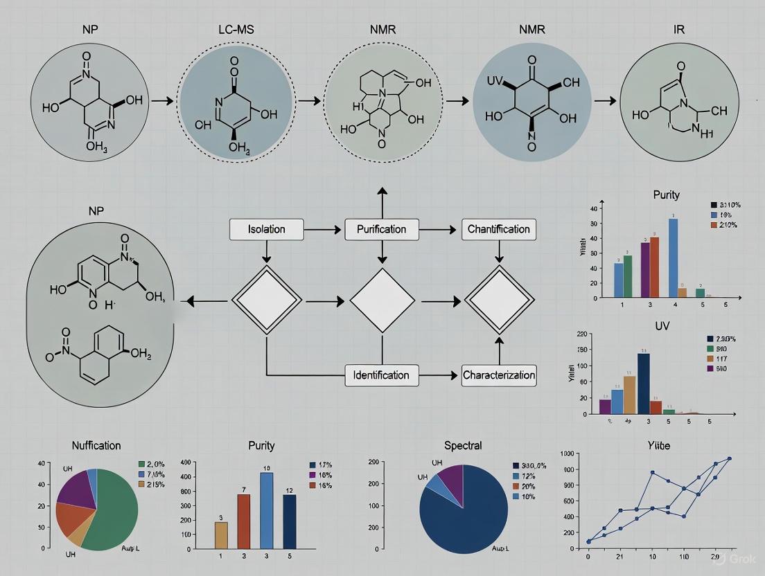
Natural Product Characterization Workflow
(Natural Product Characterization Workflow: A sequential process from source material to characterized research material)
Natural Product Lead Prioritization Framework
(Natural Product Lead Prioritization: Multi-tiered screening cascade with feedback loops)
Integrated Drug Discovery Pathway
(Integrated Drug Discovery Pathway: Conventional process with natural product integration points and challenges)
Natural products (NPs) are a cornerstone of drug discovery, distinguished by their unparalleled structural diversity and broad-spectrum bioactivity honed by millions of years of evolutionary refinement [7]. However, the path from crude extract to characterized bioactive compound is fraught with technical challenges. The core of this difficulty lies in the analytical process itself: researchers must navigate a labyrinth of complexity, from the initial screening of intricate biological mixtures to the final definitive structural characterization. This technical support center is designed to function as a strategic guide, directly addressing the specific, high-impact problems encountered in daily laboratory work. By providing clear, actionable troubleshooting protocols and foundational knowledge, we aim to empower researchers to overcome these barriers, enhance the reliability of their data, and accelerate the discovery of novel therapeutic agents.
Troubleshooting Guides & FAQs
This section provides targeted solutions for common, yet critical, technical challenges in natural product analysis using Liquid Chromatography-Mass Spectrometry (LC-MS).
Frequently Asked Questions (FAQs)
Q1: My LC-MS analysis shows inconsistent results and high background noise. What are the most likely sources of contamination? Systematic errors and artefacts are common in trace-level HPLC-MS analysis, and the mass spectrometer is the source of problems in only a minority of cases [8]. The most frequent culprits are:
- Solvents and Reagents: HPLC-grade solvents are often tested for UV transparency but not for a low MS background. Contaminants in water or organic solvents can produce a high chemical background [8].
- Sample Preparation: The sample matrix itself can introduce ions that suppress the ionization of your target analytes [8].
- HPLC System: The stationary phases in reversed-phase HPLC columns can slowly hydrolyze, releasing bound lipophilic rests that increase the background. Active sites on the phase can also cause adsorption and degradation of compounds at trace levels [8].
- Buffer Salts: Non-volatile buffers (e.g., phosphate buffers) are incompatible with MS and create severe contamination and ion suppression [8].
Q2: Why is the signal for my target compound so weak, even though I know it's present in the sample? Signal weakness or loss is often related to compound ionization or system configuration:
- Ion Suppression: Your analyte's ionization can be suppressed by a more easily ionizable background or by a co-eluting substance. This is a particularly common issue with Electrospray Ionization (ESI) [8].
- Adduct Formation: Ion-molecule adducts can form between analytes and alkaline metal ions (e.g., Na+, K+) or ammonium, which may disperse the signal for a single compound across multiple m/z values [8].
- Source Misalignment: In Atmospheric Pressure Chemical Ionization (APCI), a misaligned corona discharge needle can cause highly reproducible non-linearity in calibration curves, affecting sensitivity [8].
- In-Source Fragmentation: The compound might be fragmenting before it reaches the mass analyzer, making the molecular ion difficult to detect [9].
Q3: I have an accurate molecular mass, but I'm finding multiple structural matches in databases. How can I confidently identify the correct one? This is a fundamental challenge in natural products research. An accurate mass alone is often insufficient because many isomeric molecules share the same molecular formula [9]. To assign the correct structure, you need orthogonal information:
- Tandem MS (MS/MS): Use MS/MS to generate a fragmentation fingerprint. The fragmentation pattern can be compared to a standard run on the same instrument under identical conditions [9].
- Chromatographic Retention Time: Matching the retention time to an authentic standard provides strong evidence [9].
- Nuclear Magnetic Resonance (NMR): NMR remains the gold standard for de novo structure elucidation, especially for distinguishing between isomers and determining stereochemistry [9].
Advanced Troubleshooting Guide
For persistent or complex issues, follow this structured diagnostic approach.
Table 1: Troubleshooting Common LC-MS Performance Issues
| Observed Problem | Potential Root Cause | Diagnostic Steps | Corrective Action |
|---|---|---|---|
| High Background Noise | Contaminated mobile phase or sample [8] | Run a blank gradient with no injection. | Use MS-grade solvents and additives. Re-prepare mobile phases. Purify sample if needed. |
| Column bleed (hydrolysis of stationary phase) [8] | Check if noise increases with column temperature/age. | Replace column. Use a guard column. | |
| Poor Sensitivity | Ion suppression from co-eluting compounds [8] | Post-infuse analyte and inject sample to observe signal dip. | Improve chromatographic separation. Dilute sample. Modify extraction/cleanup. |
| Source contamination or misalignment [8] | Check instrument calibration and tuning reports. | Clean ion source. Perform mass calibration and instrument tuning. | |
| Irreproducible Quantification | Non-linear calibration due to source issues [8] | Inspect calibration curve for consistent non-linearity. | Verify and realign the corona needle (APCI). Check for active sites in flow path. |
| Adsorption to active sites in flow path or column [8] | Analyze a standard at high and low concentration; look for signal loss. | Use inert (e.g., PEEK) components. Passivate system. Add modifier to mobile phase. | |
| Inability to Identify Unknown | Lack of orthogonal data for isobaric compounds [9] | Search molecular formula in Dictionary of Natural Products; note number of hits. | Acquire MS/MS spectra. Use orthogonal separation (HILIC, etc.). Isolate compound for NMR analysis. |
Protocol: Systematic LC-MS Performance Diagnosis
Isolate the Subsystem: Begin by determining whether the problem originates from the LC system, the MS, or the sample itself.
- Step 1: Inject a pure standard with a known calibration. If the signal is normal, the problem is likely sample-specific (e.g., matrix effects). If the signal is abnormal, proceed to Step 2.
- Step 2: Run a system suitability test with a standard mix. Evaluate pressure, peak shape, and retention time consistency to diagnose LC issues.
- Step 3: Run a solvent blank. A high background indicates contaminated mobile phases or a dirty source.
Check Instrument Calibration: Regularly verify mass accuracy and sensitivity using manufacturer-recommended calibration mixes. Poor sensitivity or mass accuracy in the calibration process directly points to an MS source or analyzer problem [10].
Validate Sample Introduction: Ensure the autosampler is functioning correctly by checking for precise injection volumes and the absence of carryover, which can be a significant source of contamination and inaccurate quantification [11].
The following workflow diagrams the logical process for moving from problem observation to resolution.
The Scientist's Toolkit: Essential Research Reagents & Materials
The selection of reagents and consumables is a critical, yet often overlooked, factor in determining the success of a natural products analysis project. The following table details key materials and their functions.
Table 2: Key Research Reagent Solutions for Natural Product LC-MS
| Item | Function & Importance | Technical Notes & Pitfalls |
|---|---|---|
| MS-Grade Solvents | Provide a low chemical background for high-sensitivity detection. Using HPLC-grade solvents tested only for UV can introduce significant noise [8]. | Always use solvents and water specifically certified for LC-MS to minimize baseline artefacts and ion suppression. |
| Volatile Buffers & Additives | Enable efficient desolvation and ionization in the MS source. Non-volatile buffers (e.g., phosphates) are incompatible and will contaminate the instrument [8]. | Use ammonium formate/acetate, formic acid, or acetic acid. Avoid halides and phosphates. |
| Biocompatible LC Systems | Prevent adsorption of analytes to active metal surfaces in the flow path, which is crucial for recovering certain natural products at trace levels. | For sensitive compounds, use systems with PEEK, MP35N, gold, or ceramic flow paths [12]. |
| U/HPLC Columns with High Efficiency | Provide the chromatographic resolution needed to separate complex natural product extracts, reducing ion suppression and enabling accurate identification [13]. | Select columns with small particle sizes (e.g., sub-2µm) and appropriate stationary phases (C18, HILIC, etc.) for your compound class. |
| Stable Isotope-Labeled Internal Standards | Account for matrix-induced ion suppression/enhancement and correct for analyte loss during sample preparation, ensuring accurate quantification [13]. | Ideally, use a (^{13}\text{C}) or (^{15}\text{N})-labeled version of the analyte. If unavailable, use a closely related structural analogue. |
Experimental Workflows: From Screening to Confirmation
A robust analytical workflow is essential for navigating the complexity of natural product extracts. The process typically moves from untargeted screening to targeted characterization.
Core Protocol: Untargeted Screening for Novel Natural Products
Principle: This methodology uses high-resolution LC-MS to comprehensively profile a complex natural product extract without prior knowledge of its composition, aiming to highlight novel or unknown compounds for further investigation [9] [13].
Step-by-Step Methodology:
- Sample Preparation: Minimally, a crude extract is dissolved in an MS-compatible solvent and centrifuged or filtered to remove particulate matter. The goal is to avoid introducing unnecessary complexity or contaminants [8].
- Chromatographic Separation: Use a UHPLC system with a binary pump and a high-efficiency reversed-phase column (e.g., C18, 1.7-1.8µm particle size). Employ a long, shallow gradient (e.g., 5-95% organic modifier over 30-60 minutes) to maximize separation of the complex mixture [12] [13].
- High-Resolution Mass Spectrometry Analysis:
- Instrument: A Q-TOF or Orbitrap mass spectrometer is preferred for its high mass accuracy and resolution [12] [13].
- Acquisition: Data is collected in data-dependent acquisition (DDA) mode. The instrument continuously performs full MS scans (e.g., m/z 50-1500) and automatically selects the most intense ions from each scan for MS/MS fragmentation.
- Data Processing and Dereplication:
- Molecular Formula Assignment: Use software to deconvolute the data, assigning molecular formulas based on accurate mass and isotope patterns [9].
- Database Search: Search the assigned formulas against natural product databases (e.g., Dictionary of Natural Products, AntiMarin, COCONUT, LANaPDB). This critical "dereplication" step identifies known compounds, saving effort on re-isolation [9] [14].
- Novelty Assessment: Compounds whose formulas do not return a database match become high-priority targets for isolation and full structure elucidation.
The following diagram illustrates this multi-stage workflow.
Core Protocol: Structural Confirmation Using LC-MS/MS and NMR
Principle: Once a compound of interest is isolated, its structure must be confirmed. This protocol leverages the complementary strengths of MS/MS and NMR to achieve definitive identification [9].
Step-by-Step Methodology:
- LC-MS/MS Analysis of Pure Compound:
- Inject the purified compound.
- Acquire high-quality MS/MS spectra at multiple collision energies to generate a comprehensive fragmentation pattern.
- Interpret the fragments to propose a substructure or to match against a spectral library if available [9].
- NMR Spectroscopy:
- Prepare a purified sample (micrograms to milligrams, depending on sensitivity).
- Acquire a suite of 1D and 2D NMR experiments (e.g., (^{1}\text{H}), (^{13}\text{C}), COSY, HSQC, HMBC).
- NMR data provides definitive proof of constitution, connectivity, and stereochemistry, which MS cannot reliably achieve on its own [9].
- Data Integration:
- Correlate the MS/MS fragmentation pathways with the structural features elucidated by NMR.
- This combined approach provides a high degree of confidence in the final structural assignment.
Navigating the technical journey from screening to characterization in natural products research demands a systematic approach to troubleshooting and a deep understanding of the strengths and limitations of analytical technologies like LC-MS and NMR. By recognizing common pitfalls such as contamination, ion suppression, and the challenges of dereplication, and by implementing the detailed protocols and diagnostic guides provided herein, researchers can transform frustration into discovery. This technical support framework empowers scientists to generate more reliable and reproducible data, ultimately streamlining the path to uncovering the next generation of natural product-based therapeutics.
In the field of natural product (NP) drug discovery, a significant and persistent perception gap exists between industry and academic stakeholders regarding research strategies and their effectiveness. While natural products have undisputedly played a leading role in developing novel medicines, trends in pharmaceutical research investments indicate that NP research is neither prioritized nor perceived as fruitful in drug discovery programmes compared with incremental structural modifications and large-volume high-throughput screening (HTS) of synthetic compounds [15] [16].
This gap demonstrates a fundamental dissonance: individuals from both sectors perceive high potential in NPs as drug leads, yet simultaneously express criticism toward prevalent industry-wide discovery strategies [15]. This article explores the technical barriers underpinning this divide and provides practical troubleshooting guidance to enhance collaborative research efficacy, bridging the gap between theoretical academic research and industry's practical application needs.
Survey Insights: Quantifying the Divide
Stakeholder surveys reveal critical differences in how industry and academic professionals perceive drug discovery strategies and outcomes. A comprehensive survey of 52 industry and academic experts provides quantitative evidence of this divide [15] [16].
Table 1: Perceived Effectiveness of Drug Discovery Strategies
| Discovery Strategy | Industry Perception | Academic Perception | Effectiveness Gap |
|---|---|---|---|
| Natural Products | Lower priority despite high potential | High potential, undervalued | Significant |
| High-Throughput Screening (HTS) of Synthetics | Higher hit rates perceived | Lower hit rates perceived | Moderate |
| Incremental Structural Modifications | Prioritized in R&D investments | Less favored compared to NPs | Significant |
Table 2: Stakeholder Satisfaction with Current Strategies
| Assessment Area | Industry Satisfaction | Academic Satisfaction | Shared Concerns |
|---|---|---|---|
| Current Discovery Efforts | Not more effective than previous decades | Not more effective than previous decades | Perception of stalled progress |
| Company/Industry Strategy | Highly critical | Critical | Widespread dissatisfaction |
| Hit Rates in HTS | Higher perception | Lower perception | Methodological differences |
Survey findings indicate that industry contacts perceived higher hit rates in HTS efforts compared to academic respondents, despite neither group perceiving current discovery efforts as more effective than previous decades [16]. Surprisingly, many industry contacts expressed strong criticism toward prevalent company and industry-wide drug discovery strategies, indicating a high level of dissatisfaction within the commercial sector [15].
Technical Barriers in Natural Product Research
Characterization and Reproducibility Challenges
Botanical natural products present unique technical hurdles that contribute to the industry-academia divide. These complex mixtures differ from pharmaceutical drugs in that their composition varies depending on genetics, cultivation conditions, and processing methods [17]. This inherent variability creates significant reproducibility challenges, as many studies are conducted with poorly characterized materials, making results difficult to interpret or replicate [17].
Troubleshooting Guide: Addressing Characterization Challenges
Q: How can I ensure my botanical natural product is suitable for research?
- A: The ideal botanical natural product for research should be: representative of what consumers use; authenticated (species verified); well-characterized with known active constituents; free of contamination and adulteration; available in sufficient quantity; and consistent across the study duration with established shelf life and batch-to-batch reproducibility [17].
Q: What are the essential steps for characterizing a botanical study material?
- A: Follow this workflow: (1) Conduct thorough literature research on major metabolites and traditional use; (2) Select and authenticate botanical material using voucher specimens; (3) Characterize chemically using targeted or untargeted methods; (4) Ensure stability and batch-to-batch consistency [17].
Q: Why is a voucher specimen necessary?
- A: A voucher specimen provides a permanent, verifiable record of the plant material used in your research. It allows for taxonomic identification by a trained botanist and is essential for preserving a record of the original sample, enabling future replication studies. Major natural product journals require deposition in a herbarium [17].
Technological and Strategic Hurdles
The pharmaceutical industry's shift away from NP research stems from several perceived barriers: traditional NP screening is labor-intensive, requiring multi-step extractions and structural elucidation; production scaling of rare metabolites presents bottlenecks; and legal complexities surrounding international collaboration under frameworks like the Nagoya Protocol create additional hurdles [7]. Furthermore, the "one-drug-one-target" paradigm that dominates pharmaceutical development conflicts with the inherent multi-target nature of many natural products [18].
Bridging the Gap: Modern Methodologies and Solutions
Advanced Characterization Workflows
Implementing robust, standardized workflows for natural product characterization is fundamental to bridging the perception gap. The following methodology ensures research materials meet rigorous standards for reproducible science.
Experimental Protocol: Comprehensive Botanical Natural Product Characterization
Objective: To authenticate, characterize, and ensure quality consistency of botanical natural products for research applications.
Materials and Reagents:
- Botanical raw material or extract
- Herbarium voucher specimen materials
- Solvents for extraction (e.g., methanol, ethanol, water)
- Reference standards for marker compounds
- LC-MS grade solvents and additives for analysis
Procedure:
- Literature Review & Planning: Research traditional usage, common species, plant parts, and preparation methods. Identify known bioactive compounds or characteristic markers [17].
- Sample Acquisition & Authentication: Source material from reputable suppliers. Collect a voucher specimen from the same lot, including as many plant parts as possible (roots, leaves, flowers). Deposit the authenticated voucher in a recognized herbarium for permanent reference [17].
- Chemical Characterization:
- Targeted Analysis: If active constituents are known, use targeted methods (e.g., HPLC-DAD, LC-MS/MS) to quantify these compounds and ensure compliance with existing monographs [17].
- Untargeted Analysis: For less-defined materials, employ untargeted metabolomics (e.g., UHPLC-QTOF-MS) to generate a comprehensive metabolite profile. Compare against commercial or in-house spectral libraries [17].
- Contamination & Adulteration Screening: Test for heavy metals, pesticides, mycotoxins, and potential adulterants with synthetic drugs using validated analytical methods [17].
- Stability & Batch Monitoring: Conduct accelerated stability studies under various conditions (temperature, humidity). Analyze multiple batches to establish reproducibility ranges for key markers [17].
The following workflow diagram visualizes the key stages of this characterization process.
Fig 1. Workflow for characterizing botanical natural products.
Innovative Technologies Closing the Gap
Emerging technologies are directly addressing historical barriers to NP research, offering solutions that align with both academic and industry priorities.
Table 3: Innovative Technologies and Their Applications
| Technology | Application in NP Research | Impact on Perception Gap |
|---|---|---|
| Genome Mining & Metagenomics | Uncovers cryptic biosynthetic gene clusters, predicts novel compounds without traditional extraction [7]. | Reduces reliance on bulk biomass, addresses supply bottlenecks. |
| AI & Machine Learning | Accelerates hit discovery, de-replication, and prediction of biosynthetic pathways [7]. | Increases efficiency and success rates, making NP research more competitive with synthetic libraries. |
| Synthetic Biology | Engineers microbial hosts for sustainable production of complex NPs [7]. | Solves supply and sustainability issues, enabling scalable production. |
| Network Pharmacology | Embraces multi-target action of NPs, providing a scientific framework for traditional medicine [18]. | Bridges philosophical divide between reductionist and holistic approaches. |
The Scientist's Toolkit: Essential Research Reagents and Solutions
Table 4: Key Research Reagent Solutions for NP Screening & Characterization
| Reagent / Material | Function & Application | Technical Notes |
|---|---|---|
| Herbarium Voucher Specimen | Provides taxonomic verification and a permanent physical record of the plant material studied [17]. | Essential for publication and reproducibility. Must be deposited in a recognized herbarium. |
| Authenticated Reference Standards | Enables quantification of active constituents or marker compounds for quality control [17]. | Critical for targeted analytical methods (HPLC, LC-MS) to ensure consistency. |
| Stable Isotope-Labeled Probes | Used in metabolomic studies to trace biosynthetic pathways and identify novel metabolites [7]. | Aids in de-replication and understanding NP biosynthesis. |
| LC-MS/MS Solvents & Columns | High-purity solvents and specialized chromatography columns for separating complex NP mixtures [17]. | Required for high-resolution metabolomics and sensitive detection. |
| Genomic DNA Extraction Kits | Isolate high-quality DNA from plant or microbial sources for genome mining and barcoding [7]. | Necessary for genetic authentication and biosynthetic gene cluster analysis. |
| HTS Assay Kits & Reagents | Standardized biochemical or cell-based assays for high-throughput screening of NP libraries [15] [7]. | Allows for comparison of NP hits against synthetic libraries in industry-standard formats. |
The industry-academia perception gap in natural product research stems from tangible technical challenges and strategic differences. However, the shared recognition of NPs' inherent potential, combined with powerful new technologies and standardized methodologies, provides a clear path forward. By adopting robust characterization protocols, leveraging innovations in genomics and AI, and embracing frameworks like network pharmacology, both sectors can bridge this divide. Fostering strategic partnerships built on mutual respect, clear intellectual property agreements, and shared goals is essential to revitalize natural products as a sustainable and innovative source of next-generation medicines [7] [19].
For researchers in natural product screening, navigating the legal landscape is as crucial as mastering the laboratory techniques. The Convention on Biological Diversity (CBD) and its supplementary agreement, the Nagoya Protocol on Access and Benefit-Sharing (ABS), form the core international framework governing how genetic resources are accessed and how benefits from their utilization are shared [20] [21].
These agreements operate on the principle that countries have sovereign rights over their natural resources [20] [22]. For a scientist, this means that the plant, microbial, or animal material you are investigating is not just a biological sample; it is a genetic resource subject to specific legal requirements. The primary objective of the Nagoya Protocol is the fair and equitable sharing of benefits arising from the utilization of genetic resources, thereby contributing to the conservation and sustainable use of biodiversity [21] [23]. Adherence to these protocols is not merely a legal formality but a fundamental aspect of ethical and reproducible research in drug discovery and natural product characterization.
Troubleshooting Guides & FAQs
Frequently Asked Questions (FAQs)
Q1: As an academic researcher conducting non-commercial natural product discovery, am I obligated to comply with the Nagoya Protocol? A: Yes. The obligations of the Nagoya Protocol apply to "utilization of genetic resources," which is defined to include "research and development on the genetic and/or biochemical composition of genetic resources" [21] [22]. This definition covers both commercial and non-commercial research. Many provider countries do not differentiate between pure and applied research in their access legislation. It is critical to comply with the legal requirements of the country providing the genetic resource, regardless of your research's commercial intent.
Q2: What is the most common pitfall for researchers when sourcing genetic materials from international collaborators? A: The most common pitfall is assuming that a Material Transfer Agreement (MTA) from a research institution or culture collection automatically covers all Nagoya Protocol compliance requirements. While an MTA is essential, the protocol requires Prior Informed Consent (PIC) from the competent national authority of the provider country and the establishment of Mutually Agreed Terms (MAT) that specifically address benefit-sharing [21] [22]. You must verify that your collaborator obtained these documents at the time of original collection and that their terms permit your intended use and subsequent transfers.
Q3: Our research involves screening a library of synthetic compounds derived from a natural product scaffold. Are these compounds subject to the Protocol? A: This is a complex, evolving area. The Nagoya Protocol applies to genetic resources and "traditional knowledge associated with genetic resources" [21]. If your synthetic library is based on a chemical structure from a genetic resource covered by the Protocol, it may be considered a subsequent application and fall within its scope. The definitive factor is the national legislation of the provider country. You must carefully review the specific terms of the MAT and PIC, which may define the scope of "derivatives" and "applications." When in doubt, seek legal advice and err on the side of caution.
Q4: Where can I find reliable and official information on a country's specific access requirements? A: The Access and Benefit-sharing Clearing-House (ABSCH) is the official platform established by the Nagoya Protocol for this purpose [24]. It is a key tool for providing legal certainty and transparency. On the ABSCH, you can find:
- National focal points and competent national authorities for each country.
- Domestic regulatory ABS requirements, permits, and relevant legislation.
- Internationally Recognized Certificates of Compliance (IRCC) which serve as proof that a resource was accessed legally [24] [22].
Q5: What happens if our research project transitions from basic research to a commercial application? A: The MAT you agreed upon at the start of your research must specifically address this scenario [21] [22]. The MAT should outline the benefit-sharing obligations triggered by commercialization, which could include monetary benefits (e.g., milestone payments, royalties) or non-monetary benefits (e.g., joint development, capacity building). If your initial MAT did not cover commercialization, you must re-engage with the provider to negotiate new terms. Retroactively seeking permission is a breach of the Protocol and can lead to serious compliance issues.
Common Errors and Solutions Table
The table below outlines common procedural errors and their solutions to help you troubleshoot compliance issues in your research workflow.
| Error Stage | Common Error | Potential Consequence | Solution |
|---|---|---|---|
| Pre-Access | Assuming a country has no ABS legislation because it is not listed. | Accessing resources illegally, leading to compliance breaches. | Consult the ABS Clearing-House first, then contact the National Focal Point for definitive confirmation [24]. |
| Negotiation | Not specifying the scope of research and potential commercial applications in the MAT. | Disputes with the provider country when research evolves or commercializes. | Ensure MAT is clear, covers all phases of research (including commercialization), and includes dispute resolution clauses [21] [22]. |
| Documentation | Failing to keep detailed records of PIC, MAT, and permits and transferring materials to third parties without proper documentation. | Inability to demonstrate due diligence, breaking the chain of compliance. | Maintain a dedicated digital repository for all ABS documents. Use a Nagoya-compliant MTA for all transfers [21]. |
| Checkpoints | Not engaging with national checkpoints (e.g., at the grant application or patent filing stage). | Non-compliance is not caught early, leading to larger problems later. | Proactively declare due diligence to relevant national checkpoints as required by user country measures (e.g., in the EU) [21]. |
Experimental Protocols for Compliance
Objective: To establish the legal provenance of a genetic resource before it is acquired for research, ensuring compliance with the CBD and Nagoya Protocol.
Materials:
- Computer with internet access: For consulting online databases.
- ABS Compliance Documentation Tracker: A spreadsheet or electronic lab notebook (ELN) system with fields for key document details.
Methodology:
- Identify the Provider Country: Determine the country of origin of the genetic resource. For biological samples, this is the country from which the organism was originally collected from in-situ conditions [25].
- Consult the ABS Clearing-House:
- Access the ABSCH website.
- Search for the provider country's profile to identify its Competent National Authority (CNA) and review its national ABS laws and requirements [24].
- Determine Access Conditions:
- If the country has ABS measures, proceed to determine the process for obtaining Prior Informed Consent (PIC).
- If the country has no established measures, document this finding from the CNA or National Focal Point (NFP).
- Verify Source Compliance (if sourcing from a repository):
- When obtaining resources from a culture collection (e.g., DSMZ), request their Internationally Recognized Certificate of Compliance (IRCC) or equivalent proof that the resource was accessed in accordance with the provider country's PIC and MAT [21].
- Ensure the repository's MTA allows for your intended research use.
- Document the Process:
- Record all steps, including dates of website checks, communications with authorities, and copies of permits or certificates, in your ABS Compliance Documentation Tracker.
Protocol: Negotiating Mutually Agreed Terms (MAT)
Objective: To establish a legally sound contract that outlines the terms of access, use, and benefit-sharing for a genetic resource.
Materials:
- Draft MAT template: Often provided by the provider country's CNA.
- Research project outline: A clear description of the intended research, including possible commercial outcomes.
Methodology:
- Prepare a Research Plan: Draft a comprehensive yet clear description of your research. Specify if the work is non-commercial, has potential for commercialization, or is for taxonomic identification only.
- Identify Benefit-Sharing Options: Review the Annex of the Nagoya Protocol for a list of monetary and non-monetary benefits. Propose benefits that are appropriate and feasible for your project. Examples include [21] [22]:
- Non-monetary: Collaboration with scientists from the provider country, participation in product development, sharing of research results, and admittance to related training.
- Monetary: Upfront payments, royalties on net sales, or contributions to a conservation fund.
- Clarify Key Terms: Ensure the MAT explicitly defines:
- The genetic resource covered.
- The scope and field of use of the research.
- Provisions for third-party transfers.
- Rights to intellectual property.
- Terms for benefit-sharing triggered by specific events (e.g., publication, patenting, commercialization).
- Execute and Archive: Once negotiated and signed, store the final MAT with the PIC document. Ensure all relevant team members are aware of its terms and restrictions.
Workflow Visualization
The following diagram illustrates the logical workflow a researcher should follow to legally access and utilize a genetic resource.
The Scientist's Toolkit: Research Reagent Solutions
The following table details key resources and tools essential for navigating the regulatory framework, rather than the wet-lab reagents.
| Tool / Resource | Function & Utility in ABS Compliance | Key Considerations for Researchers |
|---|---|---|
| ABS Clearing-House (ABSCH) [24] | The official online platform for information on ABS, national focal points, and competent national authorities. | Use it as the first point of verification for any country's ABS requirements. Always check for updated information. |
| Internationally Recognized Certificate of Compliance (IRCC) [24] [21] | Proof that a genetic resource was accessed in accordance with PIC and established MAT. It is registered in the ABSCH. | When sourcing from a repository, request the IRCC. This is your primary evidence of legal provenance. |
| Competent National Authority (CNA) [21] [22] | The entity within a provider country with the legal authority to grant access (PIC) and negotiate MAT. | All official communication and applications for access must go through the CNA. Do not rely on informal agreements. |
| Mutually Agreed Terms (MAT) Document [21] [22] | The binding contract that outlines the conditions of use, benefit-sharing, and third-party transfers. | This is the most critical document to protect your research. Ensure it is precise and covers all potential future uses. |
| Material Transfer Agreement (MTA) | A contract governing the transfer of tangible research materials between two organizations. | Your MTA must be consistent with and reference the underlying PIC and MAT. It cannot override or negate them. |
Advanced Analytical and Screening Technologies: A Practical Toolkit for Researchers
FAQs and Troubleshooting Guides
HPLC-MS/MS Troubleshooting
Q1: My LC-MS analysis shows significant signal suppression and high background noise. What could be the cause?
Signal suppression and high background noise are frequently linked to mobile phase contaminants and non-volatile additives [26].
- Solution: Ensure all mobile phase additives are of the highest purity and are volatile. Avoid non-volatile buffers like phosphate; instead, use volatile alternatives such as ammonium formate or ammonium acetate at concentrations typically around 10 mM [26]. Formic acid (0.1%) is a good volatile additive for controlling pH; however, be cautious with alternatives like trifluoroacetic acid (TFA) as they can cause significant signal suppression [26]. Always use a divert valve to direct only the analyte peaks of interest into the mass spectrometer, preventing contamination from the solvent front and the high organic wash portion of the gradient [26].
Q2: How can I confirm if a performance issue is with my LC-MS method or the instrument itself?
Implement a routine benchmarking procedure [26].
- Solution: Establish and regularly run a benchmarking method using a standard compound like reserpine. Perform five replicate injections to assess key parameters like retention time reproducibility, peak height, and shape [26]. If you encounter problems with your analytical method, run the benchmark. If the benchmark performs as expected, the issue lies with your method or sample preparation. If the benchmark fails, the problem is likely with the instrument itself [26].
Q3: What is the most critical step for optimizing MS parameters for my specific analytes?
Infusion tuning is essential for compound-dependent optimization [26].
- Solution: Directly infuse your analyte into the MS and optimize source parameters like voltages, gas flows, and temperatures. Do not rely solely on an autotune function. Perform an autotune followed by a manual tune for your specific analytes to achieve the optimum signal [26]. When adjusting parameters that generate a response curve, set the value on a "maximum plateau" where small variations do not cause large changes in response, ensuring method robustness [26]. Save individual tune files for different compound groups [26].
FTIR Spectroscopy Troubleshooting
Q4: My FTIR spectrum for a plant extract has poor resolution and undefined peaks. How can I improve the sample preparation?
Poor resolution can stem from inadequate purification of the crude extract or overly thick sample preparation.
- Solution: FTIR is a fast and non-destructive method for identifying functional groups in phytochemicals [27]. However, for complex plant extracts, further purification is often necessary. Prior to FTIR analysis, employ chromatographic techniques like HPLC to separate individual compounds or fractionate the extract [27]. This reduces spectral overlap from multiple concurrent compounds, leading to cleaner and more interpretable spectra. Ensure your sample is properly prepared for your chosen technique (e.g., as a dry film for ATR-FTIR or a pellet with KBr for transmission FTIR).
Q5: Can FTIR quantitatively determine the concentration of a specific flavonoid in my extract?
FTIR is primarily a qualitative or semi-quantitative technique for functional group identification [27].
- Solution: For accurate identification and quantification of specific compounds like flavonoids, FTIR should be combined with other techniques [27]. Use HPLC-DAD or LC-MS/MS for precise quantification [27]. FTIR is excellent for "fingerprinting" and confirming the presence of major functional groups (e.g., hydroxyl, carbonyl, unsaturated bonds) associated with compound classes like flavonoids, but it is not the preferred tool for exact concentration measurement in complex mixtures [27].
SEM-EDX Troubleshooting
Q6: My SEM-EDX analysis of my plant-synthesized nanoparticles shows a very weak signal for the metal. What might be wrong?
Weak signals can be caused by insufficient nanoparticle loading, thick or charging samples, or improper analysis conditions.
- Solution: Ensure your sample is conductive. For non-conductive biological samples, coating with a thin carbon layer is often necessary. For green-synthesized nanoparticles, confirm that the biosynthesis was successful and that nanoparticles are present on the surface [28]. Refer to the established protocol for Papilionanthe teres leaf extract synthesis as a reference [28]. Make sure the sample is dry and properly mounted. Increase the counting time or beam current to improve the signal-to-noise ratio, but be mindful of potential beam damage to the sample [29].
Q7: Why can't I detect light elements like carbon, nitrogen, and oxygen in my biological sample with EDX?
This is a fundamental limitation of standard EDX systems [29] [30].
- Solution: EDX has lower sensitivity for elements with low atomic numbers (below sodium) [30]. The X-rays from light elements are of low energy and are easily absorbed by air or the detector window. While detection is possible, it requires specialized detectors and can be challenging [29]. The presence of these light elements is often inferred, and techniques like FTIR or NMR are better suited for their direct detection and characterization in organic matrices [27].
Q8: My EDX spectral peaks for phosphorus and sulfur are overlapping. How can I resolve this?
Peak overlap is a common challenge in EDX analysis, as elements with adjacent atomic numbers can have overlapping X-ray energies [29].
- Solution: Use the manufacturer's software deconvolution tools to mathematically resolve the overlapping peaks [29]. Ensure your system is well-calibrated. In some cases, using a different X-ray line for identification (e.g., K vs. L lines) might help, though this is not always possible with biological samples. For critical analysis, complement EDX with a technique like NMR, which excels at distinguishing between different chemical environments of atoms like phosphorus-31 ( [31], [27]).
Experimental Protocols for Key Techniques
- Principle: Plant extracts contain phytochemicals that act as reducing and stabilizing agents to convert metal salts into nanoparticles.
- Materials: Fresh or dried plant leaves (e.g., Papilionanthe teres), silver nitrate (AgNO₃) solution, distilled water, laboratory glassware, centrifuge.
- Procedure:
- Extract Preparation: Wash, dry, and grind plant leaves. Prepare an aqueous extract by boiling the plant material in distilled water and filtering the mixture.
- Synthesis Reaction: Mix the filtered plant extract with an aqueous solution of AgNO₃ under constant stirring. The reaction can be monitored by a color change.
- Purification: Centrifuge the resulting nanoparticle suspension at high speed to pellet the nanoparticles. Discard the supernatant and re-disperse the pellet in distilled water. Repeat this washing step 2-3 times.
- Characterization: Analyze the purified nanoparticles using SEM-EDX for morphology and elemental composition, and FTIR to identify the functional groups of phytochemicals capping the nanoparticles [28].
- Principle: To preserve the native structure and elemental composition of a biological sample for analysis under the high vacuum of an electron microscope.
- Materials: Phosphate buffer, paraformaldehyde, osmium tetroxide, ethanol series, propylene oxide, epoxidic resin, ultramicrotome.
Procedure (Standard Resin Embedding):
- Primary Fixation: Fix a small tissue sample (~1 mm³) in buffered 4% paraformaldehyde to preserve structure.
- Post-fixation: Treat with 2% osmium tetroxide, which also adds conductivity.
- Dehydration: Pass the sample through a graded series of ethanol (30%, 50%, 70%, 95%, 100%) to remove water.
- Infiltration and Embedding: Transition the sample into propylene oxide and then infiltrate with epoxidic resin. Embed in fresh resin and polymerize in an oven.
- Sectioning: Use an ultramicrotome to cut ultrathin (~100 nm) or semi-thin sections.
- Analysis: Mount the sections on a suitable stub and analyze unstained under SEM-EDX to avoid interference from heavy metal stains [29].
Note: For elemental analysis, cryo-fixation (flash-freezing) is the gold standard to prevent loss or translocation of diffusible ions, but it is more technically demanding [29].
- Principle: High-performance liquid chromatography separates compounds in a complex extract, a diode array detector provides UV-Vis spectra, and tandem mass spectrometry enables identification and structural elucidation.
- Materials: HPLC-grade solvents (e.g., methanol, acetonitrile, water), volatile acids (e.g., formic acid), solid-phase extraction (SPE) cartridges, syringe filters, plant extract.
- Procedure:
- Sample Preparation: Defat and extract plant material using a suitable solvent (e.g., methanol). Pre-purify the crude extract using SPE to remove contaminants [26]. Filter the final extract through a 0.22 µm membrane filter before injection.
- LC Separation: Use a reversed-phase C18 column. Employ a binary gradient with a mobile phase A (e.g., water with 0.1% formic acid) and phase B (e.g., acetonitrile with 0.1% formic acid). The gradient runs from low to high organic modifier [27] [26].
- DAD Detection: Acquire UV-Vis spectra throughout the run, which helps identify compound classes like flavonoids and phenolic acids based on their chromophores [27].
- MS/MS Analysis: Couple the HPLC outlet to the mass spectrometer via an electrospray ionization (ESI) source. Use tandem mass spectrometry (MS/MS) to fragment precursor ions. This fragmentation pattern is a unique fingerprint for compound identification [27].
- Data Analysis: Compare retention times, UV spectra, precursor mass, and fragmentation patterns with those of authentic standards or databases.
Comparison of Analytical Techniques in Phytochemical Profiling
| Technique | Core Function | Key Information Obtained | Sample Requirements | Key Limitations |
|---|---|---|---|---|
| HPLC-MS/MS [27] | Separation, identification, and quantification of individual compounds. | Molecular weight, structural fragments, precise quantification. | Liquid extract, purified and filtered. | Complex operation; requires volatile mobile phases [26]; high instrument cost. |
| FTIR [27] | Identification of functional groups and chemical bonds. | Presence of functional groups (e.g., -OH, C=O, C-O); compound class fingerprint. | Solid (KBr pellet) or liquid film. | Difficult for complex mixtures without prior separation; semi-quantitative at best. |
| SEM-EDX [29] [30] | Morphological imaging and elemental analysis. | Surface topography, elemental composition (atomic no. >10), spatial distribution. | Solid, dry, conductive (often requires coating). | Cannot distinguish ionic states [29]; low sensitivity for light elements [30]; vacuum required. |
| NMR [27] | Elucidation of molecular structure and dynamics. | Detailed carbon-hydrogen skeleton; unambiguous structure determination. | Solubilized extract or pure compound. | Lower sensitivity than MS; requires relatively pure samples for full structural elucidation. |
Research Reagent Solutions
| Essential Material | Function | Application Notes |
|---|---|---|
| Volatile Buffers (Ammonium formate/acetate) [26] | pH control in mobile phase without contaminating the MS ion source. | Use at concentrations ~10 mM; preferable to non-volatile phosphates. |
| C18 Solid-Phase Extraction (SPE) Cartridges | Pre-purification of crude plant extracts to remove contaminants. | Prevents column clogging and ion source contamination in LC-MS [26]. |
| HPLC-grade Solvents | Serve as the mobile phase for chromatography. | Low UV cutoff and high purity are essential for sensitive detection. |
| Formic Acid | A volatile acid additive to improve protonation and chromatography in LC-MS. | A common alternative to TFA, which can cause signal suppression [26]. |
| Silver Nitrate (AgNO₃) | Precursor salt for the green synthesis of silver nanoparticles. | Reacts with phytochemicals in plant extracts to form Ag nanoparticles [28]. |
Workflow and Conceptual Diagrams
Integrated Phytochemical Analysis
LC-MS Method Optimization
High-Throughput Screening (HTS) is an automated, robotic method that enables researchers to rapidly test thousands to millions of chemical, biological, or material samples for biological activity or specific properties [32] [33]. In drug discovery, HTS serves as a primary strategy to identify starting compounds ("hits") for small-molecule drug design campaigns, particularly when little is known about the target structure, which prevents structure-based drug design [34] [33]. The two fundamental approaches in antibacterial drug discovery are cellular target-based HTS (CT-HTS), which uses whole cells to identify intrinsically active agents, and molecular target-based HTS (MT-HTS), which uses purified proteins or enzymes to identify specific inhibitors [35].
Technical Comparison: Cellular vs. Molecular Target-Based Assays
Table 1: Fundamental Characteristics of Cellular and Molecular Target-Based HTS Platforms
| Characteristic | Cellular Target-Based HTS (CT-HTS) | Molecular Target-Based HTS (MT-HTS) |
|---|---|---|
| Screening System | Whole living cells (can be 2D or 3D cultures) | Purified proteins, enzymes, or receptors |
| Biological Context | High physiological relevance; maintains cellular environment | Low physiological relevance; isolated system |
| Primary Advantage | Identifies compounds with cellular activity and permeability | Reveals direct target binding and specific mechanism |
| Primary Disadvantage | Target identification can be challenging | Hits may lack cellular activity due to permeability issues |
| Hit Validation Needs | Secondary screening to eliminate non-specific cytotoxic compounds | Secondary screening to eliminate pan-assay interference molecules (PAINS) |
| Throughput Potential | Generally lower due to biological complexity | Generally higher due to simplified system |
| Information Obtained | Phenotypic response, cell viability, pathway modulation | Direct binding, enzymatic inhibition, binding affinity |
Table 2: Technical Performance and Output Metrics
| Performance Metric | Cellular Target-Based HTS | Molecular Target-Based HTS |
|---|---|---|
| Typical Hit Rate | Approximately 0.3% with natural products [35] | <0.001% with synthetic libraries [35] |
| False Positive Sources | General cytotoxicity, off-target effects, assay interference | Chemical reactivity, metal impurities, assay technology artifacts, autofluorescence, colloidal aggregation [33] |
| Key Quality Control Measures | Cell viability assessment, morphology checks, multiplexed readouts | Counter-screens for promiscuous inhibitors, purity verification, dose-response confirmation |
| Data Complexity | High (multiple cellular parameters possible) | Lower (typically single parameter readouts) |
Troubleshooting Guides & FAQs
Assay Development and Validation
FAQ: What statistical measures should I use to validate my HTS assay before screening? The Z'-factor is an essential statistical parameter for assessing HTS assay quality. A Z'-factor above 0.5 is generally considered excellent, indicating a robust assay with good separation between positive and negative controls [32] [36]. Plate uniformity studies should be conducted over multiple days (3 days for new assays, 2 days for transferred assays) using Max (maximum signal), Min (background signal), and Mid (intermediate signal) controls to establish signal window stability and reproducibility [36].
Troubleshooting Guide: Addressing Poor Z'-Factor Values
- Problem: Low Z'-factor (<0.5) indicating poor separation between controls.
- Potential Causes & Solutions:
- High signal variability: Optimize reagent concentrations, ensure consistent cell seeding density, verify incubation times and temperatures.
- Insufficient signal window: Increase assay dynamic range by adjusting detection parameters or using more sensitive reporters.
- Edge effects in microplates: Use plate seals to prevent evaporation, ensure proper humidity control during incubations.
- Reagent instability: Prepare fresh reagents daily, aliquot and freeze reagents properly, verify storage conditions.
Technical Artifacts and Interference
FAQ: Why do I get different results between cell-based and biochemical assays for the same compounds? Lack of concordance between cellular and enzyme activity is common [37]. In cellular assays, antimicrobial activity might actually result from off-target toxicity rather than specific target inhibition. Conversely, molecules active in molecular target assays may fail in cellular assays due to poor membrane permeability, efflux pump activity, or binding to intracellular proteins like albumin that reduce effective concentration [37]. Always confirm that enzyme hits translate to cellular target engagement.
Troubleshooting Guide: Managing Compound Interference in HTS
- Problem: High false positive rates due to compound interference.
- Common Interference Mechanisms & Solutions:
- Autofluorescence: Use red-shifted fluorophores or switch to luminescence-based detection methods [33].
- Chemical reactivity: Include counter-screens with detergent-based assays to identify promiscuous inhibitors [32] [33].
- Colloidal aggregation: Add non-ionic detergents (e.g., 0.01% Triton X-100) to assay buffer [33].
- Metal impurities: Use chelating agents in assays or pre-purify compound libraries [33].
- Pan-Assay Interference Compounds (PAINS): Apply computational filters to identify and remove known PAINS from hit lists [33] [35].
Natural Product-Specific Challenges
FAQ: What are the special considerations when screening natural product libraries? Natural product extracts present unique challenges including complex composition with molecules at varying concentrations, presence of colored compounds that interfere with detection, potential for antagonistic or synergistic effects between components, and high probability of rediscovering known compounds [35]. To address these issues: pre-fractionate complex extracts, use orthogonal detection methods (not reliant on absorbance), include dereplication strategies (early identification of known compounds), and employ mechanism-informed phenotypic screening with reporter genes [35].
Troubleshooting Guide: Natural Product Library Screening Issues
- Problem: Inconsistent activity in natural product screening.
- Potential Causes & Solutions:
- Concentration variability: Standardize extraction methods, perform bioassay-guided fractionation.
- Non-specific binding: Add carrier proteins (e.g., BSA) to reduce non-specific binding.
- Carryover of interferents: Implement wash steps in cell-based assays, use longer incubation times to distinguish specific from non-specific effects.
- Rediscovery of known compounds: Implement early dereplication using LC-MS and NMR comparison to known compound databases.
Experimental Protocols and Workflows
Protocol: Cell-Based Viability Assay (CT-HTS)
Purpose: To identify compounds that inhibit bacterial growth or kill pathogenic bacteria. Materials: Bacterial culture (e.g., S. aureus), compound library, 384-well microplates, culture medium, viability stain (e.g., resazurin), DMSO, automated liquid handler, plate reader. Procedure:
- Prepare compounds in DMSO in source plates (typical concentration: 10 mM).
- Dilute compounds in medium to working concentration (final DMSO ≤1%).
- Dispense bacterial suspension (~5×10^5 CFU/mL, 50 μL/well) into 384-well plates.
- Add compounds (0.1 μL from source plate using pintool), include controls: medium-only (background), DMSO (negative), known antibiotic (positive).
- Incubate 16-20 hours at 37°C.
- Add resazurin solution (5 μL of 0.15 mg/mL), incubate 2-4 hours.
- Measure fluorescence (Ex560/Em590).
- Calculate % inhibition = 100 × [1 - (Sample - Background)/(Negative Control - Background)].
Validation Parameters: Z'-factor >0.5, signal-to-background ratio >3, coefficient of variation <10% [36].
Protocol: Enzyme Inhibition Assay (MT-HTS)
Purpose: To identify compounds that inhibit specific enzymatic activity. Materials: Purified enzyme, substrate, compound library, assay buffer, 384-well microplates, DMSO, detection reagents. Procedure:
- Prepare enzyme solution in assay buffer (optimized concentration).
- Dispense compounds (0.1 μL from DMSO stock) to assay plates.
- Add enzyme solution (20 μL/well), pre-incubate 15 minutes.
- Initiate reaction with substrate solution (5 μL/well).
- Incubate appropriate time (determined by kinetic analysis).
- Stop reaction if needed, measure signal (absorbance, fluorescence, or luminescence).
- Include controls: no enzyme (background), no inhibitor (negative control), known inhibitor (positive control).
- Calculate % inhibition = 100 × [1 - (Sample - Background)/(Negative Control - Background)].
Validation Parameters: Z'-factor >0.5, signal-to-background >5, linear reaction progress, appropriate Km for substrate [36].
HTS Workflow and Decision Pathways
HTS Platform Selection and Hit Validation Workflow
The Scientist's Toolkit: Essential Research Reagents and Materials
Table 3: Key Research Reagent Solutions for HTS Implementation
| Reagent/Material | Function in HTS | Application Notes |
|---|---|---|
| Microtiter Plates | Assay vessel for high-density screening | Available in 96-, 384-, 1536-well formats; choice depends on throughput needs and available liquid handling capabilities [33] |
| DMSO | Universal solvent for compound libraries | Final concentration should be kept under 1% for cell-based assays unless demonstrated otherwise by compatibility testing [36] |
| Automated Liquid Handlers | Precise dispensing of nanoliter to microliter volumes | Essential for reproducibility; robots from manufacturers like Tecan or Hamilton can significantly increase throughput and accuracy [32] |
| Fluorescent Dyes/Reporters | Signal generation for detection | Include viability indicators (resazurin), calcium-sensitive dyes (Fluo-4), FRET pairs; choice depends on assay compatibility and interference potential [34] |
| Cell Lines | Cellular context for CT-HTS | Use relevant cell types (primary, engineered reporter lines); consider 3D cultures for enhanced physiological relevance [38] |
| Purified Proteins/Enzymes | Targets for MT-HTS | Require validation of functionality and purity; stability under assay conditions must be established [34] |
| Control Compounds | Assay validation and normalization | Include known inhibitors/activators for both positive and negative controls; essential for Z'-factor calculation [36] |
Advanced Applications in Natural Product Research
Natural products present both opportunities and challenges for HTS campaigns. More than 50% of currently available antibiotics are derived from natural products, yet discovering new antibiotics from natural sources has seen limited success due to the rediscovery of known compounds and the complexity of natural extracts [35]. Innovative approaches are emerging to address these challenges:
Mechanism-Informed Phenotypic Screening: This strategy uses reporter gene assays that indicate which signaling pathways hits are interacting with, providing both phenotypic information and mechanism insight [35]. For example, imaging-based HTS assays can identify antibacterial agents based on biofilm formation ability or using reporters of antibacterial activity such as adenylate cyclase that releases upon cell lysis [35].
Virulence and Quorum-Sensing Targeting HTS: This approach screens for inhibitors of virulence factors rather than essential growth pathways, potentially reducing selective pressure for resistance. Successful examples include LED209, identified by screening 150,000 molecules using CT-HTS, which demonstrated successful in vivo antibacterial activity against Salmonella typhimurium and Francisella tularensis [35].
Biomimetic Conditions: Newer screening approaches attempt to mimic real infection environments to better study ligand-target interactions, enabling the design of more effective antibacterial drugs [35]. These systems better account for factors like protein binding, pH variations, and metabolic activity that affect compound efficacy in real-world applications.
FAQs: Core Concepts and Applications
Q1: What are the main advantages of using reporter gene assays (RGAs) for screening natural products compared to traditional methods?
Reporter gene assays offer several key advantages for screening bioactive compounds from complex natural product mixtures. They are highly versatile and reliable, providing a direct, functional readout of biological activity by linking a specific cellular pathway or response to the expression of an easily measurable reporter protein [39]. Unlike assays that merely show binding, RGAs can reveal whether a compound activates or inhibits a entire biological pathway, making them ideal for phenotypic screening [40]. Furthermore, RGAs, especially those using secreted reporters, are well-suited for high-throughput screening (HTS) platforms, allowing for the efficient testing of thousands of samples [40] [41]. They are also mechanism-of-action (MOA) related, which increases their value in characterizing how a natural product exerts its effect [41].
Q2: How do anti-virulence strategies differ from conventional antibiotics, and why are they promising for natural product research?
Conventional antibiotics typically target essential bacterial processes like cell wall synthesis, exerting strong selective pressure that drives antibiotic resistance. In contrast, anti-virulence strategies aim to disarm pathogens by targeting their virulence factors (VFs)—molecules that enable the bacteria to cause disease—without inhibiting growth or killing the bacteria [42] [43]. This approach is promising because it potentially reduces the selective pressure for resistance development [42] [44]. Natural products are an excellent source for anti-virulence compounds, as many plant-derived compounds, such as polyphenols and alkaloids, have been shown to effectively disrupt virulence mechanisms like quorum sensing (QS) and biofilm formation [44].
Q3: What is bioaffinity ultrafiltration, and when should I use it in my natural product screening workflow?
Bioaffinity ultrafiltration is a straightforward and practical method for rapidly identifying ligands from a complex natural extract that bind to a specific protein target. The process involves incubating the target protein with the extract, using ultrafiltration to separate the small molecule ligands bound to the protein from unbound components, and then identifying the bound ligands with techniques like UPLC-MS/MS [45]. You should use this method when you want to quickly isolate target-specific active compounds from a complex mixture without first performing lengthy separation of inactive components [45]. It is particularly useful for initial screening to "fish out" potential hit compounds that interact with a protein target of interest, such as screening an Oroxylum indicum extract for ligands that bind to the NDUFS3 protein [45].
Troubleshooting Common Experimental Issues
Table 1: Troubleshooting Reporter Gene Assays
| Problem | Possible Cause | Solution |
|---|---|---|
| High Background Signal | Endogenous cellular enzyme activity interfering with the reporter [40]. | Use a secreted reporter (e.g., SEAP) and leverage its unique properties (e.g., heat stability) to inactivate endogenous enzymes [40]. |
| Low Signal-to-Noise Ratio | Low transfection efficiency or weak promoter activity. | Optimize transfection protocols; use a stronger or more specific promoter element to drive reporter expression. |
| High Variability Between Replicates | Inconsistent cell seeding, transfection, or assay conditions. | Standardize cell culture and assay protocols rigorously; use internal control reporters (e.g., dual-luciferase systems) to normalize for variability [40]. |
| False Positives in Screening | Compound cytotoxicity or non-specific activation of pathways. | Include cell viability assays in parallel; counterscreen against a different, non-specific reporter system to rule out general activators. |
Table 2: Troubleshooting Anti-Virulence and Bioaffinity Assays
| Problem | Possible Cause | Solution |
|---|---|---|
| Anti-virulence compound shows no efficacy in vivo | Poor pharmacokinetic properties (rapid metabolism, inadequate delivery to infection site) [44]. | Explore advanced delivery systems like nanoparticles to improve stability and targeted delivery [44]. |
| Bioaffinity screening identifies too many non-specific binders | Non-specific, hydrophobic, or low-affinity interactions with the target protein. | Include stringent wash steps with buffers containing mild detergents or competitors; use a control (denatured) protein to identify and subtract non-specific binders. |
| Inability to identify molecular target of a natural product hit | The screening was a "black-box" phenotypic screen [40]. | Employ target deconvolution strategies such as affinity chromatography, protein microarrays, or RNA-seq to identify the pathways affected. |
Detailed Experimental Protocols
Protocol 1: Implementing a Secreted Reporter Gene Assay for Pathway Screening
This protocol outlines the steps for using a secreted alkaline phosphatase (SEAP) reporter to screen for compounds that modulate a specific signaling pathway.
- Reporter Construct Design: Clone the promoter or enhancer element of interest (e.g., one containing response elements for NF-κB, antioxidant response elements, etc.) upstream of the SEAP gene in a mammalian expression vector.
- Cell Line Development: Stably transfect the reporter construct into a relevant cell line (e.g., HEK293, HeLa). Select stable clones using an appropriate antibiotic (e.g., G418) and screen for clones with high inducible SEAP expression and low background.
- Assay Setup and Compound Screening:
- Seed the reporter cells in 96- or 384-well plates and allow them to adhere overnight.
- Treat cells with test compounds (e.g., natural product fractions), positive controls, and vehicle controls (e.g., DMSO).
- Incubate for a predetermined time (e.g., 16-24 hours).
- Signal Detection:
- Collect a small aliquot of the cell culture medium.
- Heat the medium sample (e.g., 65°C for 30 minutes) to inactivate endogenous alkaline phosphatases [40].
- Add a chemiluminescent SEAP substrate and measure the light emission using a luminometer. The signal is proportional to the pathway activity induced by the test compound.
Protocol 2: Bioaffinity Ultrafiltration Screening for Protein Ligands
This protocol describes a method to identify small molecules from a natural extract that bind to a purified protein target.
- Preparation:
- Purify the recombinant target protein (e.g., with a His-tag for easy purification) [45].
- Prepare the natural product extract and dissolve in a suitable buffer.
- Ligand Binding:
- Incubate the target protein with the natural product extract at a defined temperature (e.g., 37°C) for 30-60 minutes to allow ligand-protein binding.
- Include a negative control where the protein is omitted or denatured.
- Ultrafiltration:
- Transfer the mixture to an ultrafiltration device (e.g., a centrifugal filter with a molecular weight cutoff smaller than the protein but larger than the small molecules).
- Centrifuge to separate the protein-ligand complex (retentate) from unbound compounds (filtrate).
- Washing and Elution:
- Wash the retentate multiple times with buffer to remove non-specifically bound compounds.
- Elute the specifically bound ligands by disrupting the protein-ligand interaction. This can be done by denaturing the protein with an organic solvent (e.g., methanol) or a denaturing buffer.
- Ligand Identification:
Key Signaling Pathways and Workflows
Diagram: Anti-Virulence Strategy Against Pseudomonas aeruginosa
This diagram illustrates the mechanism of anti-quorum sensing (QS) strategies, a key anti-virulence approach, for disrupting bacterial communication and pathogenicity.
Diagram: Bioaffinity Ultrafiltration Workflow
This diagram outlines the step-by-step process of using bioaffinity ultrafiltration to screen for active compounds in a natural product extract.
The Scientist's Toolkit: Research Reagent Solutions
Table 3: Essential Reagents for Featured Screening Approaches
| Reagent / Tool | Function / Application | Example in Context |
|---|---|---|
| Secreted Alkaline Phosphatase (SEAP) | An extracellular reporter protein; allows repeated, non-destructive sampling of culture medium or blood for pathway activity [40]. | Screening for NF-κB pathway inhibitors in a macrophage cell model. |
| Metridia Luciferase (MetLuc) | A secreted luciferase reporter; provides a highly sensitive, bioluminescent readout for HTS [40]. | Used in dual-reporter systems with SEAP for normalization in high-throughput screens. |
| Firefly Luciferase | An intracellular reporter for monitoring transcriptional activity; offers high sensitivity and a broad dynamic range [40]. | Measuring activation of an antioxidant response element (ARE) promoter by a natural product. |
| His-Tagged Recombinant Protein | Allows for efficient purification of the target protein using nickel-nitrilotriacetic acid (Ni-NTA) resin [45]. | Purifying NDUFS3 protein for bioaffinity ultrafiltration screening [45]. |
| Ultrafiltration Devices | Centrifugal filters with specific molecular weight cutoffs; separate protein-ligand complexes from unbound molecules [45]. | Isolating ligands bound to a target enzyme from a crude plant extract. |
| Quorum Sensing Inhibitors (QSIs) | Natural or synthetic compounds that block bacterial communication circuits (e.g., Las, Rhl systems) [44]. | Using plant-derived polyphenols to inhibit biofilm formation in P. aeruginosa. |
This technical support center is designed to help researchers tackle the most common technical barriers in natural product screening and characterization. The guidance below, framed in a question-and-answer format, is based on current best practices and emerging methodologies to assist you in building robust, efficient, and compliant natural product libraries.
FAQ 1: How can I reduce chemical redundancy in my natural product extract library to improve screening efficiency?
The Problem: Large natural product libraries often contain significant structural redundancy, leading to inefficient use of resources, increased costs, and duplicated efforts during high-throughput screening (HTS) campaigns.
The Solution: Implement a mass spectrometry-based rational reduction strategy to maximize scaffold diversity with a minimal set of extracts.
Detailed Methodology:
- LC-MS/MS Data Acquisition: Perform untargeted liquid chromatography-tandem mass spectrometry (LC-MS/MS) on all extracts in your initial library.
- Molecular Networking: Process the MS/MS fragmentation data through classical molecular networking software (e.g., GNPS) to group spectra into structural scaffolds based on fragmentation similarity [46].
- Scaffold-Based Selection: Use custom algorithms (e.g., available R code from the cited study) to iteratively select extracts. The algorithm first picks the extract with the greatest scaffold diversity, then adds the extract that contributes the most new, unrepresented scaffolds, repeating until a desired diversity threshold (e.g., 80-100%) is reached [46].
Expected Outcomes: This method can dramatically reduce library size while increasing bioassay hit rates. Testing against targets like Plasmodium falciparum showed that a rationally reduced library achieving 80% scaffold diversity contained only 50 extracts but had a 22% hit rate, compared to an 11.3% hit rate in the full 1,439-extract library [46].
Table 1: Performance of Rationally Reduced vs. Full Natural Product Library
| Metric | Full Library (1,439 extracts) | Rational Library (50 extracts, 80% diversity) |
|---|---|---|
| Library Size Reduction | Baseline | 28.8-fold reduction |
| Hit Rate vs. P. falciparum | 11.26% | 22.00% |
| Hit Rate vs. T. vaginalis | 7.64% | 18.00% |
| Hit Rate vs. Neuraminidase | 2.57% | 8.00% |
| Retention of Bioactivity-Correlated Features | Baseline (e.g., 10 features) | High (e.g., 8 out of 10 features retained) |
Diagram 1: Rational Library Reduction Workflow
FAQ 2: What are the critical quality control parameters for standardizing herbal products according to global guidelines?
The Problem: Inconsistent quality, misidentification of botanicals, and adulteration hinder the international acceptance and therapeutic reliability of herbal products.
The Solution: Adhere to a comprehensive set of quality control parameters as outlined in guidelines like the WHO's 2025 guide for herbal product standardization [47].
Required Quality Control Measures:
- Botanical Identification: Use macroscopic, microscopic, and molecular (e.g., DNA barcoding) methods to authenticate plant species and parts [47].
- Good Manufacturing Practices (GMP): Follow WHO GMP for herbal medicines, which cover facility design, process validation, and personnel training [47].
- Stability Testing: Conduct studies to determine the product's shelf life and appropriate storage conditions [47].
Table 2: Essential Quality Control Tests for Herbal Products
| Parameter | Purpose | Common Tests/Techniques | Example Application |
|---|---|---|---|
| Physicochemical Testing | Assess product consistency and chemical properties | pH, viscosity, solubility, HPLC, TLC | Quantifying curcumin in turmeric extracts via HPLC [47] |
| Microbiological Testing | Ensure absence of harmful microorganisms | Total viable count, yeast & mold, pathogen tests | Microbial safety check for Echinacea tinctures [47] |
| Heavy Metal & Pesticide Limits | Verify compliance with safety limits | ICP-MS, Atomic Absorption Spectroscopy | Testing Ashwagandha root powder for arsenic levels [47] |
| Adulteration & Contaminants | Detect non-declared or harmful substances | Visual inspection, spectroscopy, chemical markers | Identifying synthetic dyes in "natural" herbal teas [47] |
| Chromatographic Fingerprinting | Confirm identity and quantify active compounds | TLC, HPLC with reference markers | TLC fingerprinting to confirm sennosides in Senna leaves [47] |
FAQ 3: What data management strategy can help overcome the fragmentation of natural product data for AI applications?
The Problem: Natural product data is multimodal (genomic, metabolomic, spectroscopic), unstandardized, and scattered across repositories, making it difficult to train effective AI models [48].
The Solution: Move towards constructing and utilizing a Natural Product Science Knowledge Graph [48] [49].
Implementation Strategy:
- Adopt a Graph-Based Data Structure: A knowledge graph uses nodes (e.g., a chemical structure, a gene, a species) and edges (the relationships between them) to integrate disparate data types in a machine-readable format [48].
- Integrate Multimodal Data: The graph should incorporate chemical structures, mass spectra, genomic data (Biosynthetic Gene Clusters), bioassay results, and expert annotations [48].
- Leverage Community Efforts: Participate in community-driven initiatives, such as the Plant Wikipathways project, which aims to build a queryable knowledge base for plant natural product discovery by linking paired multi-omics data and curated pathway knowledge [49].
Benefits: This structure allows for complex querying and causal inference, enabling AI models to anticipate new natural product chemistry, bioactivity, or biosynthetic pathways by learning the relationships between data, much like a human expert would [48].
Diagram 2: Knowledge Graph for Data Integration
FAQ 4: Which experimental protocol can directly identify bioactive ligands from complex natural product extracts?
The Problem: Bioactivity-guided fractionation of natural product extracts is time-consuming, labor-intensive, and can lead to loss of activity through multiple fractionation steps [50].
The Solution: Employ Affinity Selection Mass Spectrometry (AS-MS), a high-throughput, label-free biophysical method that directly identifies ligands bound to a biological target [50].
Detailed AS-MS Workflow (Ultrafiltration Method):
- Incubation: Incubate your target protein (or other macromolecule) with the complex natural product extract or library at equilibrium conditions.
- Separation: Transfer the mixture to an ultrafiltration device. Centrifuge to separate the target-ligand complexes (retained) from the unbound molecules (filtrate).
- Washing: Wash the retentate with a suitable buffer to remove non-specifically bound compounds.
- Dissociation: Dissociate the ligands from the target protein by adding an organic solvent (e.g., methanol, acetonitrile) or changing the pH to denature the complex.
- Analysis & Identification: Analyze the dissociated ligands using LC-MS/MS. Identify the ligands by correlating the mass spectral data with fragmentation experiments, spectral libraries, and molecular networking [50].
Key Considerations:
- The target is typically used in molar excess of the small molecules in the library.
- Control experiments are crucial for calculating an affinity ratio and distinguishing true binders from background.
- AS-MS can identify multiple ligands with different mechanisms of action (orthosteric, allosteric) in a single experiment [50].
The Scientist's Toolkit: Research Reagent Solutions
Table 3: Essential Materials and Solutions for Featured Experiments
| Item | Function/Application | Key Features & Examples |
|---|---|---|
| LC-MS/MS System | Core analysis for library reduction (FAQ1), AS-MS (FAQ4), and metabolomics. | Enables high-resolution separation and structural elucidation via tandem mass spectrometry [46] [50]. |
| Molecular Networking Software (e.g., GNPS) | Processes MS/MS data to group molecules by structural similarity for library reduction. | Open-source platform for organizing complex metabolomic data into molecular families [46]. |
| Ultrafiltration Devices | Key for solution-based AS-MS protocols to separate target-ligand complexes. | Devices with membranes with specific molecular weight cut-offs (e.g., 10-100 kDa) [50]. |
| Chromatography Reference Standards | Essential for quality control (FAQ2) and confirming ligand identity in AS-MS. | Certified reference materials for HPLC/TLC (e.g., curcumin, sennosides) to validate methods [47]. |
| Wikidata/WikiPathways | Community platforms for building and contributing to structured biological knowledge. | Serve as foundations for creating linked open data and knowledge graphs in natural product research [49]. |
| Certified Organic Reference Materials | For ensuring regulatory compliance and validating sourcing claims. | Materials with certifications like USDA Organic or those meeting pharmacopoeial standards (USP) [51] [52]. |
Solving Practical Challenges: From Assay Interference to Compound Supply
In natural product screening and characterization research, a significant technical barrier is the prevalence of false positive results and interference from pan-assay interference compounds (PAINS). These artifacts can misdirect research efforts, consume valuable resources, and ultimately hinder drug discovery progress. PAINS are chemical compounds that react nonspecifically with numerous biological targets rather than specifically affecting one desired target, often yielding false positive results in high-throughput screens [53]. This technical support center provides targeted troubleshooting guidance to help researchers identify, mitigate, and overcome these challenges in their experimental workflows.
FAQs: Understanding False Positives and PAINS
What are Pan-Assay Interference Compounds (PAINS) and why are they problematic?
PAINS are chemical compounds that frequently produce false positive results in high-throughput screening assays due to their tendency to react nonspecifically with multiple biological targets rather than specifically interacting with a single desired target [53]. They are problematic because they can waste significant research effort and resources as scientists pursue compounds that initially appear promising but ultimately lack true specificity or therapeutic potential.
What are some common mechanisms through which PAINS interfere with assays?
PAINS employ diverse interference mechanisms. Two commonly encountered mechanisms include direct interference with detection technologies and redox activity, which can be particularly problematic when caused by compound impurities [54]. Some PAINS are known promiscuous aggregators, though interestingly, approximately 4% of FDA-approved drugs fall into this category, indicating that PAINS behavior doesn't automatically preclude therapeutic potential [54].
How can I determine if my screening hit is a genuine active compound or a PAINS-related false positive?
Rather than simply removing suspected PAINS from screening libraries, which could potentially eliminate legitimate hits, implement a systematic, target-specific strategy to identify and characterize potential false positives during hit identification and validation stages [54]. This approach includes conducting counter-screens specifically designed to detect common interference mechanisms and carefully characterizing compound behavior across multiple assay formats.
What are some specific strategies to reduce background noise in binding assays like ELISA?
Several technical factors can be optimized to minimize background noise. These include using high-quality reagents with proper storage conditions, optimizing blocking buffers and incubation times, implementing thorough but balanced washing procedures, and selecting detection systems with superior signal-to-noise ratios [55]. Additionally, running appropriate controls (blank, negative, and positive) is crucial for distinguishing true signals from background noise [55].
Troubleshooting Guides
Guide 1: Identifying and Filtering PAINS in Screening Libraries
Problem: Multiple compounds from my screening library show activity across diverse assay targets, suggesting potential PAINS behavior.
Solution: Implement a multi-tiered filtering and validation strategy:
Initial Computational Filtering: Use substructure filters to flag compounds containing known PAINS motifs in your screening library [53]. Be aware that while these filters are valuable, their accuracy has been criticized in some contexts, so they should not be used as the sole exclusion criterion [53].
Experimental Triage: For compounds that show activity, conduct the following investigations:
- Test compounds in assays utilizing different detection technologies (e.g., fluorescence, luminescence, radioactivity)
- Evaluate concentration-response relationships; PAINS often show unusual patterns
- Assess behavior in presence of interfering substances (e.g., detergents, reducing agents)
Orthogonal Assay Validation: Confirm activity using a completely different assay format that measures the same biological endpoint but relies on different detection principles.
Table: Common PAINS Mechanisms and Detection Methods
| Interference Mechanism | Characteristic Signs | Detection Methods |
|---|---|---|
| Detection Technology Interference | Inconsistent activity across different assay formats | Counter-screens with alternative detection technologies |
| Redox Activity | Oxidation/reduction-dependent signals | Redox-sensitive assays; testing with antioxidants |
| Promiscuous Aggregation | Non-linear concentration responses | Dynamic light scattering; detergent sensitivity tests |
| Protein Reactivity | Irreversible or covalent modification | Dialysis experiments; mass spectrometry analysis |
Guide 2: Reducing Background Noise in Binding Assays
Problem: High background noise is obscuring specific signal in binding assays such as ELISA, reducing sensitivity and accuracy.
Solution: Systematically optimize key assay parameters:
Reagent Quality and Selection:
Blocking Optimization:
Washing Procedure Enhancement:
Assay Condition Calibration:
Detection System Selection:
Table: Optimization Strategies for Reducing Assay Background Noise
| Parameter | Potential Issue | Optimization Approach |
|---|---|---|
| Blocking Buffer | Non-specific binding | Test different blocking agents (BSA, casein); optimize concentration and incubation time |
| Wash Stringency | Residual unbound reagent | Adjust number of washes; include mild detergent; optimize wash buffer composition |
| Incubation Conditions | Non-specific interactions | Adjust temperature and duration; longer, cooler incubations may improve specificity |
| Detection Method | Low signal-to-noise ratio | Switch to higher sensitivity detection (e.g., chemiluminescent substrates) |
| Sample Purity | Contaminant interference | Implement purification steps; use protease inhibitors to prevent degradation [55] |
Guide 3: Managing False Positives in Natural Product Screening
Problem: Natural product extracts show promising activity in initial screens but fail to yield identifiable active compounds upon follow-up.
Solution: Address the unique challenges of natural product screening:
Sample Preparation Considerations:
Analytical Triage:
- Apply bioassay-guided fractionation to track activity
- Use orthogonal analytical methods (HPLC, MS, NMR) for compound identification
- Employ computational approaches to prioritize compounds for isolation [56]
Library Design and Curation:
- For virtual screening of natural product databases, be aware of challenges with compound availability from suppliers [56]
- Consider the ecological impact of sourcing natural materials in sufficient quantities for testing [56]
- Address the technical difficulties of separating pure compounds from complex natural mixtures [56]
Experimental Protocols
Protocol 1: Counterscreen for Detection Technology Interference
Purpose: To identify compounds that interfere with assay detection systems rather than specifically modulating the biological target.
Materials:
- Test compounds (including suspected PAINS and known actives as controls)
- Assay reagents for primary detection system
- Alternative detection system (e.g., switch from fluorescence to luminescence)
- Appropriate instrumentation for both detection methods
Method:
- Prepare compound plates with serial dilutions of test compounds
- Run identical assay conditions with both detection systems
- For fluorescence-based assays, include additional controls:
- Include fluorescence quenching controls
- Test compound autofluorescence at relevant wavelengths
- Assess inner filter effects at high compound concentrations
- Compare dose-response curves and activity patterns between detection platforms
- Compounds showing significant discrepancies between systems likely represent detection technology interference
Interpretation: Genuine bioactive compounds typically show consistent activity patterns across different detection technologies, while PAINS often demonstrate technology-dependent activity.
Protocol 2: Redox Interference and Aggregation Testing
Purpose: To identify compounds that interfere with assays through redox mechanisms or non-specific aggregation.
Materials:
- Test compounds
- Redox-sensitive dyes (e.g., DTT, glutathione)
- Detergents (e.g., Triton X-100)
- Dynamic light scattering instrument
- Standard assay reagents
Method: Redox Testing:
- Perform standard activity assay with test compounds
- Repeat assay in presence of 1-5 mM DTT or other reducing agents
- Compare activity with and without reducing agents
- Compounds losing activity in presence of reducing agents may function through redox mechanisms
Aggregation Testing:
- Incubate compounds at relevant testing concentrations in assay buffer
- Analyze by dynamic light scattering to detect particles >50 nm
- Perform activity assays in presence of 0.01-0.1% Triton X-100
- Compounds losing activity in presence of detergent may be promiscuous aggregators
Interpretation: True inhibitors typically maintain activity despite these interventions, while redox-active compounds and aggregators show detergent- or DTT-dependent activity loss.
Visual Workflows and Diagrams
The Scientist's Toolkit: Research Reagent Solutions
Table: Essential Reagents for Mitigating Assay Interference
| Reagent/Material | Function | Application Notes |
|---|---|---|
| High-Quality Monoclonal Antibodies | Specific binding to single epitopes | Reduce non-specific interactions compared to polyclonals [55] |
| Protein-Based Blocking Agents (BSA, Casein) | Cover non-specific binding sites | Select agents that don't cross-react with your specific reagents [55] |
| Wash Buffers with Mild Detergents | Remove unbound reagents | Tween-20 helps disrupt weak non-specific interactions [55] |
| Protease Inhibitor Cocktails | Prevent sample degradation | Reduce background from protein degradation products [55] |
| Chemiluminescent Substrates | High sensitivity detection | Superior signal-to-noise ratio vs. chromogenic substrates [55] |
| Redox-Sensitive Reagents (DTT, Glutathione) | Identify redox-based interference | Counterscreen for redox-active compounds [54] |
| Detergents (Triton X-100) | Disrupt compound aggregation | Identify aggregator-based false positives [54] |
Optimizing Extraction and Separation to Overcome Technical Bottlenecks
Troubleshooting Guides
Common LC-MS Issues and Solutions
Broad Peaks
| Symptom | Potential Cause | Solution |
|---|---|---|
| Broad, flattened peaks | System not equilibrated [57] | Equilibrate column with 10 volumes of mobile phase [57] |
| Injection solvent too strong [57] | Ensure injection solvent is same or weaker strength than mobile phase [57] | |
| Injection volume too high [57] | Reduce injection volume to avoid column overload [57] | |
| Old or contaminated column [57] | Replace guard cartridge or wash/replace the analytical column [57] | |
| Temperature fluctuations [58] | Use a thermostatically controlled column oven [58] |
Tailing Peaks
| Symptom | Potential Cause | Solution |
|---|---|---|
| Asymmetric, tailing peaks | Old guard cartridge [57] | Replace the guard cartridge [57] |
| Injected mass too high [57] | Reduce sample concentration to avoid mass overload [57] | |
| Voided column [57] | Replace the column; avoid using outside recommended pH range [57] | |
| Strong analyte adsorption [58] | "Prime" the system with repeated injections to saturate adsorption sites [58] |
Low Detection Sensitivity
| Symptom | Potential Cause | Solution |
|---|---|---|
| Small or missing peaks | Decreased column efficiency [58] | Replace aged column; plate number decrease reduces peak height [58] |
| Analyte adsorption ("Stickiness") [58] | Prime system with analyte; use appropriate column surfaces [58] | |
| No chromophore (UV detection) [58] | Use alternative detection (e.g., MS, fluorescence) for weak UV absorbers [58] | |
| Data acquisition rate too low [58] | Increase data acquisition rate to prevent peak broadening [58] | |
| Old detector lamp [57] | Replace lamp after >2000 hours of use [57] |
Varying Retention Times
| Symptom | Potential Cause | Solution |
|---|---|---|
| Inconsistent retention times | System not equilibrated [57] | Equilibrate with more mobile phase [57] |
| Temperature fluctuations [57] | Use a column oven [57] | |
| Pump not mixing solvents properly [57] | Check proportioning valve; manually blend for isocratic methods [57] | |
| Mobile phase pH fluctuations [57] | Buffer mobile phase to control ionizable compound retention [57] |
Macroporous Resin Purification Issues
Poor Component Recovery
| Symptom | Potential Cause | Solution |
|---|---|---|
| Low recovery of target compounds | Incorrect resin type [59] | Screen resins; AB-8 showed superior recovery for Astragalus saponins [59] |
| Suboptimal elution conditions [59] | Optimize ethanol concentration (e.g., 70-75%), pH (e.g., 6.0-6.5), and sample concentration [59] | |
| Excessive elution flow rate [59] | Reduce flow rate (e.g., 2.0 mL/min) to improve resin-component interaction [59] |
Frequently Asked Questions (FAQs)
Q1: What statistical approach is recommended for optimizing a multi-component extraction with many variables? A: For initial screening of many variables, use Plackett-Burman Design (PBD) to identify significant factors with minimal experiments. Follow with a Central Composite Design (CCD) to model interactions and determine optimal parameters. For multi-component systems, apply the Entropy Weight Method (EWM) to assign objective weights to each component based on its variation, generating a single comprehensive score for optimization [59].
Q2: How can I screen for biologically active components in a natural product extract more efficiently? A: Utilize bioaffinity techniques that leverage specific biological interactions. Emerging methods include affinity selection mass spectrometry (ASMS), cell membrane chromatography (CMC), surface plasmon resonance (SPR), and affinity ultrafiltration (UF). These techniques rapidly identify components binding to targets like enzymes or receptors, streamlining the discovery pipeline [60].
Q3: My detection sensitivity is lower than expected. What are the primary chemical causes? A: Key chemical causes include:
- System Adsorption: "Sticky" analytes (e.g., biomolecules) adsorb to surfaces in the flow path. Prime the system by injecting the analyte multiple times until response stabilizes [58].
- Missing Chromophore: For UV detection, ensure your analyte has a chromophore. Sugars and aliphatics are weak UV absorbers; consider alternative detectors [58].
- Mass Overload: Reduce sample concentration if injected mass is too high [57].
Q4: Are there modern alternatives to traditional solvent screening for extraction processes? A: Yes, Machine Learning (ML) and high-throughput (HT) platforms are advanced alternatives. ML models can predict solvent-solute interactions and optimize yields by learning from large datasets. AI-assisted screening is particularly valuable for designing and screening green solvents like Ionic Liquids (ILs) and Deep Eutectic Solvents (DESs) [61].
Q5: How do I select the best macroporous resin for my purification? A: Conduct static adsorption/desorption experiments with multiple resin types. Select based on the recovery percentage of your key target compounds. For example, in purifying seven Astragalus saponins, AB-8 resin was chosen because it showed high recovery (82-99%) for most saponins compared to other resins [59].
Experimental Protocols & Data
Protocol 1: Optimizing Purification using EWM, PBD, and CCD
This protocol details the optimization for macroporous resin purification of multiple Astragalus saponins [59].
1. Define Comprehensive Score via Entropy Weight Method (EWM):
- Weigh individual components (e.g., saponins) based on the variation in their content data. Components with greater variation are assigned a higher objective weight.
- Calculate the Comprehensive Score (Z) as the sum of the product of each component's content and its assigned weight. Use Z as the evaluation index for all experiments.
2. Single-Factor Experiments:
- Identify the value range for all potential factors (e.g., elution flow rate, ethanol concentration, pH, sample concentration).
- Vary one factor at a time while holding others constant. Plot the Comprehensive Score (Z) against each factor to determine the preliminary optimal range.
3. Screen Significant Factors with Plackett-Burman Design (PBD):
- Use PBD to screen the many factors from the single-factor experiments.
- The design will identify which factors (e.g., pH, sample concentration, ethanol fraction) have a statistically significant impact on the Comprehensive Score.
4. Optimize with Central Composite Design (CCD):
- Apply CCD to the significant factors identified by PBD.
- Fit a quadratic regression model to the data to understand factor interactions and pinpoint the true optimum conditions.
Protocol 2: Resin Screening for Saponin Recovery
The table below shows recovery data for seven Astragalus saponins across different macroporous resins, leading to the selection of AB-8 resin [59].
Table 1: Macroporous Resin Screening for Astragalus Saponins Recovery (%)
| Resin Type | Astragaloside V | Astragaloside IV | Astragaloside III | Astragaloside II | Astragaloside I | Isoastragaloside I | Isoastragaloside II |
|---|---|---|---|---|---|---|---|
| AB-8 | 82% | 92% | 99% | 90% | 50% | 83% | 95% |
| D101 | 84% | 93% | 18% | 85% | 64% | 77% | 89% |
| X-5 | 70% | 86% | 42% | 56% | 66% | 97% | 96% |
| H-20 | 58% | 70% | 61% | 25% | 81% | 83% | 86% |
| HPD-300 | 57% | 78% | 71% | 30% | 71% | 83% | 76% |
| S-8 | 47% | 34% | 9% | 6% | 68% | 0% | 1% |
The Scientist's Toolkit: Key Research Reagents & Materials
Table 2: Essential Materials for Extraction and Separation Workflows
| Item | Function/Application | Example from Context |
|---|---|---|
| AB-8 Macroporous Resin | A weakly polar adsorbent for purifying medium-polarity compounds from crude extracts. | Optimal for purification of multiple Astragalus saponins, showing high recovery rates [59]. |
| Ionic Liquids (ILs) | A class of salts in liquid state, tunable for specific extraction tasks, proposed as green solvent alternatives. | Studied for solvent extraction processes, such as recovery of 2,3-butanediol or metal ions [61]. |
| Deep Eutectic Solvents (DESs) | Eutectic mixtures of HBDs and HBAs; potential green, biodegradable, and low-cost solvents. | Used for extracting carnosic acid from rosemary; screened using COSMO-RS and machine learning [61]. |
| Bioaffinity Materials | Stationary phases or magnetic beads with immobilized biological targets (enzymes, receptors, cell membranes). | Used in techniques like CMC and ASMS to screen for active components in natural product extracts [60]. |
| Berberine | An isoquinoline alkaloid used as a reference standard and active component in pharmacological research. | Studied for its hypolipidemic, hypoglycemic, and antitumor effects [62]. |
| Capsaicin | A pungent metabolite used as a reference standard and for studying TRPV1 receptor-related pathways. | Approved in a patch for neuropathic pain; studied for antitumor and metabolic effects [62]. |
Workflow and Pathway Diagrams
Experimental Optimization Workflow
Low Sensitivity Troubleshooting Logic
Sustainable Sourcing and Characterization of Natural Products
FAQ: What are the critical first steps for sourcing botanical natural products to ensure sustainable, high-quality research?
Before beginning any in vitro or in vivo studies, a rigorous pre-characterization of the botanical natural product is essential for research quality and reproducibility [17]. The ideal study material should be representative of what consumers use, authenticated, well-characterized, free of contamination, available in sufficient quantity, and consistent across the study duration [17].
Recommended Protocol: Literature and Usage Review
- Action: Conduct a thorough literature review to identify the typical plant parts used (e.g., roots, leaves), traditional preparation methods (e.g., tea, tincture, capsule), and the most common commercially available forms [17]. Consult national survey data and sales reports to understand real-world usage.
- Rationale: This ensures your research is ecologically valid and addresses materials relevant to public health.
Recommended Protocol: Authentication via Voucher Specimens
- Action: When obtaining raw plant material, collect a voucher specimen from the same lot used for research. This specimen should include as many plant parts as possible (e.g., roots, stem, leaves, flowers) and be identified by a trained botanist [17].
- Rationale: A voucher specimen deposited in a recognized herbarium provides a permanent, public record of the plant's taxonomic identity, which is crucial for replicating studies and is a mandatory requirement for publication in many major natural product journals [17].
Recommended Protocol: Comprehensive Chemical Characterization
- Action: Use a combination of analytical techniques to create a detailed chemical profile. Targeted analysis quantifies known active or marker compounds, while untargeted metabolomics (e.g., using LC-QTOF-MS) can comprehensively assess the full suite of metabolites present, detecting contaminants or adulterants [17].
- Rationale: Botanical natural products are complex mixtures whose composition can vary dramatically. Detailed characterization is the foundation for understanding biological activity and ensuring batch-to-batch reproducibility [63] [17].
FAQ: How can we ensure a sustainable and consistent re-supply of natural product study material?
Sustainability in the supply chain involves both environmental and operational dimensions, ensuring that research is not only "green" but also resilient against disruptions.
Strategy: Data-Driven Replenishment Optimization
- Action: Leverage inventory management systems that use AI and machine learning to analyze historical data, market trends, and real-time sales for highly accurate demand forecasting [64]. Establish clear inventory policies for optimal order quantities and lead times.
- Rationale: This minimizes the risk of both stockouts (which can halt research) and overstocking, which leads to waste, excessive carrying costs, and potential material degradation [64].
Strategy: Enhance Supply Chain Resilience
- Action: Foster collaboration and data sharing with suppliers. Utilize systems that provide real-time visibility into inventory levels and shipment status, often enabled by Internet of Things (IoT) sensors [64].
- Rationale: A transparent and coordinated supply chain is better prepared to navigate global disruptions, which increased 30% in the first half of 2024, ensuring a more reliable supply of research materials [64].
Strategy: Procure from Sustainable Suppliers
- Action: Choose suppliers who demonstrate a commitment to environmental sustainability through ethical manufacturing, recycling programs for packaging, and designing products that use less plastic [65].
- Rationale: This aligns laboratory procurement with broader sustainability goals, reducing the environmental footprint of the research itself and supporting a circular economy [66] [65].
Compound Optimization and Advanced Analytical Techniques
FAQ: What are the primary technological challenges in identifying unknown compounds in natural product extracts?
The core challenge is de novo structure elucidation from complex mixtures. While mass spectrometry (MS) is powerful, it has limitations. A common joke in the field illustrates this: "NMR spectroscopy is like your mother; she knows what is good for you and tells you what you need to hear. MS is like your lover, willing to say whatever you want to hear whether it is true or not" [9].
- Challenge: Distinguishing Isobars. Many different natural products share the same molecular formula. For example, hundreds of isobaric flavonoids exist in plants, making them indistinguishable by accurate mass alone [9].
- Challenge: Lack of Universal MS/MS Libraries. Unlike GC-MS with its extensive, searchable electron ionization (EI) libraries, no perfect, comprehensive database of LC-MS/MS spectra exists for natural products, making identifications tentative without orthogonal data [9].
FAQ: How do we optimize MS parameters for a newly isolated natural product?
Automatic compound optimization features in instrument software are used to determine the best precursor ions and fragmentor voltages for MS/MS analysis.
Critical Protocol: Adjusting Source/Gas Parameters in Analyst Software
- Problem: During automatic compound optimization, the software may use default source parameters (e.g., Temperature = 0, GS1 = 20) that are unsuitable for your flow rate or compound, leading to poor ionization [67].
- Solution: The default source parameter values used during automatic optimization must be changed directly in the software's settings menu. Simply changing these parameters during manual tuning will not affect the defaults, and the system will revert to them upon starting automatic optimization [67]. Always confirm the new default settings are appropriate for your LC/MS method.
Recommended Protocol: A Multi-Technique Identification Workflow
- Accurate Mass Determination: Use high-resolution MS (e.g., QTOF, Orbitrap) to obtain a precise molecular formula [9].
- MS/MS Fragmentation: Generate fragment spectra to gain structural insights and compare with available standards or literature data [9].
- Database Searching: Search the molecular formula against specialized databases like the Dictionary of Natural Products or SciFinder to get a list of candidate structures [9].
- Orthogonal Confirmation: Use NMR spectroscopy to solve the structure definitively. For pure, novel compounds, NMR remains the gold standard for structure elucidation [9].
Logical Workflow for Natural Product Identification
The following diagram illustrates the decision-making process for identifying unknown compounds, integrating both MS and NMR techniques.
Troubleshooting Common Experimental Issues
FAQ: Our natural product screening results are irreproducible. What could be the cause?
Irreproducibility is a major hurdle in natural product research, with one leading journal reporting an acceptance rate for natural product submissions of approximately 11%, compared to 22% for other topics [63]. Common causes and solutions are below.
Problem: Inconsistent Study Material.
- Cause: Chemical composition of botanical natural products varies due to genetics, growing conditions, and processing [17].
- Solution: Rigorously characterize each batch using the pre-characterization protocols outlined in Section 1. Maintain detailed records of the source, lot number, and analytical profiles for all study materials [17].
Problem: Compound Degradation During Storage.
- Cause: Natural products can be sensitive to light, heat, and oxygen.
- Solution: Conduct stability studies to establish a shelf life. Define proper storage conditions (e.g., -20°C, desiccated, under inert gas) and ensure they are consistently maintained [17].
FAQ: How can we reduce the high environmental footprint of our laboratory operations?
Laboratories are resource-intensive, consuming 5-10 times more energy per square meter than a typical office building and generating millions of tonnes of plastic waste annually [66]. The table below summarizes impactful mitigation strategies.
Table: Sustainable Laboratory Practices and Their Impact
| Practice Category | Specific Action | Key Benefit / Data Point |
|---|---|---|
| Energy Reduction [66] [65] | Close fume hood sashes when not in use. | A single fume hood consumes 3.5 times more energy than an average household [66]. |
| Turn off equipment (e.g., computers, HPLCs) when not in use. | Group equipment on power strips with timers for easy shutdown [65]. | |
| Use energy-efficient models when replacing equipment (e.g., ULT freezers). | One ULT freezer consumes 2.7 times more energy than an average household [66]. | |
| Waste Reduction & Green Procurement [65] | Switch from single-use plastic to reusable glassware where safe. | Reduces plastic waste generation at the source [65]. |
| Autoclave and reuse plastic consumables (e.g., pipette tip boxes). | Extends product life and reduces waste [68]. | |
| Choose suppliers with product take-back and recycling programs. | Supports a circular economy for lab plastics [65]. | |
| Process Optimization [65] | Perform regular equipment maintenance (e.g., freezer coil cleaning). | Ensures optimal, energy-efficient functioning and longevity [65]. |
| Implement process intensification (e.g., using HYPERFlask for cell culture). | Achieves higher output using less space, plastic, and reagents [65]. | |
| Utilize digital tools for inventory management and AI for experimental design. | Reduces over-ordering, "wasted experiments," and duplicate efforts [64] [68]. |
The Scientist's Toolkit: Key Research Reagent Solutions
Table: Essential Materials and Tools for Natural Product Research
| Item | Function / Application |
|---|---|
| Voucher Specimen [17] | Provides a permanent taxonomic record of the plant material used, which is essential for research reproducibility and a requirement for publication. |
| Chemical Standards (Authentic Markers) [17] | Used for targeted analysis to quantify known active compounds and to confirm compound identity by matching retention time and MS/MS spectra. |
| High-Resolution Mass Spectrometer (HRMS) [9] | e.g., QTOF or Orbitrap instruments. Used for determining accurate molecular mass and formula, and for performing untargeted metabolomics. |
| NMR Spectrometer [9] | The primary tool for de novo structure elucidation of unknown pure compounds, providing definitive atomic connectivity and stereochemistry. |
| Greener Choice Products [65] | Consumables (e.g., glassware, plastics) designated as environmentally preferable due to recycled content, energy-efficient manufacturing, or take-back programs. |
| Reference Databases [9] | e.g., Dictionary of Natural Products, SciFinder. Used to search molecular formulas and spectral data against known compounds for identification. |
Sustainable Natural Product Research Workflow
This workflow integrates the core strategies of sourcing, characterization, and optimization within a sustainable research framework.
Integrating Cheminformatics and Automation for Streamlined Workflows
Technical Support Center
Troubleshooting Guides
Q1: Our automated synthesis platform is producing low yields on predicted reaction pathways. What should we investigate? A: Low yields in automated synthesis often stem from discrepancies between digital predictions and physical lab conditions. Follow this diagnostic protocol:
- Step 1: Validate the Digital Reaction Prediction. Re-run your target molecule through multiple retrosynthesis tools (e.g.,
IBM RXN,AiZynthFinder, orASKCOS) [69]. Cross-reference the suggested pathways; if they consistently disagree with your chosen route, the underlying model may have been trained on limited data for your specific reaction class. - Step 2: Audit the Chemical Data Quality. Check the structures in your input file. Inaccurate representation of stereochemistry or functional groups in common line notations (SMILES, InChI) is a frequent source of error [70]. Use a toolkit like
RDKitto standardize and validate all molecular structures before submission [69]. - Step 3: Correlate with Physical Parameters. Automated systems are sensitive to parameters that may not be fully captured in silico. Create a correlation table between failed reactions and their physical conditions.
| Physical Parameter | Common Issue | Diagnostic Check |
|---|---|---|
| Mixing Efficiency | Inhomogeneous reaction mixture | Verify stirring speed/vortexing; check for precipitate formation |
| Heating/Cooling | Inaccurate temperature control | Calibrate the heating block or cooling unit; verify thermal transfer |
| Reagent Degradation | Air/moisture sensitive reagents | Audit the integrity of reagent stocks and inert atmosphere systems |
Q2: Our machine learning model for predicting bioactivity performs well on training data but poorly on new natural product compounds. What is the likely cause? A: This is a classic case of model overfitting or a data bias problem, common when models trained on synthetic compounds encounter unique natural product scaffolds [70].
- Step 1: Analyze Data Balance. Curate your training dataset to include a robust set of both active and inactive compounds. The lack of high-quality negative (inactive) data severely limits a model's ability to generalize [70]. Mine public databases like
PubChemorChEMBLfor relevant bioassay data that includes inactive results [70]. - Step 2: Enhance Feature Representation. Standard molecular fingerprints may not adequately capture the complex, stereo-dense structures of natural products. Implement alternative feature sets from
RDKitor use graph-based neural networks (Chemprop) that learn directly from molecular structure [69]. - Step 3: Perform Domain Adaptation. If your training data is primarily composed of drug-like synthetic molecules, fine-tune your model on a smaller, curated dataset of natural product structures and their properties to adapt it to the new chemical domain.
Q3: When integrating multiple cheminformatics tools (e.g., for docking, QSAR), the data transfer between them fails or produces errors. How can we standardize the workflow? A: This is an issue of data interoperability. Establishing a standardized data pipeline is crucial.
- Step 1: Implement a Canonical Molecular Representation. Convert all chemical structures to a standard representation at the start of the workflow. The
InChIidentifier is often preferred for data exchange due to its standardization, whileSMILESshould be canonicalized using a consistent algorithm [70]. - Step 2: Create a Unified Data Schema. Develop a master data table that defines the required fields and formats for each tool in the chain. Use a scripting language (e.g., Python) to automatically convert and validate the output from one tool into the correct input for the next.
- Step 3: Utilize a Centralized Database. Instead of passing files, use a centralized chemical database (e.g., an internal
PostgreSQLdatabase with a chemical extension) where each tool reads from and writes to a single source of truth, ensuring consistency [71].
Frequently Asked Questions (FAQs)
Q: What are the essential checks before submitting a large virtual screen to an automated cloud computing environment? [71] A:
- Pre-flight Checklist:
- Structure Validation: Ensure all ligands in your library are valid, synthetically accessible chemical structures. Remove salts and standardize tautomers.
- File Format Confirmation: Verify that your molecular file format (e.g., .sdf, .mol2) is compatible with the cloud platform's computational nodes.
- Cost Estimation: Run a test job on a small subset (e.g., 100 compounds) to estimate the total computational cost and time for the full library.
- Result Parsing: Confirm that your result analysis scripts can correctly parse the output files generated by the cloud platform's environment.
Q: How can we ensure the reproducibility of an automated cheminformatics-aided experiment? A: Reproducibility requires meticulous documentation of both the digital and physical aspects of the experiment.
- Digital Provenance: For all computational steps, record the exact software versions (e.g.,
RDKit 2025.03.1), parameters, and random number generator seeds used [69] [70]. - Physical Protocol: In the electronic lab notebook (ELN), document not just the reagents and volumes, but also the specific details of the automated platform: liquid handler model, tip type, labware material, and any system calibration logs.
- Data Snapshotting: Archive a complete snapshot of the input datasets and configuration files at the time the experiment was run.
Q: Our robotic liquid handler consistently deviates from specified volumes when dispensing viscous natural product extracts. How can we compensate? A: Viscous fluids present a known challenge. Implement the following:
- Calibration: Perform a gravimetric calibration specifically using the viscous solvent of interest (e.g., DMSO at the expected concentration of your extract) to create a volume correction factor.
- Protocol Adjustment: Incorporate pre-wetting steps (aspirating and dispensing the liquid 2-3 times before the final transfer) to condition the tips and improve accuracy.
- Liquid Class: If available, work with your vendor to create or adjust the "liquid class" settings for viscous solutions, which control parameters like aspiration/dispense speed and delay times.
The Scientist's Toolkit: Key Research Reagent Solutions
The following table details essential computational tools and resources for cheminformatics-driven natural product research.
| Resource Name | Type | Primary Function in Workflows |
|---|---|---|
| RDKit [69] | Software Library | Open-source toolkit for cheminformatics; used for molecular descriptor calculation, fingerprint generation, and structure standardization. |
| IBM RXN [69] | Web Service | AI-powered platform for predicting chemical reaction outcomes and planning retrosynthetic pathways. |
| PubChem [70] | Database | Public repository of chemical molecules and their biological activities; essential for data mining and model training. |
| AutoDock [69] | Software | Molecular docking simulation software for predicting how small molecules bind to a protein target. |
| ChemNLP [69] | Tool/Module | Natural Language Processing tool for mining chemical information from scientific literature and patents. |
| AiZynthFinder [69] | Software | Tool for retrosynthetic analysis, using a neural network to suggest synthetic routes for a target molecule. |
Experimental Workflow Visualization
The following diagram illustrates a streamlined, integrated workflow for the cheminformatics-guided discovery and characterization of natural products.
Diagram 1: Integrated NP Discovery Workflow
Methodology for a Key Experiment: AI-Guided Targeted Isolation
Experiment: Using predictive models to guide the automated isolation of a suspected novel natural product from a complex extract.
1. Objective: To isolate and characterize a potential novel natural product, predicted by in silico tools, from a microbial fermentation broth.
2. Prerequisites:
- Sample: Pre-fractionated crude natural extract.
- Software:
RDKit,Chempropor similar property prediction model,IBM RXNorAiZynthFinderfor synthesis planning, an electronic lab notebook (ELN) integrated with the automated platform. - Hardware: LC-MS system, automated fraction collector, robotic liquid handling system for micro-scale workup.
3. Step-by-Step Protocol:
- Step 1: Molecular Feature Prioritization.
- Analyze the LC-MS data of pre-fractionated extracts to identify molecular features (mass, retention time).
- Use
RDKitto calculate molecular descriptors for features with unknown identities. - Input these descriptors into a pre-trained
Chempropmodel to predict a bioactivity-relevant property (e.g., solubility, toxicity, or target binding probability) [69]. - Prioritize one feature with a high predicted activity score and no match in public databases for isolation.
Step 2: In Silico Structure Elucidation & Route Planning.
- Subject the high-resolution MS/MS spectrum of the prioritized feature to an in silico fragmentation tool to propose one or more candidate structures [70].
- Input the top candidate structure into a retrosynthesis planner (
IBM RXN,AiZynthFinder) [69]. Evaluate the suggested routes for feasibility. A plausible biosynthetic pathway adds confidence to the structural assignment.
Step 3: Automated, Targeted Isolation.
- Program the automated platform using the following logic table to handle the purification.
| Condition | Action Triggered by Automated System |
|---|---|
| LC-MS signal of target mass | Activate fraction collector for a defined time window around the peak. |
| Purity of collected fraction < 95% | Automatically re-inject the fraction onto a different LC method (e.g., orthogonal chemistry). |
| Fraction volume > 1 mL | Trigger liquid handler to transfer aliquot to a speed-vac for concentration. |
- Step 4: Validation & Characterization.
- Obtain NMR data on the purified compound from the automated isolation.
- Compare the experimental data with the predicted structure. If they match, proceed to biological testing. If not, use the new structural information to refine the in silico models and repeat the cycle.
Evaluating Success: Benchmarking Natural Products Against Synthetic Libraries
High-Throughput Screening (HTS) is a foundational method in early drug discovery, enabling researchers to rapidly test hundreds of thousands of compounds against biological targets to identify potential "hits" [72]. A critical metric in this process is the hit rate—the percentage of tested compounds that show a desired biological activity. This analysis compares the efficacy of natural product libraries against synthetic molecule libraries, focusing on their respective hit rates, inherent challenges, and strategies to overcome technical barriers in natural product research. Natural products, derived from organisms like fungi, plants, and bacteria, are a vital source of novel drug candidates, accounting for a significant proportion of newly approved pharmaceuticals [46] [63].
FAQs on Library Efficacy and Hit Rates
What is the typical hit rate for natural product libraries versus synthetic libraries?
Hit rates can vary significantly based on the biological target and library composition. However, natural product libraries often demonstrate higher hit rates in certain assays due to their evolutionary optimization for biological interaction.
The table below summarizes hit rate data from a screening campaign against various microbial targets, comparing a full natural product library with a rationally minimized subset [46].
Table 1: Hit Rate Comparison for Natural Product Libraries
| Activity Assay | Hit Rate in Full Library (1,439 extracts) | Hit Rate in 80% Scaffold Diversity Library (50 extracts) | Hit Rate in 100% Scaffold Diversity Library (216 extracts) |
|---|---|---|---|
| Plasmodium falciparum | 11.26% | 22.00% | 15.74% |
| Trichomonas vaginalis | 7.64% | 18.00% | 12.50% |
| Neuraminidase | 2.57% | 8.00% | 5.09% |
Synthetic compound libraries, while often having a lower baseline hit rate, provide access to vast, well-defined chemical space. For instance, Evotec's screening collection of over 850,000 compounds is carefully curated for diversity and "drug-likeness," designed to deliver tractable hit compounds [72].
Why might a rationally minimized natural product library show a higher hit rate than a full library?
A rationally minimized library, as shown in Table 1, can exhibit a significantly increased hit rate because it removes structural redundancy. Large natural product libraries often contain many extracts with overlapping chemical profiles, leading to the repeated discovery of the same bioactive compounds. A method that uses liquid chromatography-tandem mass spectrometry (LC-MS/MS) and molecular networking to select extracts based on scaffold diversity can dramatically reduce library size while concentrating the chemical novelty and bioactive potential, thereby increasing the hit rate [46].
What are the main technical challenges in screening natural product libraries?
Researchers face several key challenges when working with natural product libraries [56]:
- Structural Redundancy and Bioactive Re-discovery: Libraries can contain thousands of extracts with overlapping chemistries, wasting resources on duplicate hits.
- Complex Mixtures: Natural extracts are complex, making it difficult to separate and identify the single compound responsible for activity.
- Sample Availability and Sustainability: Obtaining sufficient quantities of a pure natural product for testing can be difficult, and over-harvesting can have ecological consequences.
- Compound Identification: Dereplicating known compounds and elucidating novel structures is time-consuming and requires specialized expertise.
Troubleshooting Guides
Issue: Low Hit Rate in High-Throughput Screening
Potential Causes and Solutions:
Cause: Low Chemical Diversity in Library.
- Solution: Rationalize your library. For natural product libraries, employ LC-MS/MS-based molecular networking to select a subset of extracts that maximizes scaffold diversity, effectively removing redundancy and increasing hit rates [46]. For synthetic libraries, ensure access to a diverse collection; for example, Evotec's library includes diverse compounds, fragments, natural products, and macrocycles [72].
Cause: Poor Assay Quality or Interference.
- Solution: Implement rigorous assay development and counter-screening.
- Assay Optimization: Optimize assays for accuracy, reproducibility, and minimal interference before miniaturization for HTS. Calculate the Z'-factor to ensure robustness (a value above 0.5 is generally good) [32].
- Counter-Screening: Run secondary assays to identify and filter out compounds that interfere with the assay read-out (e.g., auto-fluorescent compounds) [72].
- Solution: Implement rigorous assay development and counter-screening.
Issue: High Rate of False Positives or Non-Reproducible Hits
Potential Causes and Solutions:
- Cause: Assay Interference or Non-Specific Binding.
- Solution: Implement a robust hit confirmation cascade.
- Confirmatory Screening: Re-test active compounds from the primary screen using the same assay conditions to confirm reproducibility.
- Dose-Response Screening: Test confirmed hits over a range of concentrations to determine potency (e.g., EC50 or IC50).
- Orthogonal Screening: Use a different technology or assay (e.g., a biophysical method like Affinity Selection Mass Spectrometry) to confirm direct binding to the target [72].
- Solution: Implement a robust hit confirmation cascade.
Issue: Difficulty in Identifying the Active Compound in a Natural Product Extract
Potential Causes and Solutions:
- Cause: Complexity of the Natural Extract Mixture.
- Solution: Integrate advanced analytics early in the process.
- Bioactivity-Correlated Analysis: Use LC-MS data from the screening library to identify features (unique m/z and retention time) that are statistically correlated with bioactivity. This helps pinpoint the active compound(s) within a complex extract [46].
- Molecular Networking: Use tools like GNPS to cluster MS/MS spectra based on structural similarity. This allows for the dereplication of known compounds and the identification of novel scaffolds linked to bioactivity [46].
- Solution: Integrate advanced analytics early in the process.
Experimental Protocols
Protocol 1: Rational Minimization of a Natural Product Library using LC-MS/MS
This protocol outlines a method to reduce library size while increasing hit rates by minimizing structural redundancy [46].
Workflow Diagram: Rational Library Minimization
Materials and Reagents:
- Natural Product Extract Library: A collection of crude or pre-fractionated extracts.
- Liquid Chromatography-Tandem Mass Spectrometry (LC-MS/MS) System: For untargeted metabolomic data acquisition.
- GNPS (Global Natural Products Social Molecular Networking) Platform: For classical molecular networking analysis [46].
- Custom R Code: For the iterative library selection algorithm (available from the source study) [46].
Methodology:
- Data Acquisition: Perform untargeted LC-MS/MS analysis on all extracts in the library.
- Molecular Networking: Process the raw MS/MS data through the GNPS platform to create a molecular network. Spectra with similar fragmentation patterns will cluster into nodes, representing molecular scaffolds.
- Iterative Library Building:
- Using a custom algorithm, select the single extract in the library that contains the greatest number of unique molecular scaffolds.
- Iterate by subsequently selecting the extract that adds the most scaffolds not already present in the growing rational library.
- Continue this process until a pre-defined level of scaffold diversity is achieved (e.g., 80% or 100% of the total scaffolds detected in the full library).
Protocol 2: Standard High-Throughput Screening Workflow
This protocol describes a generalized HTS process applicable to both synthetic and natural product libraries [72] [32].
Workflow Diagram: High-Throughput Screening Cascade
Materials and Reagents:
- Compound Library: A diverse collection of synthetic compounds or natural product extracts.
- Assay Reagents: Purified protein, cell line, or microorganism relevant to the biological target.
- Microtiter Plates: 96, 384, or 1536-well plates.
- Automated Liquid Handling Robotics: For precise, high-speed dispensing.
- Detection System: Plate reader capable of measuring fluorescence, absorbance, or luminescence.
Methodology:
- Assay Development & Miniaturization: Develop a biologically relevant assay that is robust, reproducible, and can be miniaturized for automation. Validate using controls and calculate a Z'-factor.
- Primary Screening: Screen the entire library at a single concentration using automated systems.
- Hit Confirmation:
- Confirmatory Screening: Re-test all active compounds from the primary screen under identical conditions to remove false positives.
- Dose-Response: Re-test confirmed hits across a range of concentrations to determine potency.
- Orthogonal Screening: Subject potent hits to a different assay technology to verify the mechanism of action and target engagement.
The Scientist's Toolkit: Essential Research Reagents and Materials
Table 2: Key Resources for Hit Rate Analysis and Screening
| Item | Function in Research |
|---|---|
| LC-MS/MS System | Enables untargeted metabolomic profiling of natural product extracts, providing the data for molecular networking and library minimization [46]. |
| GNPS Platform | A computational platform for creating molecular networks from MS/MS data, crucial for dereplication and scaffold-based diversity analysis [46]. |
| Diverse Compound Library | A large, well-curated collection of compounds (e.g., >850,000 compounds) is fundamental for HTS, providing the chemical matter for hit identification [72]. |
| Automated Liquid Handling Robotics | Allows for the rapid and precise dispensing of nanoliter to microliter volumes of compounds and reagents into high-density microtiter plates, enabling HTS [72] [32]. |
| Microtiter Plates (384-/1536-well) | The standardized vessel for HTS assays, allowing for thousands of reactions to be run in parallel in a miniaturized format to reduce reagent costs and increase throughput [32]. |
| HTS-Compatible Assay Kits | Pre-optimized biochemical or cell-based assays designed for robustness in automated screening environments, reducing development time. |
| Bioinformatics & Data Analytics Tools | Software and algorithms (including AI/ML) are essential for analyzing the vast datasets generated by HTS, identifying hit patterns, and prioritizing compounds for follow-up [72]. |
For researchers in natural product-based drug discovery, Taxol (paclitaxel) and Vinblastine represent landmark successes that provide invaluable roadmaps for overcoming technical barriers. These compounds, derived from the Pacific yew tree and the Madagascar periwinkle respectively, exemplify how challenges in sourcing, mechanism elucidation, and clinical application can be addressed through rigorous scientific methodology. This guide details the experimental paradigms and troubleshooting approaches derived from these two natural products to support your research in screening and characterization.
Drug Profiles and Key Data
Table 1: Comparative profiles of Taxol and Vinblastine
| Parameter | Taxol (Paclitaxel) | Vinblastine |
|---|---|---|
| Natural Source | Pacific yew tree (Taxus spp.) [73] | Madagascar periwinkle (Catharanthus roseus) |
| Classification | Microtubule stabilizer [74] | Vinca alkaloid; Microtubule destabilizer [74] |
| Primary Mechanism | Promotes microtubule assembly and stabilizes against depolymerization [74] | Binds tubulin to disassemble microtubules [74] |
| Key Molecular Target | β-tubulin subunit on microtubules | α/β tubulin heterodimers [74] |
| Major Clinical Uses | Ovarian cancer, breast cancer, NSCLC, Kaposi's sarcoma [74] | Hodgkin's disease, testicular cancer, lymphoma, Kaposi's sarcoma [75] |
| Noted Resistance Factor | High expression of Op18/stathmin [74] | Less impacted by high Op18/stathmin levels [74] |
Table 2: Key experimental findings from combination studies
| Experimental Context | Finding | Experimental Implication |
|---|---|---|
| Sequential Administration (VBL → PTX) | Rapid tubulin polymerization occurred [76] | Order of administration critically alters cytoskeletal effects. |
| Sequential Administration (PTX → VBL) | Time-/dose-dependent reversal of PTX-induced polymerization [76] | Antagonistic effects possible depending on schedule. |
| Simultaneous Administration | Diminution of PTX-induced polymerization; maximum reduction at equal concentrations [76] | Synergistic vs. antagonistic effects are schedule-dependent. |
| Cytotoxicity (Sequential) | Synergistic cell kill [76] | Validated synergistic schedule identified. |
| Cytotoxicity (Simultaneous) | Antagonistic cell kill [76] | Highlights critical nature of administration timing. |
Experimental Protocols and Workflows
Protocol: Analyzing Tubulin Polymerization Dynamics
This protocol is adapted from in vitro studies used to characterize the opposing mechanisms of Taxol and Vinblastine [76].
- Cell Preparation: Culture adherent human carcinoma cells (e.g., KB or MCF-7 lines) in appropriate medium (e.g., RPMI-1640 with 10% FBS) until they reach 70-80% confluency [74].
- Drug Treatment:
- Single-Agent: Treat cells with a range of concentrations of Taxol (microtubule stabilizer) or Vinblastine (microtubule destabilizer) for 1-24 hours.
- Combination (Sequential): Pre-treat cells with one agent (e.g., 100 nM Taxol for 4 hours), then add the second agent (e.g., 100 nM Vinblastine) for a further incubation period [76].
- Combination (Simultaneous): Treat cells with both agents at the same time [76].
- Cell Lysis and Fractionation: Lyse cells using a microtubule-stabilizing buffer containing GTP. Separate polymerized tubulin (cytoskeleton) from soluble tubulin via centrifugation.
- Quantification: Resuspend the polymerized tubulin pellet. Quantify the amount of tubulin in each fraction using quantitative Western blotting [74] or spectrophotometric methods.
- Immunofluorescence Validation: Fix treated cells and stain with anti-α-tubulin antibodies and a fluorescent secondary antibody. Use fluorescence microscopy to visualize microtubule bundle formation (Taxol) or network disruption (Vinblastine) [76].
Protocol: Assessing Cytotoxicity and Synergy
This protocol uses the Median Dose Effect principle and Combination Index (CI) analysis to determine drug interactions [76].
- Cell Seeding: Plate cells in 96-well plates at a density of 5,000 cells/well and allow to adhere overnight [74].
- Dose-Response Treatment: Treat cells with a range of concentrations of Taxol and Vinblastine alone, and in combination at a fixed ratio (e.g., 1:1 molar ratio). Include solvent (DMSO) controls.
- Viability Assay: After 72 hours of incubation, assess cell viability using an MTT assay. Add MTT reagent (e.g., 10 µL of 5 mg/mL) to each well and incubate for 4 hours. Dissolve the resulting formazan crystals in DMSO and measure the absorbance at 490 nm [74].
- Data Calculation:
- Calculate the fraction affected (Fa) for each drug concentration.
- Use software (e.g., CalcuSyn) to determine the CI values.
- CI < 1 indicates synergy; CI = 1 indicates additivity; CI > 1 indicates antagonism [76].
Pathway and Workflow Diagrams
Troubleshooting FAQs
FAQ: Our combination study of Taxol and Vinblastine shows antagonistic effects instead of synergy. What is the most likely cause?
- Answer: The most critical factor to check is your drug administration schedule. Simultaneous administration of these two drugs is known to be antagonistic. To achieve synergy, you must use a specific sequential schedule (Vinblastine followed by Taxol). Re-optimize your protocol to test different sequences and time intervals between administrations [76].
FAQ: We are screening natural product fractions, but our cell-based assays are plagued by high background noise and nonspecific cytotoxicity. How can we improve signal-to-noise?
- Answer: This is a common challenge with complex mixtures. Implement a prefractionation step using solid-phase extraction (SPE) or HPLC to reduce complexity and separate nuisance compounds. Additionally, integrate a cytological profiling (CP) platform that uses high-content imaging and multiple fluorescent probes. This allows you to distinguish specific phenotypic changes from general cytotoxicity, providing a more robust biological signature for your active fractions [77].
FAQ: We have identified a fraction with promising bioactivity, but we are struggling to identify the specific compound responsible. What integrated approaches can we use?
- Answer: Employ an integrated platform that combines multiple data streams.
- Use mass spectrometry-based metabolomics to characterize all compounds in your active fraction.
- Screen the fraction across multiple profiling platforms (e.g., gene expression FUSION and cytological profiling CP).
- Use bioinformatic tools like Similarity Network Fusion (SNF) to integrate the datasets and link the biological activity signature to specific masses in your metabolomics data. This "Compound Activity Mapping" powerfully pinpoints the active constituent [77].
- Answer: Employ an integrated platform that combines multiple data streams.
FAQ: Our research on a Taxol-resistant cell line shows high expression of Op18/stathmin. What is an alternative therapeutic strategy?
- Answer: Vinblastine presents a viable alternative. Evidence shows that high Op18/stathmin expression negatively correlates with Taxol sensitivity but has a minor impact on Vinblastine cytotoxicity. Vinblastine inhibits malignant phenotypes by increasing the phosphorylation (thereby inactivating) Op18/stathmin, making it a candidate for treating such resistant tumors [74].
The Scientist's Toolkit: Research Reagent Solutions
Table 3: Essential reagents and resources for research
| Reagent/Resource | Function/Application | Example from Taxol/Vinblastine Research |
|---|---|---|
| Op18/Stathmin Antibodies | Detect expression and phosphorylation status of the stathmin protein. | Phospho-specific antibodies used to show VBL increases phosphorylation at Ser16, Ser25, Ser38, Ser63 [74]. |
| Cell Lines with Defined Resistance Profiles | Model drug resistance and investigate mechanisms. | NCI-H1299 NSCLC cells (Taxol-resistant, high stathmin) used to compare VBL vs. Taxol efficacy [74]. |
| Prefractionated Natural Product Libraries | Provide partially purified samples for HTS, reducing nuisance compounds. | Libraries like the NCI's (1,000,000 fractions) improve screening performance and streamline dereplication [78]. |
| Integrated Screening Platforms (FUSION & CP) | Determine mechanism of action and biological targets of novel bioactive compounds. | FUSION uses gene expression signatures; CP uses high-content imaging to profile phenotypic changes [77]. |
Frequently Asked Questions
Q1: Why is there no observed bioactivity in my natural product screening assay? Natural products often exhibit low concentration in initial extracts. Concentrate your sample using lyophilization or rotary evaporation, and resuspend in DMSO to increase compound density. Check solvent compatibility with your assay system - excessive DMSO (>1%) can inhibit biological activity. Include positive controls with known bioactive compounds to verify assay functionality.
Q2: How can I distinguish true bioactivity from non-specific binding in cell-based assays? Implement counter-screening assays targeting related but distinct biological pathways. True bioactive compounds typically show dose-dependent responses, while non-specific binders exhibit inconsistent patterns across concentrations. Include detergent controls (e.g., 0.1% Triton X-100) to identify membrane-disrupting compounds that cause false positives.
Q3: What methods improve compound stability during bioactivity testing? For oxidation-sensitive compounds, add 0.1% ascorbic acid to screening buffers. For light-sensitive compounds, use amber vials and limit light exposure during handling. Maintain samples at 4°C during extended screening protocols. Consider prodrug approaches for compounds with rapid metabolic degradation.
Q4: How do I address solubility issues affecting bioactivity results? For hydrophobic natural products, use solubilization agents like cyclodextrins (1-5 mM) or increase DMSO concentrations up to 2% with equivalent controls. Sonication and gentle heating (37°C) can improve dissolution. Monitor for precipitation by measuring optical density at 600nm before assay initiation.
Troubleshooting Guides
Problem: Inconsistent Bioactivity Results Between Replicates
Possible Causes and Solutions:
| Cause | Diagnostic Tests | Solution |
|---|---|---|
| Compound precipitation | Visual inspection; OD measurement | Optimize solvent system; use solubilizing agents |
| Cell passage variability | Check doubling time; morphology | Use consistent passage range (P3-P8); freeze multiple vials |
| Temperature fluctuations | Log incubator temperatures | Calibrate equipment; use temperature monitoring |
| Enzyme activity degradation | Test positive control compounds | Aliquot enzymes; verify storage conditions |
Step-by-Step Resolution Protocol:
- Repeat assay with fresh compound preparation and include internal controls
- Verify cell viability >95% for cell-based assays using trypan blue exclusion
- Calibrate pipettes and use master mixes to minimize volumetric errors
- Include standard curve with known bioactive compound to confirm assay sensitivity
- Document all parameters (passage number, time thawed, solvent batch) for correlation analysis
Problem: Poor Translation from In Vitro to Animal Models
Troubleshooting Steps:
- Verify compound stability in physiological conditions (37°C, pH 7.4) for 24 hours using LC-MS
- Assess plasma protein binding using equilibrium dialysis; high binding (>95%) reduces free compound available for bioactivity
- Evaluate metabolic stability in liver microsome preparations; identify major metabolites
- Optimize administration route based on compound properties:
- Intravenous for highly polar compounds
- Oral administration with absorption enhancers for permeable compounds
- Implement pharmacokinetic profiling to establish dosing regimen that maintains effective concentrations
Experimental Protocols & Methodologies
Primary Bioactivity Screening Protocol
Objective: Identify initial bioactive compounds from natural product extracts with minimal false positives.
Materials Required:
- Natural product library (crude extracts or purified fractions)
- Target-specific assay system (enzymatic, receptor binding, or cell-based)
- Positive and negative control compounds
- 384-well assay plates
- Multimode plate reader
Procedure:
- Prepare natural product samples at 100μg/mL in assay-compatible buffer
- Dispense 20μL assay components into plates using automated liquid handling
- Add 5μL test compounds to appropriate wells (final concentration: 20μg/mL)
- Include positive controls (known inhibitors/activators) and negative controls (DMSO vehicle)
- Incubate according to assay requirements (typically 1-24 hours at 37°C)
- Measure endpoint using appropriate detection method (absorbance, fluorescence, luminescence)
- Calculate percentage activity relative to controls
- Define hits as compounds showing >50% inhibition or >200% activation versus controls
Validation Parameters:
- Z-factor >0.5 indicates robust assay performance
- Coefficient of variation <15% between replicates
- Signal-to-background ratio >3:1
Secondary Specificity Profiling
Objective: Confirm target specificity and eliminate pan-assay interference compounds (PAINS).
Methodology:
- Counter-screen against related targets to establish selectivity
- Test for redox activity and aggregation potential
- Assess cytotoxicity in relevant cell lines
- Determine IC50 values for dose-response relationships
Bioactivity Assessment Parameters
| Parameter | Optimal Range | Measurement Technique | Clinical Relevance |
|---|---|---|---|
| IC50/EC50 | 1nM-10μM | Dose-response curves | Dosing frequency |
| Selectivity Index | >10-fold | Counter-screening panels | Reduced side effects |
| Therapeutic Window | >5 | Cytotoxicity vs efficacy | Safety margin |
| Plasma Stability | >4 hours | LC-MS analysis | Dosing regimen |
| Membrane Permeability | Papp >1×10⁻⁶ cm/s | Caco-2 assay | Oral bioavailability |
Natural Product Characterization Data
| Compound Class | Typical Yield (%) | Solubility Range | Stability Considerations |
|---|---|---|---|
| Alkaloids | 0.01-0.5 | Low to moderate | Light-sensitive, oxidize readily |
| Flavonoids | 0.1-2.0 | Moderate to high | pH-dependent degradation |
| Terpenoids | 0.05-1.0 | Very low | Volatile, thermally labile |
| Peptides | 0.001-0.1 | Variable | Protease susceptibility |
Research Reagent Solutions
| Reagent | Function | Application Notes |
|---|---|---|
| Cyclodextrins | Solubility enhancement | Use at 1-5mM final concentration; compatible with cellular assays |
| Protease Inhibitor Cocktails | Prevent compound degradation | Broad-spectrum for unknown extracts; specific for target classes |
| LC-MS Grade Solvents | Analytical characterization | Essential for accurate mass determination and purity assessment |
| 3D Cell Culture Matrices | Improved physiological relevance | Better predicts in vivo efficacy for tissue-penetrating compounds |
| SPE Cartridges | Rapid fractionation | C18 for most compounds; ion exchange for charged molecules |
Experimental Workflows
Natural Product Bioactivity Assessment
Bioactivity Data Analysis Pathway
Technical Barriers in Natural Product Research
Research into natural products (NPs) is a cornerstone of modern drug discovery, accounting for about half of the drugs approved by the US Food and Drug Administration (FDA), particularly for antibiotic and anticancer treatments [56]. However, scientists engaged in the screening and characterization of NPs face significant technical barriers. These challenges range from the initial identification and sourcing of compounds, navigating the complexity of NP databases, to the application of appropriate computational and experimental methods for diversity analysis and hit identification [56]. This technical support center is designed within the context of a broader thesis aimed at overcoming these specific hurdles. The following guides and FAQs provide direct, actionable solutions to common problems, enabling researchers to advance their work in NP-based drug discovery more efficiently.
Frequently Asked Questions (FAQs)
Q1: What are the main advantages of natural products over synthetic compound libraries in drug discovery? Natural products offer high structural complexity and diversity, providing unique scaffolds that cover a broader and different region of chemical space compared to synthetic compounds. They are historically proven to be a rich source of pharmacologically active lead compounds, especially for cancer and infectious diseases [56].
Q2: Why is it crucial to assess the chemical diversity of a screening library? Measuring the structural diversity of compound libraries is a critical aspect of drug discovery. It significantly impacts library acquisition, design, and selection for high-throughput screening (HTS). A comprehensive diversity assessment helps in exploring novel regions of the medicinally relevant chemical space and maintaining a balance between diversity and novelty [79].
Q3: What are the common technological barriers when working with NP databases? Researchers often face challenges such as the unavailability of listed compounds from suppliers, the difficult task of separating pure compounds from complex mixtures, limited sample quantities from natural sources, and concerns about the ecological impact of sourcing NPs. Additionally, inconsistencies in data curation and out-of-stock compounds on aggregator platforms can impede progress [56] [80].
Q4: How can in silico methods help overcome early-stage experimental barriers? In silico methods, such as virtual screening (VS) and AI-driven predictive models, can narrow down the number of candidates for experimental testing, thereby conserving often scarce and valuable natural product samples. These methods also allow for the early computational prediction of ADMET (Absorption, Distribution, Metabolism, Excretion, and Toxicity) profiles, reducing the cost and time associated with experimental profiling [56] [80].
Q5: What is a Consensus Diversity Plot (CDP) and what problem does it solve? A Consensus Diversity Plot (CDP) is a novel method that represents the global diversity of chemical libraries in two dimensions by considering multiple molecular representations (e.g., scaffolds, fingerprints, physicochemical properties) simultaneously. It solves the problem of having to analyze the structural diversity calculated with each different criterion independently, providing a unified view of "global diversity" [79].
Troubleshooting Common Experimental Issues
| Issue | Possible Cause | Solution |
|---|---|---|
| Low Hit Rate in HTS | Low library diversity; high redundancy of chemotypes. | Curate library for scaffold diversity using metrics like Shannon Entropy and CSR curves. Enrich with natural product-inspired scaffolds [79] [80]. |
| Hit Compounds are Not Synthetically Feasible | Over-reliance on complex natural product scaffolds. | Use AI-assisted retrosynthetic analysis early in the hit validation phase. Partner with synthetic chemists to assess feasibility [80]. |
| Poor ADMET Profile in Late Stages | Early screening focused solely on potency, ignoring property-based filters. | Integrate in silico ADMET prediction tools early in the screening workflow to filter out compounds with obvious liabilities [56] [80]. |
| Inconsistent Results from NP Databases | Out-of-stock compounds; poor data curation across aggregator platforms. | Verify compound availability directly with suppliers. Use platforms with rigorous stock control and data standardization [80]. |
| Difficulty Interpreting Chemical Diversity | Relying on a single molecular representation (e.g., only fingerprints). | Employ a multi-representation approach like the Consensus Diversity Plot (CDP) to get a unified view of scaffold, fingerprint, and property diversity [79]. |
Key Experimental Protocols
Protocol: Constructing a Consensus Diversity Plot (CDP) for Library Comparison
The following methodology provides a step-by-step guide for comparing the global diversity of multiple compound data sets, such as natural product libraries and synthetic drugs [79].
1. Compound Library Curation
- Action: Collect and standardize the compound libraries to be compared (e.g., a natural product library, a library of FDA-approved drugs, a commercial screening collection).
- Methodology: Use chemical data curation software (e.g., the wash module in Molecular Operating Environment - MOE). This involves disconnecting metal salts, removing simple components, and rebalancing protonation states to obtain a set of unique, standardized molecular structures [79].
- Troubleshooting Tip: Inconsistent curation leads to inaccurate results. Apply the exact same curation rules and software to all data sets.
2. Calculate Scaffold Diversity
- Action: Reduce each molecule to its molecular scaffold (cyclic system) and quantify the diversity.
- Methodology:
- Use a program like Molecular Equivalent Indices (MEQI) to derive the molecular scaffolds for all compounds in the library [79].
- Generate a Cyclic System Retrieval (CSR) curve by plotting the fraction of scaffolds (X-axis) against the fraction of compounds retrieved (Y-axis) [79].
- Quantify the curve by calculating the Area Under the Curve (AUC). A lower AUC indicates higher scaffold diversity.
- Calculate the Shannon Entropy (SE) and Scaled Shannon Entropy (SSE) to measure the distribution of compounds across scaffolds. SSE values range from 0 (minimum diversity) to 1 (maximum diversity) [79].
- Formulas:
- ( SE = -\sum{i=1}^{n} pi \log2 pi ), where ( pi ) is the probability of the ( i )-th scaffold.
- ( SSE = \frac{SE}{\log2 n} ), where ( n ) is the total number of scaffolds.
3. Calculate Fingerprint-Based Diversity
- Action: Represent the entire molecular structure and assess similarity.
- Methodology:
- Encode the molecular structures using a structural fingerprint system, such as MACCS keys or Extended Connectivity Fingerprints [79].
- Calculate the pairwise Tanimoto similarity between all compounds in the library.
- The diversity can be assessed from the distribution of these similarity values. A library with a higher proportion of low similarity scores is considered more diverse.
4. Calculate Property-Based Diversity
- Action: Assess diversity based on key physicochemical properties.
- Methodology:
- Calculate a set of six physicochemical properties frequently used in drug discovery (e.g., molecular weight, logP, number of hydrogen bond donors/acceptors) [79].
- For each compound, create a property profile vector.
- Calculate the pairwise Euclidean distance between all compound profiles in the library. A higher average distance indicates greater property diversity.
5. Generate the Consensus Diversity Plot
- Action: Integrate all three diversity metrics into a single 2D plot.
- Methodology:
- Plot the fingerprint-based diversity metric on the X-axis.
- Plot the scaffold-based diversity metric (e.g., AUC of CSR curve or SSE) on the Y-axis.
- Each data point on the graph represents one compound library.
- Map the property-based diversity onto the plot using a continuous color scale for the data points.
- Interpretation: The plot can be divided into quadrants, allowing for the direct classification of libraries as having high or low diversity based on multiple criteria simultaneously [79].
Workflow: A Strategy for Navigating NP Research Barriers
The following diagram outlines a logical workflow for a natural product-based drug discovery project, integrating strategies to overcome key technical barriers.
Natural Product Research Strategy
The Scientist's Toolkit: Research Reagent Solutions
The following table details key resources and tools essential for conducting a comparative structural analysis of chemical libraries.
| Item / Resource | Function / Application | Key Notes |
|---|---|---|
| Aggregator Platforms | Centralized platforms that consolidate commercially available compounds from multiple suppliers. | Streamlines discovery efforts by providing standardized chemical information. Note: Coverage can vary; verify stock availability [80]. |
| Molecular Diversity Analysis Tool | Software for calculating molecular scaffolds, fingerprints, and physicochemical properties. | Tools like those used to create Consensus Diversity Plots (CDPs) are vital for a unified diversity assessment [79]. |
| In silico ADMET Prediction Tools | Software for the early computational prediction of absorption, distribution, metabolism, excretion, and toxicity. | Reduces cost and time by identifying problematic compounds before experimental work [56]. |
| Natural Product Databases | Digital repositories of isolated and characterized natural product structures and data. | Essential for virtual screening. Includes comprehensive (e.g., all organisms) and focused (e.g., specific disease/region) databases [56]. |
| AI/ML Predictive Models | Artificial Intelligence and Machine Learning models for virtual screening, hit triaging, and novel compound design. | Enhances precision in selection and prioritization, helping to overcome discovery inefficiencies when used under expert supervision [80]. |
Conclusion
The integration of cutting-edge technological strategies is decisively tackling the long-standing technical barriers in natural product drug discovery. By adopting a holistic approach that combines advanced analytical characterization, innovative high-throughput and bioaffinity screening, robust troubleshooting protocols, and rigorous validation frameworks, researchers can fully leverage the unparalleled structural diversity of natural products. Future success hinges on interdisciplinary collaboration, continued investment in automation and computational tools, and the application of these integrated strategies to uncover novel leads for urgent therapeutic areas, most notably against antimicrobial-resistant pathogens. The renewed potential for natural products to fuel the drug discovery pipeline has never been more attainable.
References
- 1. How to translate a bioassay into a screening assay for natural ... [https://pubmed.ncbi.nlm.nih.gov/25221978/]
- 2. Selection and characterization of botanical natural for... products [https://pubs.rsc.org/en/content/articlehtml/2019/np/c8np00065d]
- 3. Drug Development Challenges - NCBI [https://www.ncbi.nlm.nih.gov/books/NBK195047/]
- 4. Dealing with the challenges of drug discovery [https://www.cas.org/resources/cas-insights/dealing-challenges-drug-discovery]
- 5. How to Overcome Challenges in Complex Natural Product ... [https://blog.glchemtec.ca/blog/natural-product-synthesis-challenges]
- 6. Challenges, Advances and Opportunities in Exploring ... [https://www.frontiersin.org/journals/chemistry/articles/10.3389/fchem.2021.779049/full]
- 7. Revitalizing natural products for sustainable drug Discovery [https://www.sciencedirect.com/science/article/abs/pii/S2352554125001809]
- 8. Trace analysis by HPLC-MS: contamination problems and ... [https://www.sciencedirect.com/science/article/abs/pii/S0165993602005034]
- 9. Mass Spectrometry for Natural Products Research [https://www.chromatographyonline.com/view/mass-spectrometry-natural-products-research-challenges-pitfalls-and-opportunities]
- 10. LC-MS Troubleshooting: From Frustration to Fix [https://www.chromatographyonline.com/view/lc-ms-troubleshooting-from-frustration-to-fix]
- 11. CHROMtalks 2025: Essential Troubleshooting Tips and ... [https://www.chromatographyonline.com/view/chromtalks-2025-essential-troubleshooting-tips-and-tricks]
- 12. New HPLC, MS, and CDS Products from 2024–2025 [https://www.chromatographyonline.com/view/new-hplc-ms-and-cds-products-from-2024-2025-a-brief-review]
- 13. Recent Advances in Liquid Chromatography–Mass ... [https://pmc.ncbi.nlm.nih.gov/articles/PMC12204688/]
- 14. Virtual Screening of Natural Product Databases for Drug ... [https://www.mdpi.com/journal/pharmaceuticals/special_issues/8JZF1G0H08]
- 15. Natural products and drug discovery: a survey of ... [https://pmc.ncbi.nlm.nih.gov/articles/PMC4620409/]
- 16. Natural products and drug discovery: a survey of ... [https://pubmed.ncbi.nlm.nih.gov/26578954/]
- 17. Selection and Characterization of Botanical Natural ... [https://pmc.ncbi.nlm.nih.gov/articles/PMC6658353/]
- 18. Trends and Pitfalls in the Progress of Network Pharmacology ... [https://pmc.ncbi.nlm.nih.gov/articles/PMC12030339/]
- 19. Bridge the gap between knowledge and solutions with ... [https://www.timeshighereducation.com/campus/bridge-gap-between-knowledge-and-solutions-industry-partnerships]
- 20. The Nagoya Protocol on Access and Benefit-sharing [https://www.cbd.int/abs/background]
- 21. Nagoya Protocol [https://en.wikipedia.org/wiki/Nagoya_Protocol]
- 22. Convention on Biological Diversity and the Nagoya Protocol [https://www.vliz.be/projects/marinegeneticresources/convention-biological-diversity.html]
- 23. Nagoya Protocol on Access to Genetic Resources and ... [https://www.canada.ca/en/environment-climate-change/corporate/international-affairs/partnerships-organizations/nagoya-protocol-access-genetic-resources.html]
- 24. The Nagoya Protocol on Access and Benefit-sharing [https://www.cbd.int/access-benefit-sharing]
- 25. 生物多样性公约 [https://www.mee.gov.cn/ywgz/zrstbh/swdyxbh/202404/t20240410_1070353.shtml]
- 26. How to Avoid Problems in LC–MS [https://www.chromatographyonline.com/view/how-avoid-problems-lc-ms-0]
- 27. Spectra Analysis and Plants Research 2.0 - PMC [https://pmc.ncbi.nlm.nih.gov/articles/PMC11510857/]
- 28. Phytochemical profiling and evaluation of antimicrobial ... [https://www.sciencedirect.com/science/article/pii/S2950194625002432]
- 29. Energy Dispersive X-ray (EDX) microanalysis [https://pmc.ncbi.nlm.nih.gov/articles/PMC5907194/]
- 30. EDS Analysis [https://www.creative-biostructure.com/eds-analysis.htm?srsltid=AfmBOop47DIi10eyVaHXGTpLra6tVPj5vMGw6ZM8UEug-FFgeNMD9IKV]
- 31. 1 H NMR 2D-TOCSY, ATR FT-IR and SEM-EDX for the ... [https://www.sciencedirect.com/science/article/abs/pii/S0026265X17305271]
- 32. High-Throughput Screening (HTS): A Simple Guide ... [https://www.chemcopilot.com/blog/hts-material-discovery]
- 33. High-Throughput Screening - Drug Discovery [https://www.technologynetworks.com/drug-discovery/articles/high-throughput-screening-principles-applications-and-advancements-405121]
- 34. High-Throughput Screening: today's biochemical and cell- ... [https://www.sciencedirect.com/science/article/abs/pii/S1359644620303056]
- 35. High-Throughput Screening of Natural Product and ... [https://www.mdpi.com/2218-1989/13/5/625]
- 36. HTS Assay Validation - NCBI [https://www.ncbi.nlm.nih.gov/books/NBK83783/]
- 37. Challenges in Screening - NCBI - NIH [https://www.ncbi.nlm.nih.gov/books/NBK200812/]
- 38. A review for cell-based screening methods in drug discovery [https://pmc.ncbi.nlm.nih.gov/articles/PMC10210057/]
- 39. Reporter gene assays and their applications to bioassays of natural products - PubMed [https://pubmed.ncbi.nlm.nih.gov/12748976/]
- 40. Protein Reporter Bioassay Systems for the Phenotypic Screening of Candidate Drugs: A Mouse Platform for Anti-Aging Drug Screening [https://www.mdpi.com/1424-8220/12/2/1648]
- 41. Development of reporter gene assays to determine the bioactivity of biopharmaceuticals - PubMed [https://pubmed.ncbi.nlm.nih.gov/31697994/]
- 42. VFDB 2025: an integrated resource for exploring anti ... [https://pmc.ncbi.nlm.nih.gov/articles/PMC11701737/]
- 43. VFDB 2025: an integrated resource for exploring anti- ... [https://pubmed.ncbi.nlm.nih.gov/39470738]
- 44. Anti-QS Strategies Against Pseudomonas aeruginosa ... [https://www.mdpi.com/2076-2607/13/8/1838]
- 45. Screening ligands interacting with NDUFS3 from Oroxylum ... [https://www.sciencedirect.com/science/article/abs/pii/S0006291X25007922]
- 46. Rationally minimizing natural product libraries using mass ... [https://pmc.ncbi.nlm.nih.gov/articles/PMC11915828/]
- 47. WHO 2025 Guide: Herbal Product Standardization & ... [https://www.foodresearchlab.com/insights/regulation-updates/who-herbal-labeling-2025/]
- 48. Empowering natural product science with AI - RSC Publishing [https://pubs.rsc.org/en/content/articlehtml/2025/np/d4np00008k]
- 49. FAIR DATA Fund use case: Catalyzing plant natural product ... [https://community.data.4tu.nl/2025/11/04/fair-data-fund-use-case-catalyzing-plant-natural-product-discovery-with-a-community-driven-knowledge-base/]
- 50. Affinity selection mass spectrometry (AS-MS) for ... [https://www.frontiersin.org/journals/natural-products/articles/10.3389/fntpr.2025.1562501/full]
- 51. Strengthening Organic Enforcement Frequently Asked ... [https://www.ams.usda.gov/rules-regulations/strengthening-organic-enforcement/faq]
- 52. Natural Ingredients Market Trends 2025: Quality, Testing & ... [https://njlabs.com/natural-ingredients-market-trends-2025-quality-testing-and-sustainability-innovations/]
- 53. Pan-assay interference compounds [https://en.wikipedia.org/wiki/Pan-assay_interference_compounds]
- 54. Dealing with PAINs in a drug discovery CRO [https://www.sygnaturediscovery.com/publications/posters/dealing-pains-drug-discovery-cro/]
- 55. How to Reduce Background Noise in ELISA Assays [https://synapse.patsnap.com/article/how-to-reduce-background-noise-in-elisa-assays]
- 56. Challenges in natural product-based drug discovery ... [https://pmc.ncbi.nlm.nih.gov/articles/PMC10613855/]
- 57. Chromatography Troubleshooting Guides-Liquid ... [https://www.thermofisher.com/us/en/home/industrial/chromatography/chromatography-learning-center/chromatography-consumables-resources/chromatography-troubleshooting-guides/chromatography-troubleshooting-guides-liquid-chromatography.html]
- 58. Essentials of LC Troubleshooting, Part V: What Happened ... [https://www.chromatographyonline.com/view/essentials-of-lc-troubleshooting-part-v-what-happened-to-my-sensitivity-]
- 59. Optimization of Extraction or Purification Process of Multiple ... [https://pmc.ncbi.nlm.nih.gov/articles/PMC8469851/]
- 60. Recent advances in screening active components from ... [https://www.sciencedirect.com/science/article/pii/S221138352030592X]
- 61. Solvent Screening for Separation Processes Using Machine ... [https://pmc.ncbi.nlm.nih.gov/articles/PMC12035567/]
- 62. When target identification meets single-cell multiomics - PMC [https://pmc.ncbi.nlm.nih.gov/articles/PMC9643300/]
- 63. Reproducibility of natural product research - PubMed Central [https://pmc.ncbi.nlm.nih.gov/articles/PMC7174877/]
- 64. Replenishment Optimization: The Key to Smart Inventory ... [https://stockiqtech.com/blog/replenishment-optimization/]
- 65. 10 Ways to Boost Lab Sustainability with Laboratory Best ... [https://www.corning.com/worldwide/en/products/life-sciences/resources/stories/at-the-bench/10-ways-to-boost-lab-sustainability-with-best-lab-practices.html]
- 66. The relevance of sustainable laboratory practices - PMC [https://pmc.ncbi.nlm.nih.gov/articles/PMC11078267/]
- 67. Adjusting Source/Gas Parameters for Automatic ... [https://sciex.com/support/knowledge-base-articles/how-to-change-source-gas-parameters-during-automatic-compound-optimization-in-analyst_en_us]
- 68. Sustainable laboratories [https://www.rsc.org/policy-and-campaigning/environmental-sustainability/sustainable-laboratories]
- 69. Why Cheminformatics is Important for Organic Chemists in ... [https://neovarsity.org/blogs/cheminformatics-for-organic-chemists]
- 70. the emerging impact of chemoinformatics in chemistry - PMC [https://pmc.ncbi.nlm.nih.gov/articles/PMC12333164/]
- 71. Cheminformatics Resources of U.S. Governmental ... [https://www.fda.gov/news-events/fda-meetings-conferences-and-workshops/cheminformatics-resources-us-governmental-organizations-2025-workshop-11052025]
- 72. High Throughput Screening (HTS) Services [https://www.evotec.com/solutions/drug-discovery-preclinical-development/discovery/hit-identification-services/high-throughput-screening-hts-services]
- 73. The biosynthesis and diversity of taxanes: From pathway ... [https://www.sciencedirect.com/science/article/pii/S2590346225002226]
- 74. Vinblastine differs from Taxol as it inhibits the malignant ... [https://www.spandidos-publications.com/10.3892/or.2017.5469]
- 75. VinBLAStine Monograph for Professionals [https://www.drugs.com/monograph/vinblastine.html]
- 76. Combinations of paclitaxel and vinblastine and their effects on ... [https://pubmed.ncbi.nlm.nih.gov/9426691/]
- 77. Integrated platform promises to accelerate drug discovery ... [https://news.ucsc.edu/2022/12/natural-products-screening/]
- 78. Creating and screening natural product libraries - PMC [https://pmc.ncbi.nlm.nih.gov/articles/PMC8494140/]
- 79. a global diversity analysis of chemical libraries [https://pmc.ncbi.nlm.nih.gov/articles/PMC5105260/]
- 80. The evolution of chemical collections and drug-like diversity [https://www.ddw-online.com/the-evolution-of-chemical-collections-and-drug-like-diversity-36277-202509/]