Overcoming Ion Suppression in LC-MS Dereplication: Strategies for Accurate Compound Identification in Complex Mixtures
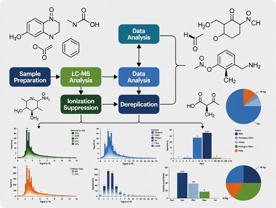
This article provides a comprehensive guide for researchers and drug development professionals on overcoming ion suppression, a major analytical challenge in LC-MS-based dereplication.
Overcoming Ion Suppression in LC-MS Dereplication: Strategies for Accurate Compound Identification in Complex Mixtures
Abstract
This article provides a comprehensive guide for researchers and drug development professionals on overcoming ion suppression, a major analytical challenge in LC-MS-based dereplication. Ion suppression from co-eluting matrix components can severely compromise signal intensity, detection limits, and the accuracy of compound identification in complex samples like natural product extracts or biological fluids. We explore the foundational mechanisms of this matrix effect, detail methodological strategies spanning from advanced sample preparation to chromatographic and instrumental optimization, and present systematic troubleshooting protocols. The article further reviews modern validation techniques and compares the efficacy of different mitigation approaches. By synthesizing current best practices and emerging solutions, this guide aims to empower scientists to develop robust, sensitive, and reliable dereplication workflows essential for accelerating drug discovery and natural product research.
Understanding the Enemy: The Fundamental Mechanisms of Ion Suppression in LC-MS Dereplication
Technical Support & Troubleshooting Center
Welcome to the technical support center for addressing ion suppression in LC-MS dereplication research. This resource provides targeted guidance for researchers encountering this pervasive matrix effect, which can compromise sensitivity, accuracy, and precision in the analysis of complex natural product or biological samples [1] [2]. The following FAQs, protocols, and strategies are framed within the critical context of developing robust, reproducible methods for compound identification and quantification.
Core Concepts & Mechanisms
What is ion suppression and why is it a critical issue in my dereplication work? Ion suppression is a matrix effect specific to mass spectrometry where co-eluting compounds from a complex sample reduce the ionization efficiency of your target analyte in the LC-MS interface [1] [3]. In dereplication, where you aim to identify known compounds in complex natural extracts quickly, suppression can lead to:
- False Negatives: Low-abundance bioactive compounds may fall below the detection limit [4].
- Quantitative Errors: Inaccurate measurement of metabolite levels, skewing biological interpretations [5] [6].
- Poor Reproducibility: Variable matrix composition causes fluctuating suppression, harming precision [1] [7].
What are the primary physical mechanisms behind ion suppression? The mechanism depends on the ionization technique, with Electrospray Ionization (ESI) being particularly susceptible [1] [3].
- In ESI: The process relies on charged droplet formation and evaporation. Co-eluting matrix components can:
- Compete for Charge: A limited excess charge is available on droplet surfaces. Compounds with higher surface activity or basicity outcompete analytes [1] [3].
- Alter Droplet Properties: Increase viscosity or surface tension, hindering solvent evaporation and ion release [3] [7].
- Cause Co-precipitation: Non-volatile materials can trap analytes in solid residues, preventing ionization [3].
- In APCI: Analytes are vaporized before ionization, generally making APCI less prone to suppression. When it occurs, it's often due to changes in charge transfer efficiency or solid formation in the vaporization region [1] [2].
How do key ionization sources compare in susceptibility? APCI often demonstrates significantly less ion suppression compared to ESI due to its different ionization mechanism [1] [3] [7]. The table below summarizes the comparison.
Table 1: Susceptibility to Ion Suppression by Ionization Source [1] [3] [2]
| Ionization Source | Mechanism | Relative Susceptibility to Ion Suppression | Primary Cause of Suppression |
|---|---|---|---|
| Electrospray (ESI) | Ion formation from charged droplets in liquid phase. | High | Competition for charge on droplet surface; altered droplet physics. |
| Atmospheric Pressure Chemical Ionization (APCI) | Vaporization followed by gas-phase chemical ionization. | Moderate to Low | Changes in charge transfer efficiency; solid formation during vaporization. |
Troubleshooting Guide: Identifying & Diagnosing Ion Suppression
Symptom: My analyte signal is much lower in a sample matrix than in a pure solvent standard.
- Diagnosis: This is a classic sign of ion suppression. The signal loss is not due to poor recovery but to interference during ionization [1] [6].
- Action: Perform a post-extraction spike experiment. Compare the signal of your analyte in a blank matrix extract spiked after preparation to the signal of an equivalent standard in neat solvent. A lower signal in the matrix indicates suppression [1] [4].
Symptom: I observe high variability in precision (%RSD) for my target analyte across different sample batches.
- Diagnosis: Variable levels of endogenous matrix components between samples (e.g., different plant extracts or patient plasma) cause varying degrees of ion suppression, leading to poor precision [7] [4].
- Action: Implement the post-column infusion experiment to map the chromatographic regions where suppression occurs. This helps pinpoint if suppression coincides with your analyte's retention time [1] [6].
Symptom: My method validation fails due to inconsistent accuracy or sensitivity.
- Diagnosis: Ion suppression directly impacts key validation parameters: accuracy (by skewing quantitation), sensitivity (by raising the limit of detection), and linearity [8] [4].
- Action: Systematically evaluate matrix effects as mandated by guidelines like the FDA's Bioanalytical Method Validation [4]. Use stable isotope-labeled internal standards (SIL-IS) where possible, as they co-elute with the analyte and best correct for suppression [5] [6].
Experimental Protocols for Validation
Protocol 1: The Post-Column Infusion Experiment (to Locate Suppression) This method visually identifies chromatographic regions affected by ion suppression [1] [6].
- Setup: Connect a syringe pump to post-column flow via a T-union. Continuously infuse a solution of your analyte (and its internal standard) at a constant, low rate.
- Run: Inject a prepared blank sample matrix (e.g., solvent-extracted control) onto the LC column and start the MS acquisition in scanning or MRM mode.
- Analysis: Monitor the signal for the infused analyte. A steady baseline indicates no suppression. Any dip in the baseline indicates the elution of matrix components that cause ion suppression. The retention time of the dip corresponds to the "danger zone" for your method [1].
Protocol 2: The Post-Extraction Spike Experiment (to Quantify Suppression) This method quantifies the magnitude of ion suppression (or enhancement) for your analyte [4] [6].
- Prepare Samples:
- (A) Neat Standard: Prepare your analyte at the target concentration in pure mobile phase or reconstitution solvent.
- (B) Spiked Matrix: Take several aliquots of a blank matrix extract (post-preparation), spike them with the same concentration of analyte.
- (C) Extracted Spiked Matrix (optional, for recovery): Spike the analyte into the blank matrix before sample preparation, then carry through the entire protocol.
- Analyze: Inject all samples and record the peak response (area or height).
- Calculate:
- Ion Suppression/Enhancement (%) =
(Response of B / Response of A) x 100 - A value <100% indicates suppression; >100% indicates enhancement.
- Compare (C) to (B) to separate ionization effects from recovery losses.
- Ion Suppression/Enhancement (%) =
Strategies for Mitigation & Correction
What is the most effective first step to reduce ion suppression? Optimize Sample Cleanup. Improving sample preparation is often the most effective strategy [8] [4].
- Switch Techniques: If you use protein precipitation (PP), consider more selective methods like Solid-Phase Extraction (SPE) or Liquid-Liquid Extraction (LLE) to remove more non-target matrix components [3] [4].
- Dilute and Inject: For concentrated samples, dilution can reduce the absolute amount of suppressors entering the source. This trades off sensitivity for robustness [3] [7].
How can I adjust my chromatographic method to minimize impact? Improve Separation. The goal is to shift your analyte's retention time away from the major suppression zones identified via the infusion experiment [3] [9].
- Modify the Gradient: Adjust the organic phase ramp to move your analyte.
- Change Chromatographic Selectivity: Use a different column chemistry (e.g., switch from C18 to phenyl-hexyl or HILIC) to alter the elution profile of both analyte and matrix interferences [5] [9].
When should I consider changing the ionization mode or source? If sample and chromatographic optimization are insufficient.
- Switch Ionization Polarity: If your analyte can ionize in both modes, try negative if you started in positive ESI, as fewer matrix compounds ionize in negative mode [1] [7].
- Change Ionization Source: If instrumentally feasible, switch from ESI to APCI (or atmospheric pressure photoionization, APPI). APCI is generally less susceptible to ion suppression from many matrix types [1] [2] [7].
Are there advanced correction techniques suitable for non-targeted dereplication? Yes, isotopic labeling workflows are emerging as a powerful solution. The IROA (Isotopic Ratio Outlier Analysis) Workflow uses a library of stable isotope-labeled internal standards spiked into every sample. By comparing the signal of the endogenous (light) analyte to its co-eluting, labeled (heavy) counterpart, the workflow can algorithmically calculate and correct for metabolite-specific ion suppression in non-targeted studies, greatly improving quantitative accuracy [5].
Visual Guide: Strategies to Overcome Ion Suppression
The following diagram illustrates the logical decision pathway for diagnosing and addressing ion suppression in method development.
The Scientist's Toolkit: Key Reagents & Materials
Table 2: Essential Research Reagents and Materials for Ion Suppression Management
| Item | Function & Role in Mitigating Ion Suppression | Key Consideration |
|---|---|---|
| Stable Isotope-Labeled Internal Standard (SIL-IS) | Gold standard for correction. Co-elutes with analyte, experiences identical suppression, allowing for accurate ratio-based quantification [5] [6]. | Ideally, the label (²H, ¹³C, ¹⁵N) should be metabolically inert and not alter chromatography. |
| IROA Internal Standard Library | A mixture of many ¹³C-labeled metabolites. Enables detection and correction of ion suppression across many analytes in non-targeted studies [5]. | Used with specialized software (e.g., ClusterFinder) to decode patterns and calculate corrections. |
| Solid-Phase Extraction (SPE) Cartridges | Selective cleanup to remove classes of matrix interferents (e.g., phospholipids, salts) prior to LC-MS, reducing source competition [8] [4]. | Select phase (C18, ion-exchange, mixed-mode) based on analyte and interference properties. |
| High-Purity LC-MS Solvents & Additives | Minimize background noise and source contamination, which can exacerbate suppression and cause artifacts [8] [9]. | Use volatile additives (ammonium formate/acetate, formic acid) and avoid non-volatile salts or ion-pairing agents. |
| Post-Column Infusion Kit (T-union, syringe pump) | Enables the critical diagnostic experiment to map chromatographic regions of ion suppression [1] [6]. | Ensure connections are leak-free and do not add significant dead volume. |
| Alternative Chromatography Columns | Different stationary phases (e.g., HILIC, phenyl, pentafluorophenyl) alter selectivity to shift analyte retention away from suppression zones [5] [9]. | Have a small portfolio of columns for method troubleshooting and optimization. |
Ion suppression is a critical matrix effect in liquid chromatography-mass spectrometry (LC-MS) where co-eluting compounds reduce the ionization efficiency of target analytes, leading to decreased signal intensity and compromised accuracy [1]. This phenomenon is a major challenge in quantitative bioanalysis, metabolomics, and dereplication research, where complex samples contain numerous endogenous and exogenous components that can interfere with analyte detection [1] [5].
The mechanisms and severity of suppression differ significantly between the two most common atmospheric pressure ionization (API) techniques: Electrospray Ionization (ESI) and Atmospheric Pressure Chemical Ionization (APCI). Understanding these differences is essential for developing robust analytical methods, troubleshooting sensitivity issues, and ensuring reliable data in drug development and natural product research [1] [10].
Troubleshooting Guide: FAQs on Ionization Suppression
Q1: My analyte signal is unexpectedly low and unstable. How can I determine if ion suppression is the cause? A: Conduct a post-column infusion experiment. This is a diagnostic protocol to map suppression zones in your chromatographic method [1].
- Experimental Protocol:
- Prepare a standard solution of your analyte at a moderate concentration (e.g., 1 µg/mL) in the starting mobile phase.
- Connect a syringe pump to the LC effluent line via a low-dead-volume T-union after the chromatographic column but before the MS ion source.
- Infuse the analyte solution at a constant, low flow rate (e.g., 5-10 µL/min) to establish a steady baseline signal.
- Inject a blank, prepared sample matrix (e.g., precipitated plasma, extracted solvent control) onto the LC column and run the analytical gradient.
- Observe the MS signal. A dip or reduction in the steady baseline indicates the elution of matrix components that suppress your analyte's ionization. The chromatographic trace of these dips reveals the retention time windows affected [1].
Q2: I'm developing a new method. Should I choose ESI or APCI to minimize suppression issues? A: The choice depends on your analyte's properties, but APCI generally experiences less severe ion suppression than ESI for small molecules (< 1000 Da) [1] [3].
- Key Mechanistic Difference:
- ESI: Ionization occurs from charged liquid droplets. Co-eluting compounds can compete for limited charge (space-charge effect) or space at the droplet surface. High concentrations of non-volatile or surface-active matrix components increase droplet viscosity/surface tension, hindering solvent evaporation and ion release [1] [3].
- APCI: The analyte is vaporized in a heated nebulizer before gas-phase chemical ionization. This eliminates competition for droplet space and charge. Suppression in APCI is less common and often related to changes in solvent evaporation efficiency or gas-phase proton transfer reactions [1] [10] [11].
- Recommendation: For non-polar, thermally stable small molecules, APCI is often more robust. For polar, thermally labile, or larger molecules, ESI is necessary. If suppression is observed in ESI, switching to APCI (if analytically feasible) can be an effective strategy [10] [3].
Q3: My chromatography looks good, but I still see suppression. What sample preparation or chromatographic strategies can help? A: Improved selectivity in sample cleanup and separation is the most direct way to reduce suppression [12] [8].
- Sample Preparation:
- Move beyond simple protein precipitation. Implement selective techniques like Solid-Phase Extraction (SPE) or Liquid-Liquid Extraction (LLE) to remove more classes of interfering matrix components [8] [3].
- Use a stable isotope-labeled internal standard (SIL-IS) for each analyte. Since the IS co-elutes with the analyte and experiences identical suppression, it corrects for the loss in signal [5].
- Chromatographic Optimization:
- Adjust Selectivity: Change column chemistry (e.g., from C18 to phenyl, pentafluorophenyl, or HILIC) to alter the retention of analytes versus matrix interferences [12].
- Extend Gradient Time: A shallower gradient improves resolution, increasing the chance of separating the analyte from suppressing compounds [12].
- Use Ion-Pairing (with caution): For highly polar acidic metabolites, volatile ion-pairing agents like tributylamine can improve retention on reversed-phase columns, aiding separation from early-eluting interferences [12].
Q4: Are there advanced methods to systematically correct for ion suppression in complex studies? A: Yes, novel workflows like the IROA (Isotopic Ratio Outlier Analysis) TruQuant have been developed for non-targeted metabolomics [5].
- Experimental Protocol (IROA Workflow):
- Spike a constant amount of a 13C-labeled internal standard (IROA-IS) library into all samples. This library contains a wide array of metabolites uniformly labeled with >95% 13C.
- Analyze samples by LC-MS. Each true metabolite will generate a doublet: the endogenous "light" (12C) peak and the spiked-in "heavy" (13C) peak.
- The heavy standard experiences the same suppression as the light analyte. Software algorithms use the signal of the suppressed heavy standard to calculate and mathematically correct the suppression effect on the light analyte signal [5].
- This approach can correct for suppression levels ranging from 1% to >90%, restoring linearity and quantitative accuracy [5].
Q5: What routine instrument maintenance practices prevent ion suppression caused by source contamination? A: Contaminant buildup in the ion source exacerbates suppression and signal instability [8] [13].
- Essential Maintenance Protocol:
- Regular Cleaning: Follow the manufacturer's schedule for cleaning the ion source components (e.g., sprayer capillary, cone, desolvation plates) with appropriate solvents (e.g., water, methanol, acetonitrile, 50:50 water:isopropanol).
- Use a Divert Valve: Install a valve to divert the LC flow to waste during column equilibration and the elution of strongly retained matrix components (e.g., lipids, salts). This prevents unnecessary contamination of the source [13].
- Employ Guard Columns: Use an inline guard column of the same chemistry as your analytical column to trap particulates and highly retained compounds.
- Monitor Source Performance: Track the signal intensity and stability of a reference compound in routine quality control samples. A gradual decline often indicates contamination.
Comparative Data: ESI vs. APCI
Table 1: Comparison of Ion Suppression Mechanisms in ESI and APCI [1] [10] [3]
| Aspect | Electrospray Ionization (ESI) | Atmospheric Pressure Chemical Ionization (APCI) |
|---|---|---|
| Ionization Phase | Late droplet phase / Gas-phase from charged droplets | Gas-phase only (after complete vaporization) |
| Primary Suppression Mechanisms | 1. Competition for limited charge in droplets (charge saturation). 2. Competition for droplet surface area (surface activity). 3. Increased droplet viscosity/surface tension slowing evaporation. 4. Gas-phase proton transfer reactions. | 1. Alteration of solvent vaporization efficiency (co-precipitation of analyte with non-volatiles). 2. Competition for gas-phase reagent ions (e.g., H3O+). |
| Susceptibility to Non-Volatile Salts/Matrix | High (disrupts droplet formation/evaporation) | Lower (analytes are vaporized) |
| Typical Analyte Suitability | Polar, thermally labile, large molecules (peptides, proteins, drugs with pre-existing charge) | Less polar, thermally stable, small to medium molecules (< 1000 Da, lipids, steroids, many natural products) |
| Relative Susceptibility to Ion Suppression | Generally higher | Generally lower |
Table 2: Effectiveness of Common Mitigation Strategies for Ion Suppression [12] [1] [5]
| Strategy | Effectiveness | Key Considerations / Trade-offs |
|---|---|---|
| Improved Chromatography | High | Most direct solution. Longer run times reduce throughput. |
| Selective Sample Cleanup (SPE, LLE) | High | Increases method development time and cost. Potential for analyte loss. |
| Switching Ionization: ESI → APCI | Medium to High | Not applicable for all analytes (e.g., large, labile molecules). |
| Dilution of Sample Extract | Medium | Simple but reduces sensitivity for low-abundance analytes. |
| Stable Isotope-Labeled Internal Standards | High (per analyte) | Gold standard for targeted quantitation. Expensive; requires synthesis for each analyte. |
| Post-Infusion Diagnosis | Diagnostic only | Essential for troubleshooting, but not a correction method. |
| Advanced Correction (e.g., IROA) | Very High (global) | Powerful for non-targeted studies. Requires specialized standards and software. |
Essential Visualizations
Diagram 1: Comparative Mechanisms of Ion Suppression in ESI vs. APCI [1] [10] [3]
Diagram 2: Workflow for Post-Column Infusion Experiment to Diagnose Suppression [1]
Diagram 3: IROA Workflow for Global Ion Suppression Correction [5]
The Scientist's Toolkit: Key Reagents & Materials
Table 3: Essential Research Reagents and Materials for Overcoming Ion Suppression
| Item / Reagent | Function in Mitigating Ion Suppression | Example / Note |
|---|---|---|
| Stable Isotope-Labeled Internal Standards (SIL-IS) | Co-elutes with analyte, experiences identical suppression, and corrects for signal loss. The gold standard for targeted quantitation [5]. | Deuterated (d-), 13C-, or 15N-labeled analogs of target analytes. |
| IROA Internal Standard Library | A mixture of many 13C-labeled metabolites used for global suppression correction and peak identification in non-targeted metabolomics [5]. | IROA TruQuant kit. Enables the workflow in Diagram 3. |
| HILIC Columns | Provides orthogonal separation to reversed-phase (RP) LC. Retains polar metabolites that elute in the void volume on RP, separating them from early-eluting salts and matrix [12]. | Aminopropyl, bare silica, or zwitterionic chemistries. |
| Volatile Mobile Phase Additives | Ensures compatibility with MS by preventing source contamination. Allows precise pH control to optimize ionization [12] [13]. | Formic acid, acetic acid, ammonium formate, ammonium acetate, ammonium hydroxide. |
| Solid-Phase Extraction (SPE) Cartridges | Selective sample cleanup to remove broad classes of matrix interferences (lipids, proteins, pigments) before LC-MS analysis [8] [3]. | C18, mixed-mode cation/anion exchange, hydrophilic-lipophilic balance (HLB). |
| Heated Nebulizer (APCI Probe) | The key hardware component for APCI. Vaporizes the LC eluent for gas-phase ionization, reducing susceptibility to suppression from non-volatiles [10] [11]. | Standard component on APCI or multimode ion sources. |
| Post-Column Infusion Kit | Hardware to perform the diagnostic infusion experiment. Critical for method development and troubleshooting [1]. | Syringe pump, PEEK low-dead-volume T-union, and fittings. |
Technical Support Center: FAQs and Troubleshooting Guides
This technical support center is designed to help researchers diagnose and resolve the critical challenge of ionization suppression in LC-MS dereplication workflows. Ion suppression occurs when co-eluting matrix components interfere with the ionization efficiency of your target analytes, leading to reduced signal fidelity, elevated detection limits, and an increased risk of false negatives—ultimately derailing natural product discovery and metabolomics studies [14] [1].
Section 1: Understanding Ion Suppression in Dereplication
Q1: My dereplication screening suddenly has lower sensitivity for known metabolites. The peaks are there, but the signal is weaker. What's happening? This is a classic symptom of ion suppression. Co-eluting matrix components, such as phospholipids, salts, or endogenous polymers from your biological extract, are competing with your analytes for charge during the electrospray ionization process [14] [1]. This competition reduces the ionization efficiency of your targets. Even if chromatographic separation looks adequate, these invisible interferents in the ion source can cause significant, variable signal loss. The problem often worsens over an injection sequence as matrix components accumulate on the column and in the ion source [14].
Q2: How can I tell if my false negatives are due to ion suppression or simply that the compound is not present? Distinguishing a true negative from a suppression-induced false negative is critical. Implement a post-column infusion experiment [14] [1]. By continuously infusing a standard of your target analyte post-column while injecting a blank matrix extract, you create a stable baseline signal. Any dip in this baseline corresponds to a retention time window where co-eluting matrix components suppress ionization. If your analyte's expected retention time falls within a suppression "dip," you have strong evidence for a false negative. Furthermore, spiking your sample with a known concentration of the target and observing a lower-than-expected recovery confirms the issue [1].
Q3: Are some ionization techniques more prone to suppression than others? Yes. Electrospray Ionization (ESI) is notoriously susceptible because ionization occurs in the liquid phase, where analytes compete for limited charge on the droplet surface [1]. Atmospheric Pressure Chemical Ionization (APCI) is generally less prone as analytes are vaporized before ionization, though it is not immune [1]. For dereplication of small molecules, testing both sources can be insightful. If sensitivity improves significantly with APCI, ion suppression in ESI is a likely culprit.
Section 2: Practical Troubleshooting and Mitigation
Q4: My initial "dilute-and-shoot" method was fast, but now my data is unreliable. What's the best first step to fix it? "Dilute-and-shoot" is a major risk factor as it introduces the full complexity of the matrix into your LC-MS system [14]. The most effective first step is to enhance sample clean-up. Move to a technique that selectively removes the interferents causing your specific problem:
- For phospholipids (a common suppressor eluting in mid-gradient), use Solid-Phase Extraction (SPE) with phospholipid removal cartridges [14].
- For proteins and peptides, optimize protein precipitation (though it's incomplete) or combine it with filtration [14].
- For broad-spectrum clean-up, evaluate liquid-liquid extraction (LLE). The optimal choice depends on your analyte's chemical properties and the dominant interferent in your matrix [8].
Q5: I've cleaned my sample, but I still see signal instability and high background. What should I check in my LC-MS system? This points to carryover or accumulation of non-volatile matrix in the instrument. Follow this checklist:
- Inspect and clean the ion source: Remove the ESI probe and thoroughly clean the capillary, cone, and other lenses with appropriate solvents (e.g., water, methanol, acetonitrile, 1% formic acid) [14].
- Evaluate the chromatographic column: Perform a post-column infusion experiment. If suppression regions have widened or shifted, phospholipids or other materials may have built up on the column head. Flush with strong solvents (e.g., 95% organic) or replace the guard/analytical column [14].
- Check for system contamination: Run strong blank gradients and inspect the baseline. High background can indicate contamination in the autosampler, tubing, or mobile phase reservoirs.
Q6: How can I adjust my chromatographic method to minimize suppression? The goal is to separate your analytes from the major suppression zones. Use the post-column infusion map as a guide to:
- Adjust the gradient: Modify the organic solvent ramp to shift your analytes' retention times away from major suppression dips (e.g., those caused by phospholipids around 4-8 minutes or salts at the void volume) [14].
- Optimize the column: Switch to a column with different selectivity (e.g., from C18 to phenyl-hexyl or HILIC) to change the elution order of analytes and matrix components.
- Extend run time: If your method is very short (<5 min), highly retained phospholipids may not elute and will accumulate, causing progressive suppression. A longer wash at high organic at the end of each run can help clear the column [14].
Section 3: Advanced Strategies and Validation
Q7: For untargeted dereplication, how do I validate that my workflow is robust against ion suppression? Incorporate these assessments into your method validation:
- Post-extraction spike experiment: Spike your target analytes into final extracted blank matrix and compare the response to neat standards in solvent. Signal recovery <85% or >115% indicates significant suppression or enhancement [1].
- Use of stable isotope-labeled internal standards (SIL-IS): For quantitative precision, SIL-IS are the gold standard as they co-elute with the native analyte and experience identical suppression, perfectly compensating for it [15].
- Dilution integrity test: Perform serial dilutions of a matrix sample. Response should be linear. Non-linearity often indicates matrix effects that are concentration-dependent [15].
Q8: Can computational tools help overcome identification gaps caused by suppression? Absolutely. When suppression leads to weak or missing MS/MS spectra, traditional library matching fails. Next-generation machine learning foundation models like LSM-MS2 can help. Trained on millions of spectra, these models learn a "chemical semantic space" and can identify compounds with higher accuracy from noisy or low-intensity spectra, effectively mitigating the impact of suppression on identification rates [16]. They are particularly valuable for distinguishing challenging isomers, a common task in dereplication [16].
Quantitative Impact of Ion Suppression on Data Quality
The following tables summarize the measurable consequences of ionization suppression and matrix effects on analytical outcomes, drawing from recent experimental data.
Table 1: Impact of Matrix Effects on Metabolite Quantification Linearity [15] This study on untargeted metabolomics highlights how non-ideal instrument response directly increases the risk of false negatives.
| Analysis Context | Key Finding | Implication for False Negatives |
|---|---|---|
| Broad Dilution Series (9 levels) | 70% of 1327 detected metabolites showed non-linear response in at least one dilution level. | Signal intensity does not reliably reflect concentration, complicating relative quantification across samples. |
| Focused Linear Range (4 consecutive levels, 8-fold range) | 47% of metabolites demonstrated linear behavior. | Over half of metabolites have a limited usable quantitative range; outside this range, data is unreliable. |
| Direction of Error | Abundances in dilute samples were mostly overestimated, rarely underestimated. | Statistical analysis is more likely to miss true differences (increase false negatives) than to create false positives. |
Table 2: Performance of Advanced Spectral Models in Overcoming Spectral Gaps [16] Machine learning models can recover identifications from sub-optimal data, partially counteracting the identification loss caused by ion suppression.
| Model & Benchmark | Performance Gain | Relevance to Dereplication |
|---|---|---|
| LSM-MS2 vs. Traditional Cosine Similarity | 30% improvement in accuracy for identifying challenging isomers. | Dramatically improves confidence in distinguishing structurally similar natural products. |
| LSM-MS2 in Complex Biological Samples | 42% more correct identifications. | Directly addresses the identification bottleneck caused by low signal-to-noise and interfering backgrounds. |
| LSM-MS2 under Low-Concentration Conditions | Maintains robust identification performance. | Mitigates the impact of sensitivity loss due to ion suppression, reducing false negatives. |
Detailed Experimental Protocols
Protocol 1: Post-Column Infusion Experiment to Map Ion Suppression Zones
Objective: To visually identify chromatographic regions where matrix components suppress (or enhance) ionization [14] [1].
Materials:
- LC-MS/MS system with a syringe pump or secondary HPLC pump.
- Tee-union for post-column mixing.
- Standard solution of a representative analyte (e.g., 100 ng/mL in mobile phase).
- Prepared blank matrix extract (e.g., from fermentation broth, plant extract).
- Mobile phase (solvent blank).
Procedure:
- Setup: Connect the syringe pump containing the analyte standard via the tee-union between the column outlet and the MS ion source.
- Establish Baseline: Start the LC gradient and the syringe pump at a constant flow (e.g., 10 µL/min). Inject a mobile phase blank. The MS signal for the infused analyte should be relatively stable, rising and falling gently with the organic modifier gradient [14].
- Inject Matrix: Without changing any parameters, inject the blank matrix extract. The analyte signal will now show dips or peaks corresponding to suppression or enhancement zones caused by co-eluting matrix.
- Identification: To identify common suppressors like phospholipids, simultaneously monitor the MRM transition m/z 184 → 184 (a signature fragment of phosphatidylcholines and lyso-phosphatidylcholines) [14]. Its chromatographic trace will align with major suppression regions.
- Analysis: Overlay the two chromatograms. Regions where the matrix injection trace falls below the solvent baseline are ionization suppression zones. Adjust your method to elute critical analytes away from these regions.
Protocol 2: Optimization of Sample Clean-up using Solid-Phase Extraction (SPE)
Objective: To selectively remove phospholipids and other interferents prior to LC-MS analysis [14].
Materials:
- SPE cartridges (e.g., 30 mg, designed for phospholipid removal).
- Conditioning solvents (e.g., methanol, water).
- Wash solvents (e.g., water, 5% methanol).
- Elution solvent (e.g., methanol, acetonitrile, or a mixture with a volatile acid/base).
- Centrifuge and vacuum manifold.
Procedure:
- Sample Preparation: Precipitate proteins from your biological sample (e.g., with cold acetonitrile). Centrifuge and collect the supernatant. Dilute with water if necessary to match the loading condition of the SPE sorbent.
- SPE Conditioning: Condition the cartridge with 1 mL of methanol, then equilibrate with 1 mL of water. Do not let the sorbent dry out.
- Sample Loading: Load the prepared supernatant onto the cartridge slowly. Collect the flow-through if analytes are unretained.
- Washing: Wash with 1 mL of a weak solvent (e.g., 5% methanol in water) to remove salts and polar interferents. Discard wash.
- Elution: Elute retained analytes with 1-2 mL of a strong organic solvent (e.g., methanol containing 1% formic acid). Collect the eluate.
- Evaluation: Evaporate the eluate under nitrogen or vacuum and reconstitute in initial mobile phase. Analyze alongside a "dilute-and-shoot" sample and a neat standard. Compare signal intensity, background noise, and the results of a post-column infusion test to assess clean-up efficiency.
Protocol 3: Implementing a Machine Learning Model for Enhanced Spectral Identification
Objective: To use a foundation model (e.g., LSM-MS2) to improve compound identification rates from low-quality spectra [16].
Procedure:
- Data Preparation: Export your MS/MS spectral data in a standard format (e.g., .mzML). Ensure metadata is included.
- Model Access: Access a cloud-based API or local installation of a spectral foundation model like LSM-MS2 [16].
- Spectral Encoding: Input your experimental spectra into the model. The model converts each spectrum into a high-dimensional embedding vector that represents its chemical features in a semantic space.
- Similarity Search: Instead of traditional library matching, perform a similarity search (e.g., cosine similarity) between your experimental spectrum's embedding and a library of embeddings generated from known reference spectra.
- Ranking & Annotation: The model returns a ranked list of candidate compounds based on embedding similarity, which often proves more accurate than spectral cosine similarity, especially for low-abundance or noisy spectra.
- Validation: Manually review top candidates, checking for consistency with retention time, precursor m/z, and plausible fragmentation.
Visual Workflows and Pathways
The Scientist's Toolkit: Key Reagents and Materials
Table 3: Essential Research Reagent Solutions for Mitigating Ion Suppression
| Item | Primary Function | Application in Dereplication |
|---|---|---|
| Phospholipid Removal SPE Cartridges | Selective sorbent to bind and retain phospholipids (lyso-PC, PC) from biological extracts [14]. | Critical for cleaning up crude fermentation broths, plant extracts, or tissue homogenates prior to LC-MS to eliminate a major source of mid-gradient ion suppression. |
| Stable Isotope-Labeled Internal Standards (SIL-IS) | Chemically identical to the analyte but with heavier isotopes (e.g., ¹³C, ²H); co-elutes and experiences identical matrix effects [15]. | Gold standard for compensating for ion suppression in quantitative dereplication and for method validation. Enables accurate recovery calculations. |
| Post-Column Infusion Tee Assembly | Low-dead-volume union to mix column effluent with a continuously infused standard solution [14] [1]. | Enables the experimental visualization of ion suppression zones in a chromatographic run, which is fundamental to method development and troubleshooting. |
| Volatile Mobile Phase Additives | Ammonium formate, ammonium acetate, formic acid, acetic acid. Promote ionization and are compatible with MS detection [8]. | Used instead of non-volatile salts (e.g., phosphate buffers) to prevent source contamination and salt-adduct formation, simplifying spectra and improving sensitivity. |
| U-¹³C Labeled Biological Reference Material | Fully isotopically labeled extract from an organism grown on ¹³C substrate (e.g., ¹³C-labeled wheat ears) [15]. | Serves as a comprehensive internal standard for untargeted metabolomics. Enables filtering of true metabolites from background and correction for global matrix effects. |
| Machine Learning Spectral Model (e.g., LSM-MS2) | A computational foundation model trained on millions of spectra for advanced spectral matching and embedding [16]. | Used to boost identification rates from low-intensity or noisy MS/MS spectra resulting from ion suppression, reducing false negatives. |
Technical Support & Troubleshooting Center
Troubleshooting Guide: Common LC-MS Ionization Suppression Issues
Q1: Why do I observe a sudden, significant drop in signal intensity for my target analytes when analyzing a new natural product extract? A: This is a classic symptom of ionization suppression caused by co-eluting matrix components. Natural product extracts are complex mixtures containing salts, organic acids, polyphenols, and phospholipids that can co-elute with your analyte, competing for charge and droplet surface during electrospray ionization (ESI). To troubleshoot:
- Dilute and Re-inject: A simple 5-10 fold dilution of the sample can reduce suppression. If signal recovers proportionally, matrix effects are confirmed.
- Analyze a Post-Column Infusion Sample: Infuse a constant flow of your analyte into the LC effluent post-column while injecting a blank matrix extract. A dip in the baseline at the retention time of your analyte visually confirms suppression.
- Modify the Chromatography: Increase gradient time or adjust mobile phase pH to shift the retention time of your analyte away from the "matrix cloud" (typically eluting in the solvent front or at mid-polarity ranges).
Q2: My internal standard (IS) signal is suppressed, but it's a stable isotope-labeled version of my analyte. What could be wrong? A: While stable isotope-labeled IS are the gold standard for compensating for suppression, they are not immune if the suppression is extremely severe or if the IS co-elutes with a high-concentration suppressor. Ensure your IS is added prior to extraction to correct for recovery issues. If suppression persists, consider:
- Using an Analog IS with Different Retention: Choose an IS that is structurally similar but has a slightly shifted retention time to avoid the exact same co-eluting interferents.
- Improving Sample Cleanup: Implement a more selective extraction or purification step (e.g., SPE with mixed-mode sorbents) prior to LC-MS analysis.
Q3: I see high background noise and inconsistent signals in biological plasma samples. What are the likely sources? A: Phospholipids and non-esterified fatty acids are the primary culprits in plasma/serum. They are ubiquitous, ionize efficiently, and cause severe, variable suppression, particularly in positive ESI mode.
- Phospholipids: Typically elute in a broad band in reversed-phase chromatography. Use a Phospholipid Removal Plate or SPE cartridge (e.g., HybridSPE, Ostro) during sample preparation.
- Proteins & Salts: Ensure complete protein precipitation and removal. Centrifuge samples thoroughly and avoid injecting the pellet or any particulate matter.
Q4: How can I quickly assess the degree of matrix effect in my method? A: Perform a quantitative matrix effect experiment as per Matuszewski et al. (2003). Compare the analyte response in three different sets:
- Set A: Analyte in pure solvent.
- Set B: Analyte spiked into post-extraction matrix.
- Set C: Analyte spiked into pre-extraction matrix. Calculate the Matrix Factor (MF = B/A) and process efficiency (PE = C/A). An MF ≠ 1 indicates ionization suppression/enhancement.
Frequently Asked Questions (FAQs)
Q: What is the single most effective sample prep step for reducing suppression in plant extracts? A: Solid-Phase Extraction (SPE) with a selective sorbent. For acidic interferents (e.g., phenolics), use a mixed-mode anion-exchange cartridge. For basic interferents, use a mixed-mode cation-exchange cartridge. This selectively retains your analyte or the interferents, dramatically cleaning the sample.
Q: Are there mobile phase additives that can help minimize suppression? A: Yes, but with caution. Additives like formic acid (0.1%) or ammonium formate (2-10 mM) can improve ionization efficiency and reproducibility. However, avoid non-volatile buffers (e.g., phosphate, Tris) at all costs, as they cause severe suppression and instrument contamination.
Q: Does switching from ESI to APCI help with suppression? A: Often, yes. Atmospheric Pressure Chemical Ionization (APCI) is less susceptible to many common matrix effects because ionization occurs in the gas phase rather than in the charged droplet. If your analyte is thermally stable and amenable to APCI, it can be a viable solution.
Q: How critical is column choice in managing matrix effects? A: Very critical. Using a UPLC column with smaller particle size (<2 μm) provides better chromatographic resolution, helping to separate analytes from co-eluting matrix components. Also, consider alternative selectivity (e.g., HILIC, phenyl-hexyl) to shift problematic interferents away from your analytes.
Experimental Data & Protocols
Table 1: Key Sources of Ionization Suppression and Mitigation Strategies
| Matrix Type | Primary Suppressing Compounds | Typical Retention Time (RP-C18) | Impact on Signal (ESI+) | Recommended Mitigation Strategy |
|---|---|---|---|---|
| Plant/ Natural Product Extract | Polyphenols, Organic Acids, Terpenoids, Chlorophyll | Early eluting (0.5-3 min), Broad bands | High to Severe | SPE (Polyamide, HLB); Dilution; Improved Chromatographic Gradient |
| Blood Plasma/Serum | Phospholipids (LPC, PC), Fatty Acids | 2-8 min (depending on chain length) | Severe & Variable | Phospholipid Removal SPE; LLE with MTBE/Hexane; 2D-LC |
| Microbial Broth/Fermentation | Salts (Na+, K+), Sugars, Peptides, Media Components (e.g., PEG) | Solvent Front, Various | Moderate to Severe | Desalting (SPE, TCA precipitation); Dialysis; Dilution |
| Urine | Urea, Salts, Metabolic Acids | Solvent Front | Moderate | Dilution; Sample Dielectric Barrier Discharge (DBD) Plasma Treatment |
Detailed Protocol: Post-Column Infusion Experiment for Visualizing Suppression
Objective: To visually identify regions of ionization suppression/enhancement in a chromatographic run. Materials: LC-MS system, syringe pump, T-union, your analytical column, blank matrix extract. Procedure:
- Prepare a solution of your target analyte at a concentration that gives a stable signal (~100 ng/mL in starting mobile phase).
- Set up a syringe pump connected via a low-dead-volume T-union placed between the column outlet and the MS ion source.
- Start the infusion of the analyte solution at a constant, low flow rate (e.g., 5-10 μL/min).
- While infusing, program the LC-MS to inject your blank matrix extract (e.g., extracted control plasma or solvent-only reconstituted plant extract) using your standard analytical method.
- Acquire data in selected ion monitoring (SIM) mode for your analyte.
- Interpretation: A stable baseline indicates no matrix effect. A dip in the baseline indicates suppression at that retention time; a peak indicates enhancement.
Objective: To calculate the Matrix Factor (MF) and Process Efficiency (PE) for a validated bioanalytical method. Procedure:
- Prepare three sets of samples (n=6 each) at Low, Mid, and High QC concentrations.
- Set A (Neat Solution): Analyte in reconstitution solvent (no matrix).
- Set B (Post-extraction Spike): Blank matrix extracted, then analyte spiked into the cleaned extract.
- Set C (Pre-extraction Spike): Analyte spiked into blank matrix, then carried through the entire sample preparation process.
- Analyze all samples by LC-MS.
- Calculate the peak area for each injection.
- Compute for each QC level:
- Matrix Factor (MF) = Mean Peak Area (Set B) / Mean Peak Area (Set A).
- Process Efficiency (PE) = Mean Peak Area (Set C) / Mean Peak Area (Set A).
- A MF of 1.0 indicates no matrix effect. <1.0 = suppression; >1.0 = enhancement.
- PE combines the effects of extraction recovery and matrix effect.
Visualizations
Troubleshooting Path for LC-MS Suppression
Mechanisms of Ionization Suppression by Matrix
The Scientist's Toolkit: Key Reagent Solutions
Table 2: Essential Materials for Mitigating Ionization Suppression
| Item/Category | Function & Rationale | Example Product(s) |
|---|---|---|
| Mixed-Mode SPE Sorbents | Selective retention of acidic/basic/neutral interferents while allowing analyte to pass (or vice versa). Crucial for cleaning complex extracts. | Oasis MCX (mixed-mode cation exchange), Oasis MAX (mixed-mode anion exchange), Strata-X-CW (weak cation exchange). |
| Phospholipid Removal Plates | Selectively binds phospholipids via zirconia-coated or other proprietary chemistry. Essential for clean plasma/serum analysis. | Waters Ostro Plate, Phenomenex Phree, HybridSPE-Phospholipid. |
| Volatile Buffers & Additives | Maintain required pH or ionic strength without causing source contamination or signal suppression. Must be MS-compatible. | Ammonium formate, Ammonium acetate, Formic acid, Trifluoroacetic acid (use with care). |
| Stable Isotope-Labeled Internal Standards (SIL-IS) | Gold standard for compensating for both matrix effects and extraction variability. Co-elutes with analyte but distinct m/z. | [13C] or [15N] labeled versions of target analytes. |
| UPLC/HPLC Columns with Alternative Selectivity | Provides orthogonal separation to shift analyte retention away from common matrix interferent bands. | HILIC columns (for polar analytes), Phenyl-Hexyl, PFP, Charged Surface Hybrid (CSH) columns. |
| Protein Precipitation Plates | High-throughput removal of proteins from biological samples, reducing a major source of suppression and column fouling. | 96-well filter plates (e.g., Agilent Captiva), using ACN or MeOH with additives. |
Building a Robust Workflow: Methodological Strategies to Minimize Ion Suppression
Technical Support & Troubleshooting Center
This center addresses common challenges encountered when implementing Solid-Phase Extraction (SPE) and Liquid-Liquid Extraction (LLE) to mitigate ionization suppression in LC-MS dereplication. The guidance is framed within the critical need for robust sample cleanup to ensure accurate metabolite identification and quantification in complex biological matrices [17].
Troubleshooting Guide: Ion Suppression and Sample Cleanup
Problem: Inconsistent analyte recovery and high %RSD during quantitative LC-MS analysis.
- Potential Cause: Incomplete removal of phospholipids, a major source of ion suppression, leading to buildup in the LC system and variable matrix effects [14].
- Diagnostic Test: Perform a post-column infusion experiment. Infuse a constant stream of your analyte into the MS while injecting a cleaned sample extract. Signal dips in the baseline indicate ion suppression zones caused by co-eluting matrix [14].
- Solution: Implement a more selective cleanup. Switch from generic protein precipitation to SPE designed for phospholipid removal (e.g., hybrid or polymeric phases). For lipid-rich matrices, consider Enhanced Matrix Removal-Lipid (EMR-Lipid) cartridges, which have shown removal efficiencies of 42-58% by weight [18].
Problem: Gradual loss of sensitivity and increased system backpressure over a batch of samples.
- Potential Cause: Accumulation of non-volatile matrix components (proteins, salts, lipids) in the ion source or on the head of the analytical column [17] [14].
- Diagnostic Test: Inspect the ion source for visible contamination. Monitor the signal of a system suitability standard over the course of a batch. A steady decline indicates buildup.
- Solution: Re-evaluate sample cleanup stringency. Ensure wash steps in SPE protocols are optimized to remove salts and polar interferences. Consider automating cleanup using online SPE or turbulent flow chromatography, which efficiently excludes high molecular weight interferences and reduces source contamination [19].
Problem: Poor recovery of a specific, ionizable analyte class (e.g., tetracyclines, beta-lactams) from a complex matrix.
- Potential Cause: The sample preparation pH or solvent strength does not favor the stability or solubility of the target analytes, leading to degradation or poor extraction efficiency [18] [20].
- Diagnostic Test: Perform a recovery test with stable isotope-labeled internal standards (SIL-IS) if available. Low and inconsistent recovery for specific analytes points to method incompatibility.
- Solution: Optimize the extraction protocol. For multi-class residue analysis, a two-step extraction with tailored buffers may be necessary [18]. For ionizable compounds, use SPE sorbents with mixed-mode interactions (e.g., reverse-phase plus cation exchange) to improve selectivity and recovery [21].
Frequently Asked Questions (FAQs)
Q1: How can I definitively prove that my sample cleanup method is effectively reducing ion suppression for my specific assay? A: The most direct way is to perform a post-column infusion experiment and compare the results from a "dilute-and-shoot" sample versus your cleaned extract [14]. Additionally, you can quantify the matrix effect (ME) by comparing the MS response of an analyte spiked into a post-extracted blank matrix to the response in a pure solvent. An ME close to 100% (or apparent recovery between 85-115%) indicates successful suppression mitigation [20].
Q2: Protein precipitation is fast and simple. Why should I consider moving to more complex SPE or LLE methods? A: While protein precipitation removes proteins, it leaves behind small molecules, salts, and most phospholipids, which are a primary cause of ion suppression in ESI-MS [14] [21]. SPE and LLE provide selective cleanup and analyte enrichment, leading to cleaner extracts, reduced source contamination, lower limits of quantification, and more robust methods suitable for regulated bioanalysis [17] [22].
Q3: What is the key to developing a successful SPE method for a new analyte? A: The core principle is orthogonality: the retention mechanism in sample preparation should differ from the separation mechanism in LC. If you use a C18 analytical column, consider a mixed-mode (e.g., cation-exchange) SPE sorbent. This approach maximizes the removal of interferences that would otherwise co-elute with your analyte [21]. Method optimization should focus on the conditioning, loading, washing, and elution solvents' pH and strength to balance high recovery with maximal cleanup [20].
Q4: For high-throughput labs, are there ways to automate advanced sample cleanup? A: Yes. Online SPE and turbulent flow chromatography (TurboFlow) are effective automated solutions. Online SPE integrates extraction directly with the LC system, improving reproducibility and sensitivity by eliminating manual steps [21] [19]. TurboFlow uses high flow rates and large-particle columns to achieve cleanup based on chemical affinity and size exclusion, allowing for direct injection of complex samples and significant time savings [19].
Performance Data & Comparative Analysis
Quantitative Outcomes of Advanced Cleanup Techniques
Table 1: Performance Metrics of SPE and EMR-Lipid Cleanup in Multi-Residue Analysis
| Cleanup Technique | Matrix | Key Performance Indicator | Reported Outcome | Source |
|---|---|---|---|---|
| Enhanced Matrix Removal-Lipid (EMR-Lipid) | Porcine/Bovine/Chicken Meat | % Analyte Recovery (at 3 spiking levels) | >90% of analytes in 60-120% range | [18] |
| Method Precision (%RSD) | >97% of analytes with RSD < 20% | [18] | ||
| Matrix Co-extractive Removal | 42-58% removal by weight | [18] | ||
| Frequency of Significant Ion Suppression (>30%) | Reduced to <15% of compounds | [18] | ||
| Polymeric SPE | Shellfish (Mussel, Scallop, Oyster) | Recovery for Lipophilic Toxins | ~90% for all toxins studied | [20] |
| Matrix Effect (Apparent Recovery) | 85-115% (ME < ±15%) with optimized LC-MS method | [20] |
Comparative Guide: Selecting a Sample Preparation Strategy
Table 2: Strategic Selection of Sample Preparation Techniques for Ion Suppression Mitigation
| Technique | Best For | Primary Mechanism | Advantages | Limitations for Dereplication |
|---|---|---|---|---|
| Protein Precipitation (PPT) | Rapid screening; high-throughput initial assessment. | Denaturation and pelleting of proteins. | Fast, simple, low-cost, universal [22] [21]. | Poor removal of phospholipids and small molecules; high ion suppression risk; less sensitivity [14] [21]. |
| Liquid-Liquid Extraction (LLE) | Non-polar to moderately polar analytes; mid-level cleanup. | Partitioning between immiscible solvents. | Excellent cleanup for certain classes; can be automated [22] [21]. | Emulsion risk; uses large solvent volumes; not ideal for very polar or ionic compounds. |
| Solid-Phase Extraction (SPE) | Targeted or class-specific analysis requiring high sensitivity. | Selective adsorption/desorption from a sorbent. | High selectivity and enrichment; clean extracts; variety of sorbents; automatable [22] [20]. | Requires method development; can be more expensive per sample. |
| Enhanced Matrix Removal (EMR) | Lipid-rich matrices (tissue, food, plasma). | Size-exclusion and chemical interaction for lipid removal. | Highly selective lipid removal; maintains good recovery for many drug-like molecules [18]. | Specific to lipid removal; may require optimization for different lipid classes. |
Detailed Experimental Protocols
This protocol demonstrates a robust approach for complex, lipid-rich matrices.
- Homogenization & Extraction: Homogenize meat sample (e.g., bovine muscle). Perform a two-step solid-liquid extraction using a tailored buffer/solvent system to efficiently cover a wide polarity range, including difficult classes like tetracyclines.
- Cleanup: Load the extract onto a pre-conditioned Enhanced Matrix Removal-Lipid (EMR-Lipid) cartridge. The sorbent selectively retains lipids via a chemical affinity mechanism.
- Wash & Elute: Pass a wash solvent to remove remaining matrix interferences. Elute the target veterinary drugs with a suitable organic solvent. The process removes 42-58% of co-extractive lipids by weight.
- Analysis & Validation: Analyze by LC-MS/MS. Validate by assessing recovery (60-120% for >90% of analytes), precision (RSD <20% for >97%), and matrix effect. The method achieves LOQs of 1-5 μg/kg.
This protocol highlights method optimization for ionization efficiency.
- Standard & Extract Prep: Prepare toxin standards (e.g., Okadaic Acid, Azaspiracid-1) in methanol. Obtain a crude methanolic extract from shellfish tissue.
- SPE Optimization: Test an array of SPE sorbents. Polymeric sorbents (e.g., Strata-X) show best retention for most toxins. Optimize loading, washing, and elution conditions (pH, solvent composition) to maximize recovery (~90%) and cleanliness.
- Orthogonal LC-MS Analysis: Analyze cleaned extracts using two orthogonal LC methods (acidic vs. alkaline mobile phase) coupled to MS. This tests the robustness of cleanup under different conditions.
- Matrix Effect Assessment: Compare the MS signal of toxins in the cleaned extract to signals in pure solvent. The optimal combination (polymeric SPE + alkaline LC) reduces matrix effects to less than ±15% (apparent recovery 85-115%).
Visual Workflows and Relationships
Cleanup Strategy Impact on LC-MS Dereplication
Post-Column Infusion to Detect Ion Suppression [14]
The Scientist's Toolkit: Essential Reagents & Materials
Table 3: Key Research Reagent Solutions for Advanced Sample Cleanup
| Item | Function in Cleanup | Application Context |
|---|---|---|
| Polymeric SPE Sorbents (e.g., Strata-X, Oasis HLB) | Retain analytes via hydrophobic and polar interactions over a wide pH range. Provide cleaner extracts than C18 silica for many biological matrices [20] [21]. | General sample cleanup for a broad spectrum of drug-like molecules in plasma, urine, or tissue extracts. |
| Mixed-Mode SPE Sorbents (Cation/Anion Exchange + RP) | Provide orthogonal selectivity by retaining analytes via ionic + hydrophobic interactions. Wash steps can remove neutral and opposite-charge interferences [21]. | Selective isolation of ionizable analytes (e.g., basic drugs) from complex matrices to minimize ion suppression. |
| Enhanced Matrix Removal (EMR) Cartridges | Selectively remove lipids from samples through a chemical affinity mechanism without retaining mid-polarity analytes [18]. | Cleanup of lipid-rich matrices (e.g., meat, liver, brain homogenate, avocado) prior to multi-residue analysis. |
| Phospholipid Removal Plates (e.g., HybridSPE) | Selectively precipitate and remove phospholipids from biological fluids using a proprietary zirconia-coated sorbent [14]. | Rapid de-lipidation of plasma or serum samples to mitigate a major source of ion suppression in ESI-MS. |
| Volatile Buffers (Ammonium Formate, Ammonium Acetate) | Provide pH control during extraction and chromatography without leaving non-volatile salts that cause ion suppression and source contamination [17]. | Used in SPE conditioning/loading solutions and as mobile phase additives in LC-MS. |
Technical Support Center: Overcoming Ionization Suppression in LC-MS Dereplication
This technical support center provides targeted troubleshooting and methodological guidance for researchers focused on overcoming ionization suppression—a major matrix effect that compromises detection capability, precision, and accuracy in LC-MS [1]. In the context of dereplication research, where the goal is to efficiently identify known compounds in complex natural product or metabolomic extracts, ionization suppression caused by co-eluting matrix components is a critical bottleneck [23]. The following guides and protocols detail how advanced chromatographic strategies, including Microflow LC, UHPLC, and orthogonal separations, can be leveraged to enhance resolution, reduce suppression, and improve the fidelity of your analyses.
Troubleshooting Guide: Common LC-MS Symptoms & Solutions in Dereplication
Use this guide to diagnose and resolve common issues that directly impact resolution and contribute to ionization suppression.
| Symptom | Primary Cause in Dereplication | Recommended Solution | Prevention Strategy |
|---|---|---|---|
| Broad Peaks | Column overload from high-concentration matrix components; extra-column volume [24]. | Reduce injection volume/mass; use narrower i.d. tubing [24]. For critical separations, switch to a UHPLC system with smaller particle columns (<2µm) to increase peak capacity [23]. | Implement sample dilution or cleaner extraction. Use Microflow LC (column i.d. ≤ 0.5mm) to improve ionization efficiency and reduce background interference [25] [26]. |
| Tailing Peaks | Secondary interactions with active sites on a contaminated or old column [24]; mismatch between injection solvent and mobile phase strength. | Replace guard cartridge; wash analytical column with strong solvent [24]. Ensure injection solvent is same or weaker strength than starting mobile phase. | Use high-quality, pH-stable columns. For basic analytes common in natural products, consider a charged surface hybrid (CSH) column. Perform regular column maintenance. |
| High Backpressure | Blockage from non-volatile or particulate matrix components [27]. | Replace in-line filter or guard column frit [27]. If pressure remains high, the analytical column frit may be blocked—consider reversing and flushing the column if permitted. | Always centrifuge or filter (0.2µm) crude extracts prior to injection. Use a guard column. |
| Low/No Signal for Expected Analytic | Severe Ionization Suppression from co-eluting matrix [1]. | First, confirm suppression: Perform a post-column infusion experiment [1]. Remediate: 1) Improve chromatography: optimize gradient for better resolution. 2) Switch ionization mode (e.g., from positive to negative ESI) [1]. 3) Implement Microflow LC: Lower flow rates (1-200 µL/min) create smaller ESI droplets, reducing competitive ionization and can lower detection limits significantly [25] [26]. | Develop methods using UHPLC for higher peak capacity (~1000 in 1 hr) [23]. Employ orthogonal sample cleanup (SPE, liquid-liquid extraction). |
| Irreproducible Retention Times | System not equilibrated; mobile phase composition or pH fluctuation [24] [27]. | Equilibrate column with at least 10 column volumes of initial mobile phase [24]. Prepare fresh, buffered mobile phases. Use a column oven for stable temperature. | Standardize mobile phase preparation. Allow sufficient system equilibration time between runs, especially after gradient methods. |
| Carryover | Particularly problematic in Microflow LC due to low flow rates and small system volumes [25]. | Implement extensive needle and injection port washing steps with strong wash solvent. Use a dedicated, longer flush gradient between samples. | Dilute viscous or concentrated samples. Ensure the autosampler wash solvent is compatible with and stronger than the sample solvent. |
Frequently Asked Questions (FAQs)
Q1: What is ionization suppression, and why is it particularly problematic for dereplication? Ionization suppression is a matrix effect where co-eluting compounds interfere with the ionization efficiency of your target analyte in the LC-MS interface, leading to reduced or inconsistent signal [1]. In dereplication, you are screening complex, unknown mixtures (e.g., plant extracts, fermentation broths) for known bioactive compounds. Suppression can cause you to miss low-abundance targets (false negatives) or mis-quantify compounds, leading to incorrect prioritization of leads for further study [1] [23].
Q2: How do Microflow LC and UHPLC help mitigate ionization suppression? They address the problem through different, complementary mechanisms:
- Microflow LC (1-200 µL/min): Operates at much lower flow rates than analytical LC (≥400 µL/min). This generates smaller initial electrospray droplets, which leads to more efficient desolvation and ionization. The increased surface area-to-volume ratio reduces the competition for charge among molecules, thereby minimizing suppression [25] [26]. Studies show sensitivity gains of 10x to over 240x for some compounds compared to analytical flow, directly lowering limits of detection [26].
- UHPLC: Uses columns packed with sub-2µm particles at high pressures to achieve superior chromatographic resolution (peak capacity). By separating the analyte from more ionizable matrix components, it prevents them from co-eluting and competing in the ion source. A peak capacity of ~1000 allows for the detection of significantly more features in a complex metabolomic sample than a peak capacity of ~350 [23].
Q3: Can I simply switch my existing HPLC method to a Microflow LC system? Not directly. Switching requires careful method translation and system considerations:
- Column Geometry: You must use a column with a smaller internal diameter (e.g., 0.3-0.5 mm vs. 4.6 mm).
- Gradient Re-scaling: The gradient profile must be re-optimized to account for the significantly reduced volumetric flow rate and column void volume.
- System Compatibility: You need an LC system capable of delivering precise, low flow rates with minimal extra-column volume and a mass spectrometer equipped with or adapted for a microspray or nano-spray ion source [25] [26].
- Carryover Management: Be prepared to implement more stringent washing protocols, as carryover can be more pronounced in microflow systems [25].
Q4: What are orthogonal separations, and when should I use them? Orthogonal separations use two distinct (orthogonal) separation mechanisms in tandem. A common example is combining Reversed-Phase LC (RPLC) with Hydrophilic Interaction Chromatography (HILIC) in a 2D setup. If your target analytes in dereplication cover a wide polarity range (e.g., both non-polar terpenes and polar glycosides), a single RPLC method may leave early-eluting polar compounds unresolved and susceptible to suppression. An orthogonal HILIC method can separate these polar compounds effectively. Using them sequentially, either offline or via 2D-LC, dramatically increases the total resolving power of your analysis for the most complex samples.
Q5: How do I test if my method suffers from ionization suppression? Two standard experimental protocols are recommended [1]:
- Post-Extraction Spike Experiment: Compare the MS response of an analyte spiked into a blank, processed matrix extract to its response in pure solvent. A lower signal in the matrix indicates suppression.
- Post-Column Infusion Experiment: Continuously infuse a standard analyte solution into the column effluent while injecting a blank matrix extract. A dip in the baseline signal on the mass spectrometer corresponds to the retention time window where ion-suppressing matrix components are eluting, providing a "suppression profile" of the chromatogram [1].
Detailed Experimental Protocols
Protocol 1: Post-Column Infusion for Mapping Ion Suppression Zones [1] Objective: To visually identify regions of the chromatogram where matrix-induced ion suppression occurs. Materials: LC-MS system, syringe pump, T-union, blank matrix extract, standard solution of target analyte. Procedure:
- Connect the syringe pump loaded with a constant concentration of your analyte (e.g., 100 ng/mL) via the T-union to the flow path after the analytical column and before the MS ion source.
- Start the infusion at a low, constant flow rate (e.g., 5-10 µL/min).
- Start your LC-MS method and inject the blank matrix extract (e.g., purified solvent extract from your biological source).
- Monitor the selected ion trace for the infused analyte. A stable baseline indicates no suppression. Any significant, reproducible decrease in signal (>20%) indicates the elution of ion-suppressing matrix components. Interpretation: The resulting chromatogram is a "suppression map." You must then adjust your chromatographic method (gradient, column chemistry) to shift your target analytes' retention times away from these suppression zones.
Protocol 2: Implementing a Microflow LC-MS Method for Enhanced Sensitivity [26] [28] Objective: To translate or develop an LC-MS method on a microflow platform to gain sensitivity and reduce matrix effects. Materials: Microflow LC system (capable of 1-200 µL/min), microspray ion source, column with ≤ 0.5 mm internal diameter, standard and matrix samples. Procedure:
- Column Selection: Choose a column with the same stationary phase chemistry as your original method but with a 0.3-0.5 mm internal diameter.
- Flow Rate Calculation: Scale the linear velocity from your original method. A rough starting point is to scale the flow rate proportionally to the square of the column radius ratio.
- Example: From a 4.6 mm i.d. column at 1.0 mL/min to a 0.5 mm i.d. column: New Flow ≈ 1.0 mL/min * (0.5/4.6)² ≈ 12 µL/min.
- Gradient Re-scaling: Scale the gradient time table proportionally to the column void volume. The gradient profile (e.g., %B vs. time) should remain similar, but the duration will be shorter.
- Example: If the original method on a 4.6x150mm column (≈2.5 mL void) uses a 20-min gradient, the new method on a 0.5x150mm column (≈30 µL void) should use a gradient duration of ~(30 µL / 2.5 mL) * 20 min ≈ 0.24 min. In practice, a slightly longer gradient (e.g., 5-15 min) is used for practical control and separation.
- Source Optimization: Optimize ion source parameters (gas flows, temperatures, voltages) specifically for the microflow rate. The optimal temperature may differ significantly from analytical flow; some compounds show better signal at lower source temperatures in microflow [26].
- Carryover Check: After running a high-concentration sample, immediately run a blank injection to assess carryover. If significant, increase wash solvent strength and flush time in the autosampler method.
The Scientist's Toolkit: Essential Reagents & Materials
| Item | Function in Combating Ionization Suppression | Application Notes |
|---|---|---|
| Solid-Phase Extraction (SPE) Cartridges (C18, HLB, Ion Exchange) | Selective sample cleanup to remove major classes of interfering matrix components (salts, lipids, proteins, humic acids) before LC-MS analysis. | Choose the SPE phase orthogonal to your analytical column. For example, use a mixed-mode cation exchanger to remove basic interferences before a C18 analysis. |
| Guard Columns & In-Line Filters (0.5µm or 0.2µm) | Trap particulate matter and strongly retained compounds that could foul the analytical column, causing peak broadening and changing retention. | Essential for analyzing crude extracts. Replace the guard cartridge regularly as part of preventative maintenance [24]. |
| High-Purity, LC-MS Grade Solvents & Volatile Buffers | Minimize background chemical noise and the buildup of non-volatile deposits in the ion source, which can destabilize the spray and contribute to suppression. | Use ammonium formate or acetate instead of phosphate or Tris buffers. Prepare mobile phases fresh daily. |
| UHPLC Columns (Sub-2µm particle size, 2.1 mm i.d.) | Provide high peak capacity separations to resolve analytes from matrix interferences, physically preventing co-elution. | Operate at high pressure (≥10,000 psi). Use with a compatible UHPLC system to achieve maximum resolution benefit [23]. |
| Microflow LC Columns & Chip-Based Devices (≤ 0.5 mm i.d.) | Enable operation at ultra-low flow rates, enhancing ionization efficiency and reducing competitive charge transfer in the ESI plume [25] [26]. | Require a dedicated or adapted microflow LC system and ion source. Ideal for sample-limited analyses. |
| Quality Control Materials: Pooled Blank Matrix, Stable Isotope-Labeled Internal Standards (SIL-IS) | Blank matrix is used in suppression tests (see Protocol 1). SIL-IS co-elute with the analyte, correcting for variability in ionization efficiency and sample preparation losses. | The ideal internal standard is a deuterated or 13C-labeled version of the analyte. If unavailable, use a structurally similar analog as a surrogate IS. |
Visual Workflows for Method Development
Flowchart: LC-MS Dereplication Method Robustness
Flowchart: Microflow LC vs. UHPLC Strategy Selection
Core Concepts: Ionization and Suppression
What is ion suppression and why is it a critical challenge in LC-MS dereplication? Ion suppression is a matrix effect where co-eluting compounds from complex samples reduce the ionization efficiency of target analytes in the mass spectrometer source [1]. In dereplication, which involves identifying known compounds in complex natural product or drug discovery extracts, this effect is particularly detrimental. It can lead to decreased sensitivity, poor reproducibility, and even false negatives, masking the presence of key bioactive molecules [1] [8]. The suppression occurs early in the ionization process, making even sensitive MS/MS systems vulnerable [1].
How do ionization mechanisms differ between ESI and APCI in relation to suppression? The mechanisms and susceptibility to ion suppression differ significantly between the two common ionization techniques:
- Electrospray Ionization (ESI): Ionization occurs in the liquid phase. Suppression is often caused by competition for limited charge on the droplet surface or for space at the droplet-gas interface [1]. High concentrations of surfactants or salts can also increase droplet viscosity/surface tension, hindering evaporation [1]. ESI is generally more susceptible to ion suppression from matrix components.
- Atmospheric Pressure Chemical Ionization (APCI): Analytes are vaporized before gas-phase ionization via a corona discharge. Suppression typically involves competition for charge transfer from reagent ions or the formation of solid precipitates [1]. APCI often experiences less ion suppression for low-to-medium polarity, low molecular weight compounds [1].
What is the role of source parameters in controlling ionization efficiency? Source parameters directly govern the formation and transfer of ions from the LC eluent to the mass analyzer. Key parameters include:
- Capillary/Sprayer Voltage: Applied potential that drives electrospray formation; optimal voltage is analyte- and flow rate-dependent [29].
- Nebulizing Gas: Breaks the liquid stream into droplets; flow rate must be optimized for the eluent composition [29].
- Drying Gas/Temperature: Evaporates solvent from droplets to release ions; critical for aqueous mobile phases [29].
- Source Position: The axial and lateral alignment of the sprayer relative to the sampling orifice dramatically affects ion sampling efficiency [29].
Table 1: Common Ionization Sources and Their Optimization for Dereplication
| Ionization Source | Best For Analytes | Key Tunable Parameters | Common Suppression Causes in Dereplication |
|---|---|---|---|
| Electrospray (ESI) [29] | Polar, ionizable, medium to high molecular weight | Capillary Voltage, Nebulizer Gas, Drying Gas Temp/Flow, Probe Position | Phospholipids [14], salts, co-eluting metabolites, polymeric contaminants |
| Atmospheric Pressure Chemical Ionization (APCI) [29] | Less polar, low molecular weight | Corona Discharge Current, Vaporizer Temperature, Nebulizer Gas | Non-volatile matrix components, high concentration of competing analytes |
| Atmospheric Pressure Photoionization (APPI) [29] | Non-polar, aromatic compounds | Lamp Energy, Dopant Type/Flow | Less studied, but matrix effects can still occur |
Troubleshooting Guides: Symptoms and Solutions
Sensitivity and Signal Issues
Q: My analyte signal has dropped significantly compared to previous runs. Where should I start troubleshooting? Follow a systematic diagnostic path, beginning with the simplest explanations [30] [31].
- Verify the Sample & Standard: Confirm sample preparation steps and standard integrity. Re-inject a recently successful standard or calibration mix [31].
- Check for Instrument Alarms/Errors: Review pressure profiles and system logs.
- Inspect the Source: Visually check for contamination on the sprayer needle, orifice, or cones. Clean if necessary [8].
- Assess Chromatography: Look for peak broadening or retention time shifts, which may indicate column degradation or mobile phase issues [31].
- Test for Ion Suppression: Perform a post-column infusion experiment (see Advanced Protocols section) to identify suppression zones [1] [14].
Q: My baseline is noisy, or I see regular oscillations in the signal. What does this indicate? Patterned baseline instability often points to instrumental rather than chemical issues [31].
- Erratic, Unpatterned Noise: Often caused by an electrical leak, a large air bubble in the system, or a failing detector lamp (for UV detectors). Check fittings, purge pumps, and degasser operation [31].
- Regular, Rhythmic Oscillations: Typically linked to pump malfunctions, such as a failing seal or a stuck check valve. Perform routine pump maintenance [31].
- General Baseline Rise or Drift: Can be caused by a contaminated flow cell, column bleed, or a significant change in laboratory ambient temperature [31].
Chromatographic Performance Issues
Q: My peaks are tailing, fronting, or splitting. How can I correct this? Poor peak shape affects integration, sensitivity, and resolution. Diagnose based on the specific symptom [31].
Table 2: Troubleshooting Guide for Poor Peak Shape [31]
| Symptom | Likely Cause | Corrective Action |
|---|---|---|
| Peak Tailing | 1. Column Overloading2. Active Silanol Sites (for basic compounds)3. Column Degradation | 1. Dilute sample or reduce injection volume.2. Add a volatile buffer (e.g., ammonium formate) to mobile phase.3. Replace guard column or analytical column. |
| Peak Fronting | 1. Solvent Incompatibility (sample solvent stronger than mobile phase)2. Column Overloading3. Column Degradation | 1. Dilute sample in a solvent matching the initial mobile phase.2. Dilute sample or reduce injection volume.3. Replace guard column or analytical column. |
| Peak Splitting | 1. Solvent Incompatibility2. Poor Sample Solubility (precipitation in flow path)3. Faulty Connection (void in column) | 1. Dilute sample in a solvent matching the initial mobile phase.2. Ensure sample is soluble in both injection solvent and mobile phase.3. Check and re-make all column connections. |
| Broad Peaks | 1. Excessive Extra-Column Volume2. Low Flow Rate3. Column Temperature Too Low4. Loss of Column Efficiency | 1. Use shorter, smaller internal diameter tubing.2. Optimize flow rate for column dimensions.3. Increase column oven temperature.4. Replace aged column; consider column with smaller particle size. |
Q: Analyte retention times are shifting unpredictably. Is this a method or instrument problem? Retention time instability can originate from multiple parts of the system [31].
- Gradual Drift Over Many Runs: Often due to column aging or gradual change in mobile phase composition (evaporation, degradation).
- Sudden, Large Shifts: Likely indicates a significant change in mobile phase composition (wrong bottle, improper preparation), a pump malfunction (flow rate inaccuracy), or a leak [31].
- Minor Fluctuations with Each Run: Can be caused by inadequate column equilibration, especially in gradient methods, or by fluctuations in column oven temperature.
Action: First, prepare fresh mobile phase from new solvent/buffer stocks. Verify pump flow rate accuracy. Ensure the column is fully equilibrated (typically 10-15 column volumes). Check for leaks, especially at the pump heads [31].
Advanced Protocols for Method Optimization and Validation
Protocol 1: Design of Experiments (DoE) for Systematic Source Optimization
A one-factor-at-a-time (OFAT) approach to source optimization is inefficient and can miss parameter interactions. A DoE approach is statistically rigorous and more effective [32].
Objective: To simultaneously optimize multiple ESI source and collision cell parameters for a class of compounds (e.g., oxylipins) to maximize signal intensity [32].
Experimental Design (Based on a 2025 Oxylipin Study) [32]:
- Select Critical Factors (Parameters): Common factors include Drying Gas Flow, Drying Gas Temperature, Nebulizing Gas Flow, and Collision Cell Gas Pressure.
- Define Levels: Set a high (+1) and low (-1) value for each factor based on instrument range and practical experience.
- Execute a Screening Design: Use a fractional factorial design (e.g., Resolution IV) to run a minimal set of experiments that identifies which factors have significant effects.
- Perform Optimization Design: Use a response surface methodology (e.g., Central Composite Design) on the significant factors to model their interaction and find the optimum setting.
Table 3: Example DoE Results for Oxylipin Optimization (Summary of Published Data) [32]
| Oxylipin Class | Key Parameter Influence | Optimal Condition Trend | Sensitivity Gain vs. Baseline |
|---|---|---|---|
| Prostaglandins (PGs), Lipoxins (LXs) | Highly sensitive to CID Gas Pressure and Temperature | Higher CID Gas Pressure, Lower Source Temperature | Signal-to-Noise increased 2-fold for LXs |
| Leukotrienes (LTs), HETEs | Sensitive to multiple source and collision factors | Requires balanced optimization | Signal-to-Noise increased 3 to 4-fold |
| HODEs, HETEs (Apolar) | Less sensitive to CID Gas Pressure | Moderate Source Temperature | Modest improvement, dependent on specific species |
Diagram 1: DoE Parameter Optimization Workflow (Max 760px)
Protocol 2: Post-Column Infusion for Diagnosing Ion Suppression
This protocol visually maps ion suppression zones in your chromatographic method [1] [14].
Materials: LC-MS/MS system, syringe pump, T-connector, standard solution of target analyte.
Procedure:
- Setup: Connect a syringe pump containing your analyte standard (e.g., 50-100 ng/mL) via a T-connector between the column outlet and the MS source.
- Baseline Signal: Start the LC gradient and syringe pump infusion. Inject a blank solvent (mobile phase). This produces a stable baseline signal showing how ionization efficiency changes with mobile phase composition over time (orange trace in diagram) [14].
- Matrix Injection: Next, inject a processed blank matrix sample (e.g., extracted plasma, plant extract). Co-eluting matrix components will cause dips in the steady analyte signal where suppression occurs [14].
- Phospholipid Monitoring (For Bio-samples): Simultaneously, monitor a characteristic phospholipid transition (e.g., m/z 184 for phosphatidylcholines) in a separate channel to identify if lipids cause the suppression (purple/red trace) [14].
Diagram 2: Post-Column Infusion Setup (Max 760px)
Interpretation: Regions where the infused signal drops during the matrix injection correspond to ion suppression zones. You must then adjust your method to move your analyte's retention time away from these zones or remove the suppressive components via sample cleanup [14].
Protocol 3: Stepwise Compound-Dependent Parameter Optimization
For tuning parameters for a specific new analyte during dereplication [33].
- Standard Preparation: Prepare a pure standard (50 ppb - 2 ppm) in a solvent compatible with your mobile phase [33].
- MS/MS Optimization (Direct Infusion):
- Introduce the standard via infusion or loop injection.
- Parent Ion: Identify the optimal precursor ion ([M+H]⁺, [M-H]⁻, [M+NH₄]⁺, etc.) and tune the Declustering Potential (or analogous voltage) for maximum intensity [33].
- Product Ions: For each precursor, ramp the Collision Energy (CE) to fragment the ion. Select 2-3 abundant product ions. Optimize the CE for each selected transition (precursor → product) to maximize response [33].
- Chromatographic Integration: Introduce the standard via the LC system. Optimize gradient, flow rate, and column temperature to achieve a sharp, well-resolved peak, ensuring it elutes away from suppression zones identified in Protocol 2 [33].
- Final Verification: Run a calibration curve to confirm linearity and sensitivity under the optimized conditions [33].
The Scientist's Toolkit: Key Reagents and Materials
Table 4: Essential Research Reagent Solutions for Ionization Optimization
| Reagent/Material | Function in Ionization Optimization | Key Considerations |
|---|---|---|
| Volatile Buffers (Ammonium Formate, Ammonium Acetate) [29] [31] | Controls mobile phase pH to keep analytes in ionized form without leaving non-volatile residues that suppress ionization. | Use at pKa ± 1 unit for desired pH. Essential for reproducible ESI of acids/bases [29]. |
| LC-MS Grade Solvents (Water, MeOH, ACN, IPA) [32] [31] | Minimizes background noise and source contamination from non-volatile impurities. Isopropanol (IPA) can be used as a dopant to reduce droplet surface tension [29]. | Higher purity reduces need for frequent source cleaning and maintains stable baseline. |
| Chemical Standards (Pure Analytes & Stable Isotope-Labeled IS) [33] | Required for tuning compound-dependent parameters (DP, CE) and for assessing sensitivity. Labeled internal standards correct for matrix effects [1]. | Use at appropriate concentration for instrument sensitivity (e.g., 50 ppb-2 ppm for tuning) [33]. |
| Phospholipid Removal Sorbent Plates (e.g., HybridSPE-Phospholipid) | Selective removal of phosphatidylcholines and lysophosphatidylcholines, a major cause of ion suppression in biological matrices [14]. | Dramatically reduces ion suppression in the mid-retention time region and prevents source contamination. |
| Solid-Phase Extraction (SPE) Sorbents (C18, Polymer, Mixed-Mode) [8] [14] | Clean up complex samples to remove salts, proteins, and other interfering matrix components before LC-MS analysis, thereby reducing ion suppression. | Choice of sorbent depends on analyte chemistry. More selective cleanup than protein precipitation. |
| Instrument Qualification/Calibration Mixes | Contains compounds with known masses and responses across a wide range. Used to verify instrument sensitivity, mass accuracy, and resolution after maintenance or optimization. | Regular use establishes a performance baseline for troubleshooting [34] [31]. |
Technical Support Center: Troubleshooting Ionization Suppression in LC-MS Dereplication
This support center provides targeted solutions for common ionization suppression issues encountered during the LC-MS analysis of complex polyherbal extracts. Ion suppression is a matrix effect where co-eluting compounds reduce the ionization efficiency of target analytes, compromising sensitivity, accuracy, and precision in quantitative and qualitative analysis [14] [1].
Frequently Asked Questions (FAQs)
Q1: My analyte peaks have decreased in area over consecutive batches, though my standards are stable. What is happening? This is a classic symptom of cumulative matrix effects. Lipids and phospholipids from the herbal matrix, especially when sample cleanup is incomplete, can build up on the LC column and in the ion source over multiple injections [14]. This buildup progressively suppresses ionization. A post-column infusion experiment can visually confirm this by showing signal drops in regions where matrix components elute [14].
Q2: How can I determine if my target compound is eluting in a region of ion suppression? Perform a post-column infusion experiment [14] [1]. Continuously infuse a standard of your analyte post-column while injecting a prepared blank herbal extract. Monitor the MS signal for your analyte. A stable signal indicates no suppression, while a dip in the signal indicates the retention time window where co-eluting matrix components are suppressing your analyte's ionization [14].
Q3: Is "dilute-and-shoot" a valid sample prep strategy for polyherbal extracts to save time? For reliable dereplication, no. Dilute-and-shoot only dilutes interfering compounds but does not remove them [14]. Phospholipids, salts, sugars, and other endogenous materials will enter the LC-MS system, causing ion suppression, contaminating the ion source, shortening column life, and leading to poor data quality and method failure over time [14] [8].
Q4: Can switching from Electrospray Ionization (ESI) to Atmospheric Pressure Chemical Ionization (APCI) solve my suppression problems? It may significantly reduce suppression. APCI is generally less susceptible to ion suppression from non-volatile salts and phospholipids than ESI because the ionization process occurs in the gas phase after evaporation, rather than in the liquid droplet [1]. Testing both ionization sources for your specific analytes is recommended during method development.
Q5: My chromatographic peaks are broad or show shoulders. Could this be related to ionization issues? Yes. Visible interferences in the chromatogram, such as split peaks, shoulders, or broadened peaks, often indicate co-elution of matrix components that can directly cause ion suppression [14]. This points to a need for improved chromatographic separation or more selective sample clean-up.
Step-by-Step Troubleshooting Guides
Issue: Sudden Loss of Sensitivity or Signal Instability
- Step 1: Perform a System Suitability Check. Inject a pure standard in neat solvent. If sensitivity is restored, the issue is sample/matrix-related, not instrumental.
- Step 2: Inject a Prep Blank. Inject your sample preparation blank (matrix processed without analytes). A noisy baseline or large ghost peaks indicate carryover or inadequate clean-up, flooding the system with interferents [8].
- Step 3: Inspect and Clean the Ion Source. If the blank is clean but sensitivity for the matrix sample is low, ion source contamination is likely. Follow manufacturer protocols to clean the spray needle, cone, and other source components [14] [8].
- Step 4: Evaluate the Column. Check system backpressure. Unusually high pressure suggests column blockage from matrix accumulation [14]. Replace or clean the column as needed.
Issue: Poor Reproducibility of Peak Areas (%RSD too high)
- Step 1: Verify Sample Preparation Consistency. Ensure all extraction, evaporation, and reconstitution steps are highly consistent. Manual protein precipitation is a common source of variability.
- Step 2: Check for Matrix Effect Variability. Prepare calibration standards in both neat solvent and in blank matrix extract (post-extraction spike). Compare the slopes of the calibration curves. A significant difference confirms a consistent matrix effect. Poor parallelism or highly variable response for the matrix-spiked standards indicates variable ion suppression [1].
- Step 3: Employ a Stable Isotope-Labeled Internal Standard (SIL-IS). This is the most effective compensation for variable ion suppression. The SIL-IS co-elutes with the analyte, experiences identical suppression, and normalizes the response [8].
Issue: Identifying the Source of Suppression in a New Method
- Step 1: The Post-Column Infusion Diagnostic. This is the definitive experiment. Set up a syringe pump to infuse your analyte into the mobile post-column. First, inject a neat solvent blank while infusing; you should see a stable signal. Then, inject your prepared polyherbal extract. Any dips in the baseline correspond to the retention times of ion-suppressing agents [14] [1].
- Step 2: Profile Phospholipids. Monitor the characteristic positive-mode MRM transition m/z 184 → 184 during the analysis of a blank extract. Phospholipids (lysophosphatidylcholines and phosphatidylcholines) are major suppressors and elute in specific regions (often mid-to-late gradient in reversed-phase) [14]. If your analyte elutes in this region, you have identified the likely cause.
- Step 3: Test Alternative Sample Prep. Compare the post-column infusion chromatogram from a "dilute-and-shoot" sample to one cleaned by Solid-Phase Extraction (SPE). Effective SPE (e.g., using phospholipid depletion plates) will remove the suppression zones, confirming the solution [14].
Core Experimental Protocols for a Suppression-Reduced Workflow
Objective: To visually identify the retention time windows in a chromatographic method where ion suppression occurs. Materials: LC-MS/MS system, syringe pump, T-union connector, analytical column, infusion standard (1 µg/mL of target analyte in starting mobile phase), blank polyherbal extract. Procedure:
- Connect the syringe pump containing the infusion standard to a T-union placed between the column outlet and the MS ion source.
- Start the LC gradient and the syringe pump at a constant flow rate (e.g., 10 µL/min). Allow the system to stabilize.
- Inject a sample of neat mobile phase. The MS signal for the infused analyte should be relatively stable, rising and falling slightly with the organic solvent gradient's ionization efficiency.
- Next, inject your prepared blank polyherbal extract.
- Overlay the two MRM chromatograms for the infused analyte. Signal drops (negative peaks) in the second chromatogram indicate regions of ion suppression caused by co-eluting matrix components. Interpretation: Use this map to adjust your method—either shift the analyte's retention time away from suppression zones or modify clean-up to remove the interfering compounds eluting in that window.
Objective: To physically remove a high-interference, hard-to-evaporate matrix like glycerin from a polyherbal extract prior to LC-MS analysis. Principle: CPC, a liquid-liquid separation technique, uses a biphasic solvent system. The interfering matrix (glycerin) is partitioned into the stationary phase, while metabolites of interest are eluted in the mobile phase. Materials: CPC instrument, biphasic solvent system (e.g., Ethyl Acetate/Acetonitrile/Water 3:3:4 v/v/v), glycerinated herbal extract. Procedure:
- System Selection: Equilibrate the CPC column with the stationary phase of the chosen solvent system.
- Sample Injection: Dissolve the glycerinated extract in a 1:1 mixture of both phases and inject.
- Elution: Rotate the column and pump the mobile phase. Glycerin, due to its high polarity, is largely retained in the aqueous stationary phase.
- Fraction Collection: Collect fractions based on elapsed volume or time.
- Analysis: Pool and evaporate fractions that are glycerin-free for direct LC-MS analysis. Fractions containing residual glycerin require further spectroscopic suppression (NMR) or can be analyzed with techniques tolerant to glycerin. Note: This protocol is adapted from dereplication strategies for glycerinated cosmetic extracts and is applicable to other problematic matrices [35].
Protocol 3: Solid-Phase Extraction (SPE) for Phospholipid Depletion
Objective: To selectively remove phospholipids, a primary cause of ion suppression in ESI [14]. Materials: Specialized phospholipid removal SPE cartridges (e.g., hybrid phospholipid), polyherbal extract in aqueous/organic load solution, vacuum manifold. Procedure:
- Conditioning: Condition the cartridge with methanol followed by water or a weak aqueous load solution.
- Loading: Dilute the herbal extract to a high-water content (>70%) and load onto the cartridge. Phospholipids are selectively retained by the functionalized sorbent.
- Washing: Wash with a mild aqueous/organic solution to remove salts and polar interferences without eluting analytes.
- Elution: Elute target analytes with a strong organic solvent (e.g., acetonitrile or methanol).
- Evaporation & Reconstitution: Evaporate the eluent and reconstitute in starting mobile phase for LC-MS. Validation: Monitor the MRM 184 → 184 in a post-column infusion test before and after SPE to confirm phospholipid removal.
Table 1: Impact of Sample Preparation on Ion Suppression and System Performance [14]
| Sample Preparation Method | Phospholipid Removal | Typical Ion Suppression Reduction | Impact on LC-MS System Lifetime | Recommended for Dereplication? |
|---|---|---|---|---|
| Dilute-and-Shoot | None | None | Severe: Rapid source and column contamination | No |
| Protein Precipitation | Partial | Moderate (fails for peptides/phospholipids) | Moderate: Requires frequent maintenance | For high-level screening only |
| Liquid-Liquid Extraction | Good (depends on solvent) | High for selected analytes | Low | Yes, for non-polar metabolites |
| Solid-Phase Extraction | Excellent (with dedicated phases) | Very High | Minimal with proper washing | Yes, optimal for robust methods |
Table 2: Comparison of Ionization Techniques Susceptibility to Suppression [1]
| Ionization Technique | Mechanism | Susceptibility to Non-Volatile Salts | Susceptibility to Phospholipids | Best For |
|---|---|---|---|---|
| Electrospray (ESI) | Ion formation in charged liquid droplets | High | Very High | Polar, thermally labile compounds |
| Atmospheric Pressure Chemical Ionization (APCI) | Gas-phase chemical ionization after evaporation | Low | Moderate | Less polar, semi-volatile compounds |
Table 3: Summary of the CPC-Based Dereplication Protocol for Glycerinated Extracts [35]
| Protocol Step | Key Parameter | Outcome/Measurement | Success Metric |
|---|---|---|---|
| CPC Fractionation | Biphasic System: EtOAc/CH3CN/Water (3:3:4) | Physical separation of glycerin (stationary phase) from metabolites (mobile phase) | Glycerin confined to early/few fractions |
| 13C-NMR with Presaturation | Presaturation pulse on glycerin 13C signals | Spectroscopic suppression of residual glycerin signals in fractions | 97% reduction in glycerin signal intensity |
| Dereplication Efficiency | Model mixture of 23 standards in 5% glycerin | Number of metabolites correctly identified | 20 out of 23 compounds detected |
Essential Research Reagent Solutions
Table 4: Key Materials for a Suppression-Reduced Dereplication Pipeline
| Reagent / Material | Function in the Pipeline | Key Consideration |
|---|---|---|
| HybridSPE-Phospholipid or Similar SPE Cartridges | Selective depletion of phospholipids from crude extracts, dramatically reducing a major source of ion suppression in ESI [14]. | Cartridge capacity must match expected load of phospholipids from the sample weight. |
| Stable Isotope-Labeled Internal Standards (SIL-IS) | Compensates for variable ion suppression and losses during sample prep by mimicking the analyte's chemical behavior [8]. | Ideal for quantitative work. For dereplication, a few key representative compounds can be used to monitor process efficiency. |
| Biphasic Solvent Systems for CPC (e.g., EtOAc/CH3CN/Water) | Enables physical removal of interfering matrices like glycerin prior to LC-MS, solving problems posed by non-volatile carriers [35]. | The system must be carefully selected to retain the interference while eluting metabolites of interest. |
| Volatile LC-MS Buffers (Ammonium Formate/Acetate) | Provides pH control and ionic strength for chromatography without causing source contamination and subsequent suppression [8]. | Always use LC-MS grade. Avoid non-volatile buffers like phosphate. |
| Post-Column Infusion Standard Solution | Diagnostic tool for mapping ion suppression zones in a chromatographic method [14] [1]. | Should be a stable, easily ionized compound relevant to the analysis, prepared in starting mobile phase. |
Workflow and Troubleshooting Diagrams
LC-MS Ion Suppression Troubleshooting Logic Flow
Suppression-Reduced Dereplication Pipeline Workflow
Diagnosis and Remedy: Practical Troubleshooting and Workflow Optimization
Technical Support Center: Troubleshooting Ion Suppression in LC-MS Dereplication
Understanding Ion Suppression: Core Concepts
What is ion suppression and why is it a critical problem in LC-MS dereplication? Ion suppression is a matrix effect where co-eluting compounds interfere with the ionization efficiency of your target analytes in the mass spectrometer source [1]. In dereplication research, which involves identifying known compounds in complex mixtures like natural product extracts or polyherbal formulations, ion suppression can lead to missed detections, inaccurate quantification, and false negatives [36]. It negatively impacts key analytical figures of merit: detection capability, precision, and accuracy [1].
How does the complexity of a sample, like a polyherbal formulation, exacerbate ion suppression? Complex samples contain hundreds of interacting metabolites (e.g., flavonoids, alkaloids, terpenes), excipients, and additives like artificial sweeteners [36]. These components can co-elute with your target compounds, competing for charge during ionization (in ESI) or affecting droplet formation and evaporation. This is especially problematic in dereplication where you are profiling many unknowns simultaneously, as suppression can vary drastically across different metabolites and retention times [5].
What are the common, often hidden, symptoms of ion suppression in my routine analysis? Symptoms are not always obvious. You may initially see good peak shapes and linear calibration curves [14]. Over time, you might observe:
- A gradual, unexplained drop in sensitivity (peak area counts).
- Increased variability in precision (%RSD) between sample runs.
- Shifting retention times or changes in system backpressure.
- The need for more frequent ion source cleaning or column replacement [14] [17].
Detection & Diagnosis: Post-Column Infusion and Post-Extraction Spiking
FAQ: Which technique should I use—Post-Column Infusion or Post-Extraction Spiking? The choice depends on what you need to learn.
- Use Post-Column Infusion when you need to map the chromatographic landscape of ion suppression. It visually shows you where in the chromatogram suppression occurs by revealing zones of signal loss when a blank matrix is injected [37] [1] [14].
- Use Post-Extraction Spiking when you need to quantify the absolute magnitude of suppression for your specific analytes at their specific retention times. It provides a numerical matrix effect (ME) value by comparing response in neat solution versus matrix [1].
Troubleshooting Guide: My post-column infusion baseline is unstable or noisy.
- Problem: High background noise or drifting baseline.
- Check 1: Infusion Solution Concentration. The concentration may be too low (amplifying noise) or too high (causing its own ion suppression). Optimize concentration to achieve a stable, robust signal with a clean baseline [37].
- Check 2: Infusion System. Ensure there are no leaks or bubbles in the syringe pump and connection tee. Use a high-quality infusion syringe pump.
- Check 3: Mobile Phase/System Compatibility. Run an infusion with a pure solvent injection. If the baseline is unstable, the issue may be with mobile phase compatibility or electrical noise, not the matrix.
- Problem: No signal during infusion.
- Check 1: Flow Path. Verify the infusion line is correctly connected post-column and pre-MS source. Confirm the infusion pump is running and the line is not blocked.
- Check 2: MS Tuning. Ensure the MS is tuned to detect the infused compound(s). Use a compound with reliable ionization properties.
Troubleshooting Guide: My post-extraction spiking results show high variability.
- Problem: High %RSD in calculated matrix factor (MF) across different lots of blank matrix.
- Check 1: Matrix Homogeneity. Biological matrices (plasma, urine) have natural variation. Use a pooled matrix if possible, and ensure the blank matrix is truly free of your analytes and interferences.
- Check 2: Sample Preparation Consistency. Any variability in the extraction efficiency of matrix components will directly affect MF. Strictly standardize your sample prep protocol [14].
- Check 3: Internal Standard. Use a stable isotope-labeled internal standard (SIL-IS) for each analyte. It corrects for variability in both sample prep and ionization suppression, improving accuracy and precision [5].
Protocol 1: Post-Column Infusion Experiment to Map Suppression Zones [37] [14]
Objective: To visualize regions of ion suppression/enhancement across the entire chromatographic run time by infusing a constant stream of analyte into the column eluent.
Materials: LC-MS/MS system, syringe pump, mixing tee, infusion standard solution.
Procedure:
- System Setup: Connect the syringe pump output to a mixing tee placed between the LC column outlet and the MS ionization source inlet.
- Prepare Infusion Solution: Select a set of probe compounds covering a range of physicochemical properties (polarities). Isotopically labeled analogs are ideal as they are distinguishable from endogenous components [37]. Dissolve in appropriate solvent (e.g., methanol/water with 0.1% formic acid) and optimize concentration (~0.025-0.25 mg/L as a starting point) [37].
- Establish Baseline: Start the LC gradient and the syringe pump at a constant flow rate (e.g., 10 µL/min). Inject a pure solvent blank (mobile phase). Monitor the selected ion traces for your infused probes. You should obtain a stable or smoothly gradient-varying signal.
- Inject Blank Matrix: Inject a processed sample of your blank biological matrix (e.g., extracted plasma, urine, or a dereplication matrix like a placebo herbal extract). Use your standard chromatographic method.
- Data Analysis: Overlay the ion trace from the blank matrix injection onto the solvent injection trace. Regions where the signal drops indicate ion suppression. Signal increases indicate ion enhancement.
Interpretation: Correlate suppression zones with specific matrix components. For example, in reversed-phase LC-MS of plasma, a major suppression zone around 7-8 minutes often corresponds to lysophosphatidylcholines, while later zones (9-23 min) correspond to phosphatidylcholines [14].
Protocol 2: Post-Extraction Spiking Experiment to Quantify Matrix Effect [1]
Objective: To calculate the absolute matrix factor (MF) for target analytes, quantifying the extent of ion suppression or enhancement.
Materials: Blank matrix, analyte stock solutions, SIL-IS.
Procedure:
- Prepare Sample Set A (Post-Extraction Spiked):
- Process multiple aliquots of blank matrix (e.g., n=6) through your entire sample preparation protocol (e.g., protein precipitation, SPE).
- After extraction and reconstitution, spike a known concentration of your target analyte(s) and SIL-IS into the cleaned matrix extract.
- Prepare Sample Set B (Neat Solution):
- Prepare the same concentration of analyte(s) and SIL-IS in neat reconstitution solvent (no matrix).
- LC-MS/MS Analysis: Analyze all samples from Sets A and B.
- Calculation: For each analyte, calculate the Matrix Factor (MF) and IS-Normalized MF.
MF = Peak Area (Set A: in matrix) / Peak Area (Set B: in neat solution)IS-Normalized MF = (Analyte Area / IS Area) in Set A / (Analyte Area / IS Area) in Set B- An MF of 1.0 indicates no matrix effect. <1.0 indicates suppression; >1.0 indicates enhancement.
Acceptance Criteria: For quantitative bioanalysis, guidelines often recommend that the IS-normalized MF has a precision (%CV) of ≤15% across different matrix lots. Significant deviation from 1.0 necessitates method modification.
Quantitative Data on Ion Suppression
Table 1: Measured Ion Suppression Across Different LC-MS Conditions in Metabolomics [5]
| Chromatographic System | Ionization Mode | Ion Source Condition | Range of Ion Suppression Observed | Key Observation |
|---|---|---|---|---|
| Reversed-Phase (C18) | Positive (ESI+) | Cleaned | 1% - >90% | Suppression varies dramatically by metabolite. |
| Reversed-Phase (C18) | Positive (ESI+) | Uncleaned | Significantly Higher | Unclean source exacerbates suppression. |
| Ion Chromatography (IC) | Negative (ESI-) | Cleaned | Up to 97% (e.g., Pyroglutamylglycine) | Severe suppression possible even in "clean" systems. |
| HILIC | Both | Both | Present across all | Suppression is a universal challenge, not limited to RP. |
Table 2: Impact of Column Chemistry on Ion Suppression from Column Bleed [38]
| Column Type | Stationary Phase Base | Recommended pH Range | Matrix Factor Range (Positive/Negative Mode) | Implication for Dereplication |
|---|---|---|---|---|
| Hybrid Organic/Inorganic | Ethylene-bridged hybrid particles | pH 2 – 10 | 0.74 – 1.16 (Minimal suppression) | Robust for broad, unknown metabolite screening. |
| Traditional Silica-Based | Silica particles with bonded phase | Limited (e.g., pH 2.5–7.5) | 0.04 – 1.86 (Severe suppression/enhancement) | Can cause unpredictable analyte loss or false positives. |
Table 3: Effectiveness of Sample Preparation in Reducing Ion Suppression [14] [36]
| Sample Prep Method | Principle | Effectiveness for Ion Suppression Removal | Suitability for Dereplication |
|---|---|---|---|
| Dilute-and-Shoot | Simple dilution | Poor. Reduces analyte and matrix equally; no removal. | Not recommended. Leads to source fouling and variable data [14]. |
| Protein Precipitation | Denatures & removes proteins | Moderate. Leaves many phospholipids and salts, causing major suppression zones [14]. | Risky for complex extracts; critical metabolites may be suppressed. |
| Solid-Phase Extraction (SPE) | Selective retention/elution | High. Can remove sugars, salts, phospholipids, and other interferences [36]. | Highly suitable. SPE C-18 used in polyherbal dereplication significantly improved signal clarity [36]. |
| Phospholipid Removal Plates | Selective binding of phospholipids | High for lipids. Specifically targets a major class of suppressors [37]. | Useful as a complementary clean-up step for lipid-rich samples. |
Visualizing the Workflows and Data
Diagram 1: Workflows for Detecting Ion Suppression (80 characters)
Diagram 2: How Sample Prep Affects Ion Suppression (68 characters)
The Scientist's Toolkit: Essential Materials & Reagents
Table 4: Key Research Reagent Solutions for Ion Suppression Studies
| Item | Function in Suppression Experiments | Recommendation for Dereplication |
|---|---|---|
| Stable Isotope-Labeled Internal Standards (SIL-IS) | Gold standard for correcting variable ion suppression. Spiked into every sample to compensate for analyte-specific matrix effects [5]. | Use for key marker compounds. For non-targeted work, consider an IROA Internal Standard library, which uses a 95% 13C-labeled mix to correct suppression across all detected metabolites [5]. |
| Post-Column Infusion Probe Mix | A cocktail of compounds infused to visualize suppression zones. Should cover a range of polarities and ionization behaviors [37]. | Include probes with properties relevant to your field (e.g., natural product-like scaffolds). Isotope-labeled probes avoid confusion with sample components [37]. |
| Blank/Placebo Matrix | The sample matrix without the target analytes. Essential for post-extraction spiking and creating suppression maps [1] [14]. | For dereplicating a herbal formula, prepare a "placebo" extract from excipients and inactive plant material [36]. |
| Phospholipid Removal Sorbents | Specialized sorbents (e.g., Ostro plates) that selectively bind phospholipids, a major source of late-eluting suppression in biological extracts [37]. | Crucial for cleaning up plant or animal tissue extracts, which are often rich in lipids. |
| Solid-Phase Extraction (SPE) Cartridges | Provide selective cleanup to remove a wide range of interferences (salts, sugars, pigments, phospholipids) prior to LC-MS [14] [36]. | C-18 SPE successfully removed interfering sugars and enhanced signal clarity in polyherbal formulation analysis [36]. |
| High-Purity, LC-MS Grade Solvents & Additives | Minimize background noise and prevent the introduction of exogenous suppressors (e.g., polymer ions from plasticizers) [1] [38]. | Non-negotiable for sensitive dereplication work to avoid artifact peaks and baseline noise. |
In LC-MS dereplication research, where the goal is the rapid identification of known compounds within complex natural product extracts, data integrity is paramount. Ionization suppression stands as a primary obstacle, often manifesting as subtle signal loss, elevated baseline noise, or inconsistent peak areas, which can lead to false negatives or inaccurate quantitation [14]. This guide provides a systematic framework for diagnosing and resolving these issues, ensuring robust and reliable results that are essential for confident compound identification and downstream drug development decisions.
FAQs on Common LC-MS Issues in Dereplication
Q1: My chromatogram has a very noisy or elevated baseline, particularly in certain regions. What could be causing this, and how do I diagnose it? A noisy or elevated baseline often indicates the presence of co-eluting, ionizable matrix components that are not your target analytes. This is a classic symptom of ongoing ionization competition [14]. To diagnose:
- Perform a blank injection: Inject your sample preparation solvent or a processed blank matrix. A persistent noisy baseline points to system contamination or a dirty ion source [8].
- Conduct a post-column infusion experiment: This is the definitive diagnostic tool. It will visualize ion suppression zones as dips in a constant analyte signal when a blank matrix sample is injected, directly showing where and how severe the suppression is occurring in your chromatographic run [14] [1].
Q2: Why are my peak areas for the same standard inconsistent (high %RSD) across multiple injections, even though peak shape looks good? Inconsistent peak areas with good peak shape suggest a variable matrix effect. The ionization efficiency of your analyte is changing because the concentration of co-eluting interferents is not constant [14] [1].
- Primary Cause: Inadequate or inconsistent sample clean-up. Techniques like simple "dilute-and-shoot" do not remove phospholipids and other endogenous materials, whose levels can vary between samples [14].
- Solution: Implement a more rigorous sample preparation protocol, such as solid-phase extraction (SPE), to remove more interferents consistently. Also, ensure your internal standard is stable-isotope labeled and co-elutes with the analyte to correct for these variable suppression effects [8].
Q3: I've developed a good method, but over time sensitivity has dropped and backpressure increased. What's happening? This indicates accumulation of non-volatile matrix components in your LC system and ion source [14].
- Mechanism: Phospholipids and proteins not fully removed during sample prep or eluted in each run can build up on the guard column, analytical column head, and the ion source sampling cone [14]. This buildup physically blocks analyte transmission and disrupts spray formation.
- Action: Implement a rigorous, longer column wash step with a strong solvent at the end of your gradient to elute these retained materials. Regularly clean or replace guard columns and follow manufacturer protocols for ion source maintenance [8].
Systematic Troubleshooting Guides
Guide 1: Diagnosing and Mitigating Ion Suppression
Ion suppression occurs when co-eluting matrix components interfere with the droplet formation or charge transfer processes during ionization, reducing the signal of your target analyte [14] [1].
- Step 1: Confirm with Post-Column Infusion. This experiment visually maps suppression zones [14].
- Protocol: Continuously infuse your analyte into the column effluent post-detector via a tee-union and syringe pump. First, inject a neat solvent blank; you should see a stable signal. Then, inject a prepared blank matrix sample. Signal drops correspond to ion suppression regions caused by matrix components eluting at that time [14] [1].
- Step 2: Identify the Interferent Class. Match suppression zones to common culprits using the table below [14]:
| Suppression Region (Typical Rt) | Likely Cause | Characteristic Evidence |
|---|---|---|
| Very early (near t0) | Salts, polar metabolites | Signal dip at void time; observed in post-column infusion. |
| Early-Mid (e.g., 1-4 min) | Proteins, Peptides | Broad suppression zone; persists even after protein precipitation [14]. |
| Mid (e.g., 4-8 min) | Lyso-Phospholipids (LPCs) | Strong suppression; monitor MRM transition m/z 184 → 184 for phosphatidylcholine fragments [14]. |
| Mid-Late (e.g., 8-25 min) | Phospholipids (PCs) | Multiple suppression zones; also detected via m/z 184 → 184 [14]. |
- Step 3: Apply Targeted Mitigation.
- Chromatographic: Adjust the gradient to move the analyte's retention time away from major suppression zones.
- Sample Prep: Switch to a more selective clean-up. For phospholipids, consider SPE with a phosphatidylcholine removal sorbent [14].
- Instrumental: For ESI methods, evaluate if switching to APCI is feasible, as it is generally less susceptible to ion suppression from phospholipids [1].
Guide 2: Resolving Inconsistent Retention Times and Peak Shape
Shifting retention times (RT) or distorted peaks (tailing, fronting, splitting) compromise identification and integration.
- Step 1: Check Mobile Phase and Column.
- Prepare fresh mobile phases daily with high-purity solvents and volatile buffers (e.g., ammonium formate/acetate). Check pH.
- If peak tailing is specific to certain analytes, consider mobile phase pH adjustment or using a different column chemistry (e.g., polar-embedded phase).
- Step 2: Assess System Contamination.
- Step 3: Review Sample Solvent Strength.
- The injection solvent should be weaker than the starting mobile phase. If the sample solvent is too strong, it will cause peak splitting or fronting. Reconstitute or dilute samples in a solvent that matches or is weaker than the initial mobile phase composition.
Experimental Protocols for Key Diagnostic Experiments
Protocol 1: Post-Column Infusion for Ion Suppression Mapping
This protocol is adapted from established methodologies [14] [1].
Objective: To visually identify the chromatographic regions where ion suppression occurs. Materials: LC-MS/MS system, syringe pump, low-dead-volume tee-union, analytical column, blank biological matrix. Reagents: Analyte standard solution (e.g., 100 ng/mL in mobile phase), sample preparation solvents, mobile phases.
Procedure:
- Setup: Connect the syringe pump loaded with the analyte standard via the tee-union between the column outlet and the MS ion source.
- Establish Baseline: Start the LC gradient and the syringe pump at a constant flow (e.g., 10 µL/min). Inject a pure solvent blank. The MS signal for the infused analyte should be relatively stable, rising and falling slightly with the organic solvent gradient.
- Inject Matrix: Without changing any conditions, inject a sample of your blank biological matrix processed using your standard method.
- Data Analysis: Overlay the MRM trace from the infusion during the matrix injection onto the trace from the solvent injection. Significant downward deviations (dips) in the signal during the matrix injection indicate ion suppression zones. Note their retention times.
- Correlate with Interferents (Optional): Simultaneously, monitor a characteristic transition for a common interferent (e.g., m/z 184 for phospholipids [14]). Correlate the appearance of these interferent peaks with the observed suppression zones.
Protocol 2: Preparation of a Complex QC Sample for System Monitoring
Regular injection of a consistent, complex quality control (QC) sample is critical for monitoring system stability over time [39]. The following is a generic protocol based on the preparation of a proteomic digest [39].
Objective: To create a reproducible QC sample that tests chromatographic performance, ionization stability, and mass accuracy. Materials: Cell pellet or tissue, homogenizer, centrifuge, SpeedVac, HPLC vials. Reagents: Lysis buffer (e.g., 100 mM ammonium bicarbonate), urea, dithiothreitol (DTT), iodoacetamide (IAA), trypsin, C18 solid-phase extraction cartridges, formic acid, acetonitrile.
Procedure:
- Lysis and Denaturation: Homogenize cells/tissue in lysis buffer. Determine protein concentration. Denature with 7M urea and reduce with 5mM DTT at 60°C for 30 minutes [39].
- Alkylation and Digestion: Dilute urea concentration to <1M. Alkylate with 15mM IAA in the dark for 30 min. Digest with trypsin (1:50 enzyme-to-protein ratio) at 37°C for 3-4 hours [39].
- Clean-up: Acidify digest with formic acid (~1% final). Desalt using a C18 SPE cartridge: condition with methanol and water, load sample, wash with 0.1% formic acid, elute with 50-70% acetonitrile in 0.1% formic acid.
- Preparation for Storage: Dry the eluent using a SpeedVac. Reconstitute in 0.1% formic acid to a final concentration of 0.5 µg/µL. Aliquot and store at -80°C [39].
- Use: Inject a fixed amount (e.g., 2.5 µg [39]) regularly. Track key metrics like total ion current (TIC) profile, base peak intensity, retention time stability of key peaks, and number of identifiable features to assess system health.
Visualizing the Troubleshooting Workflow
Diagram: Systematic LC-MS Troubleshooting Decision Tree
The Ion Suppression Mechanism in ESI and APCI
Diagram: Ion Suppression Mechanisms in ESI vs. APCI Sources [1]
Sample Preparation Decision Logic for Clean Extracts
Diagram: Sample Preparation Protocol Selection Logic
Quantitative Performance Metrics & Thresholds
Monitoring these metrics is essential for objective troubleshooting and preventative maintenance.
| Performance Metric | Ideal Value / Profile | Warning Threshold | Corrective Action |
|---|---|---|---|
| Retention Time Shift | < ±0.1 min across batch | > ±0.2 min | Check column temperature, mobile phase consistency, and column degradation [8]. |
| Peak Area %RSD (QC samples) | < 15% (ideally < 10%) | > 20% | Investigate injection precision, sample stability, and ion suppression via post-column infusion [14]. |
| Signal-to-Noise (S/N) Ratio | > 10:1 for LLOQ | Drop > 30% from baseline | Clean ion source, check nebulizer gas flows, and inspect spray needle [8]. |
| Chromatographic Peak Width | Consistent at half height | Increase > 20% | Flush column, check for void formation at column inlet, or replace guard column. |
| System Backpressure | Stable at baseline level | Sustained increase > 15% | In-line filter and guard column replacement; flush system [14]. |
The Scientist's Toolkit: Essential Reagents & Materials
| Item | Function & Role in Troubleshooting | Key Considerations |
|---|---|---|
| High-Purity, LC-MS Grade Solvents | Form mobile phases and sample solvents. Minimize background ions and contamination that cause noise and ghost peaks. | Use fresh bottles; avoid stabilizers (e.g., ethanol in chloroform) that can cause interference. |
| Volatile Buffers (Ammonium formate/acetate) | Provide pH control in the mobile phase without leaving non-volatile residues in the ion source. | Prepare fresh daily; typical concentration 2-10 mM. |
| Phospholipid Removal SPE Cartridges | Selectively remove phosphatidylcholines (PCs) and lyso-PCs from biological extracts, targeting a major source of ion suppression [14]. | Validate recovery for your analytes after using this selective clean-up step. |
| Stable Isotope-Labeled Internal Standards (SIL-IS) | Correct for variability in sample prep, injection volume, and ion suppression by co-eluting with the analyte. | Essential for reliable quantitation when matrix effects cannot be fully eliminated. |
| Guard Column / In-line Filter | Protects the expensive analytical column from particulates and strongly retained contaminants that cause backpressure and peak broadening. | First item to replace during troubleshooting of pressure or peak shape issues [14] [8]. |
| Post-Column Infusion Tee & Syringe Pump | Enables the key diagnostic experiment for visualizing ion suppression zones in the chromatogram [14] [1]. | Ensure the tee-union has minimal dead volume to avoid peak broadening. |
| Shewanella oneidensis or other QC Digest [39] | A consistent, complex sample for longitudinal monitoring of system performance, retention time stability, and sensitivity. | Run at regular intervals (e.g., start of each batch) to track system health over time. |
Technical Support Center: Troubleshooting Ionization Suppression in LC-MS Dereplication
Welcome to the Technical Support Center. This resource is designed within the context of a broader research thesis focused on overcoming ionization suppression—a major obstacle to sensitivity and accuracy in LC-MS dereplication for natural product and drug discovery research. The following guides and FAQs provide targeted strategies to troubleshoot and optimize three critical experimental levers: injection volume, mobile phase composition, and ionization mode.
Understanding the Core Challenge: Ionization Suppression
Q1: What is ionization suppression, and why is it a critical problem in LC-MS dereplication? Ionization suppression is a matrix effect where co-eluting substances from a complex sample reduce the ionization efficiency of your target analytes in the mass spectrometer source. This leads to diminished signal intensity, poor reproducibility, and inaccurate quantification [1] [3]. In dereplication, where the goal is to rapidly identify known compounds in complex biological extracts (e.g., from plants or microbial fermentations), suppression can cause you to miss minor constituents or misidentify compounds, fundamentally compromising the research [4].
Q2: What are the primary causes of ion suppression in my experiments? The main causes are:
- Co-elution of Matrix Components: Endogenous compounds (e.g., salts, phospholipids, organic acids) or sample prep artifacts that elute in the same retention window as your analyte [1] [4].
- Non-Volatile Substances: These can alter droplet formation and evaporation in the ESI source [1].
- Inappropriate Mobile Phase Additives: Use of non-volatile buffers (e.g., phosphate) or ion-pairing agents can severely suppress signals and contaminate the source [4] [13].
- Instrumental Factors: Contaminated ion sources, inappropriate source parameters, or even metal-ion interactions from stainless steel column hardware for certain analytes [40] [13].
Q3: How can I quickly diagnose if my method suffers from ion suppression? Two standard diagnostic protocols are recommended:
Post-Extraction Spike-In Experiment:
- Protocol: Prepare a blank matrix sample (e.g., solvent-extracted plant material) and process it through your full sample preparation workflow. After preparation, spike your target analyte into this "clean" matrix extract. Compare the LC-MS response of the analyte in this post-extraction spiked sample to its response in a pure solvent standard at the same concentration [1] [4].
- Interpretation: A significantly lower signal (e.g., >20% decrease) in the matrix sample indicates ion suppression.
Post-Column Infusion Experiment:
- Protocol: Continuously infuse a standard solution of your analyte directly into the LC effluent post-column via a tee-union while injecting a blank matrix extract onto the LC system [1].
- Interpretation: Monitor the baseline signal of your analyte in MRM or SIM mode. A dip in the constant baseline corresponds to the retention time where co-eluting matrix components are causing suppression, providing a "suppression profile" of your chromatogram [1].
Table 1: Key Diagnostic Protocols for Ion Suppression
| Diagnostic Method | Experimental Description | Key Outcome | Complexity |
|---|---|---|---|
| Post-Extraction Spike-In [1] [4] | Compare analyte response in spiked blank matrix vs. pure solvent. | Quantifies the overall extent of suppression for the method. | Low |
| Post-Column Infusion [1] | Infuse analyte during LC run of blank matrix. | Maps the chromatographic regions where suppression occurs. | Moderate |
Diagram 1: Systematic Workflow for Diagnosing Ion Suppression
Optimization Levers: Troubleshooting Guides & FAQs
Lever 1: Modifying Injection Volume
Q4: How does changing the injection volume help mitigate ion suppression? Reducing the injection volume decreases the absolute amount of matrix components introduced onto the column. This can prevent overloading of the chromatographic system and reduce the concentration of suppressors reaching the ion source, thereby lessening their impact [3] [41]. Conversely, for very dilute samples with low absolute matrix, increasing injection volume (with care to avoid overloading) can improve the analyte signal-to-noise ratio [42].
Q5: What is the risk of simply injecting a larger volume to boost my analyte signal? Injecting a larger volume directly increases the mass of matrix components. If these components co-elute with your analyte, they can cause more severe ion suppression, potentially negating any signal gain or making it worse. It can also lead to peak broadening and distortion if the volume exceeds the column's capacity [42] [41].
Q6: What is a safe starting point for method development, and how do I optimize?
- Starting Point: For a standard 2.1 mm ID column, 1-5 µL is a common starting volume [42].
- Optimization Protocol:
- Fix all other chromatographic conditions.
- Inject a fixed concentration of your analyte in the presence of a representative matrix.
- Vary the injection volume (e.g., 1, 2, 5, 10, 20 µL).
- Plot the peak area/height versus injection volume.
- Identify the point where the response curve deviates from linearity. The optimal volume is typically just below this threshold, maximizing signal without introducing non-linearity or excessive suppression [42].
Table 2: Guidelines for Injection Volume Optimization
| Column Internal Diameter (ID) | Typical Volume Range | Primary Consideration |
|---|---|---|
| 4.6 mm | 10 - 50 µL | Avoid volume overloading which distorts peak shape. |
| 2.1 mm | 1 - 10 µL | Balance sensitivity with matrix introduction; most common for LC-MS. |
| 1.0 mm (microbore) | 0.5 - 3 µL | Minimize extra-column band broadening; requires low-dispersion instrumentation. |
Lever 2: Optimizing the Mobile Phase
Q7: Which mobile phase additives are most compatible with LC-MS to minimize suppression? Always use volatile additives. Formic acid (0.05-0.1%), acetic acid, ammonium hydroxide, ammonium formate, and ammonium acetate (typically 2-10 mM) are standard. They provide pH control and are easily removed in the ion source, preventing contamination and suppression [17] [13]. Avoid non-volatile buffers like phosphates, and use trifluoroacetic acid (TFA) with caution as it is a known strong ion-pairing agent and signal suppressor [13].
Q8: How does mobile phase pH influence ion suppression? pH critically affects the analyte's charge state and chromatographic retention. An optimal pH can shift the retention time of your analyte away from the region of major matrix interference (as identified by the post-column infusion test). For basic compounds, a low pH (~3) promotes protonation and earlier elution on reversed-phase columns, potentially moving it away from late-eluting, hydrophobic matrix lipids [4] [13].
Q9: Can changing the column type help, even if I keep the mobile phase the same? Absolutely. Column chemistry is a powerful tool. If your analyte co-elutes with interferences on a standard C18 column, switching to an embedded polar group phase (e.g., amide, pentafluorophenyl) can alter selectivity and retention, effectively separating the analyte from suppressors [42] [40]. For compounds that chelate metals (e.g., phosphates, carboxylates), using a metal-free LC column pathway can prevent adsorption and metal-adduct formation that cause signal loss and suppression [40].
Experimental Protocol: Systematic Mobile Phase Optimization
- Identify Suppression Zone: Use the post-column infusion method to find where suppression occurs [1].
- Adjust pH: Vary mobile phase pH in 0.5-unit increments (using volatile buffers) to attempt to move your analyte's retention time out of the suppression zone.
- Change Selectivity: If pH adjustment is insufficient, test columns with orthogonal chemistry (e.g., C18 → Phenyl-Hexyl → HILIC) under your pH-optimized conditions.
- Fine-Tune Additive Concentration: Once retention is resolved, fine-tune the buffer concentration (e.g., 2 mM vs. 10 mM ammonium formate) to find the minimum required for consistent pH control, as lower concentrations often yield better sensitivity [13].
Diagram 2: Decision Logic for Mobile Phase & Column Optimization
Lever 3: Selecting and Optimizing Ionization Mode
Q10: When should I consider switching from ESI to APCI, or vice versa? Electrospray Ionization (ESI) is more susceptible to ion suppression from polar, non-volatile, and charged matrix components that compete for charge in the droplet [1] [3]. Atmospheric Pressure Chemical Ionization (APCI) is less prone to this type of suppression because ionization occurs in the gas phase from neutral molecules. If you experience severe suppression in ESI, especially for less polar, thermally stable compounds, testing APCI is a highly recommended troubleshooting step [1] [3].
Q11: Can switching polarity (positive to negative mode) solve suppression? Yes. Fewer compounds ionize efficiently in negative mode. If your analyte can be detected as a deprotonated molecule [M-H]⁻, switching to negative ion mode may drastically reduce the number of competing matrix ions, thereby alleviating suppression [1] [3]. This is particularly effective for acidic compounds like phenols, carboxylic acids, and phosphorylated molecules.
Q12: What are the key source parameters to tune after selecting a mode? Optimize these parameters for your specific analyte-matrix combination, not just for the pure standard [13]:
- Source Temperatures (Desolvation/Gas): Ensure efficient solvent evaporation.
- Nebulizer and Desolvation Gas Flows: Influence droplet formation and desolvation.
- Capillary/Needle Voltage: Critical for efficient ion formation.
- Cone Voltage/Orifice Potential: Affects ion focusing and fragmentation.
- Curtain Gas: Acts as a barrier between the source and analyzer; optimizing can improve robustness [41].
Experimental Protocol: Ionization Mode Comparison
- Baseline in ESI(+): Develop your initial method in the most common ESI positive mode.
- Diagnose Suppression: Confirm suppression via spike-in or infusion tests.
- Test APCI: If the analyte is suitable (moderate polarity, thermally stable), analyze the same extract under APCI conditions with tuned source parameters. Compare S/N ratios.
- Test Polarity Switch: If the analyte has acidic groups, analyze the extract in ESI(-) with optimized voltages. Compare response and background.
- Implement Scheduled Ionization: Once optimal conditions are found, use instrument software to activate the ion source only during the elution window of your analyte(s). This prevents contamination and suppression from matrix eluting at other times [41].
The Scientist's Toolkit: Essential Research Reagent Solutions
Table 3: Key Reagents and Materials for Mitigating Ion Suppression
| Tool/Reagent | Function/Description | Application Note |
|---|---|---|
| LC-MS Grade Solvents & Water | Minimize background ions and contamination from solvent impurities that can contribute to chemical noise and suppression [41]. | Use freshly prepared mobile phases weekly; do not top off old bottles [41]. |
| Volatile Buffers (Ammonium formate/acetate) | Provide controllable pH without leaving non-volatile residues in the ion source [17] [13]. | Start at low concentrations (e.g., 2-5 mM); "if a little works, a little less probably works better" [13]. |
| Solid-Phase Extraction (SPE) Cartridges | Selectively remove matrix interferences (e.g., phospholipids, salts) during sample clean-up [4] [17]. | Choose chemistries (C18, HLB, ion-exchange) orthogonal to your analyte retention. |
| Metal-Free (PEEK-lined) LC Columns | Eliminate analyte adsorption and metal-catalyzed reactions for chelating compounds (phosphates, acids), recovering signal [40]. | Essential for analyzing glyphosate, nucleotides, certain antibiotics, and metal-sensitive natural products. |
| Post-Column Infusion Tee-Union | Enables the post-column infusion diagnostic experiment to visually map suppression zones in the chromatogram [1]. | A critical hardware item for method development and troubleshooting. |
| In-Line Divert Valve | Routes initial column effluent and late-eluting, non-interesting material to waste, preventing contamination of the MS source [41] [13]. | Strongly recommended for analyzing dirty samples; extends source cleaning intervals. |
Diagram 3: Mechanism Comparison of ESI vs. APCI Ion Suppression
Overcoming ionization suppression in dereplication is not a single-step fix but a systematic process of diagnosis and targeted optimization. Begin by diagnosing the extent and location of suppression using spike-in or infusion tests. Then, apply the optimization levers in order: first, try to separate the analyte from suppressors by modifying the mobile phase pH and column chemistry. Second, reduce the mass of introduced matrix by optimizing injection volume. Third, consider making the ionization process more selective by switching to APCI or negative ion mode if applicable. Throughout this process, employing high-quality, MS-compatible reagents and hardware (like divert valves and metal-free columns for specific applications) is essential for developing a robust, sensitive, and reliable LC-MS dereplication method.
Proactive Maintenance Schedules to Prevent Source Contamination and Performance Drift
In LC-MS dereplication research for natural product discovery, the goal is to efficiently identify known compounds within complex biological matrices. This process is fundamentally hampered by ionization suppression, a matrix effect where co-eluting substances interfere with the ionization efficiency of target analytes, leading to reduced sensitivity, inaccurate quantification, and potential false negatives [1]. Ion suppression directly undermines the reliability of dereplication by causing unpredictable fluctuations in signal intensity, complicating spectral matching and library comparisons [43].
Proactive, scheduled maintenance is not merely operational but a critical scientific strategy to combat this issue. Contamination buildup in the ion source and chromatographic system is a primary contributor to ion suppression and analytical drift [8] [14]. A rigorous maintenance protocol ensures system stability, minimizes the introduction of variability from the instrument itself, and is therefore essential for generating reproducible, high-fidelity data required to overcome the challenges of ionization suppression in complex sample analysis [44].
Technical Support Guide: Symptom-Based Troubleshooting for LC-MS Systems
A proactive maintenance program is built on recognizing early warning signs of contamination or wear. The following table outlines common symptoms, their likely causes rooted in maintenance lapses, and targeted corrective actions.
Table 1: Common LC-MS Performance Issues: Symptoms, Causes, and Corrective Actions
| Symptom | Likely Cause (Maintenance-Related) | Corrective Action |
|---|---|---|
| Retention Time Shifts | - Degraded pump seals causing slight flow rate changes [45].- Mobile phase degradation or evaporation due to improper storage [46].- Column aging from contamination buildup [45]. | - Replace pump seals per schedule [44].- Prepare fresh mobile phase; ensure solvent reservoirs are sealed [45].- Clean or replace the analytical column; use and regularly change a guard column [46]. |
| Peak Tailing or Fronting | - Active sites on column from contaminated/matrix-loaded stationary phase [45].- Column void from clogged inlet frit due to particulate contamination [46].- Sample solvent mismatch with mobile phase (method issue) [45]. | - Flush column with strong solvents; replace guard column [45].- Reverse-flush column if allowed; replace inline filters [46].- Dilute sample in a solvent weaker than or equal to the starting mobile phase [45]. |
| Increased Baseline Noise or Drift | - Contaminated ion source (ESI probe, cone) [44].- Air bubbles in system or failing degasser [45].- Detector lamp (UV) nearing end of life [45]. | - Perform scheduled cleaning of the ion source components [44].- Purge system thoroughly; check degasser operation [45].- Replace UV lamp as per manufacturer's lifetime guidelines. |
| Loss of Sensitivity (Signal Intensity) | - Severe ion source contamination (a major cause of suppression) [8] [14].- Clogged nebulizer or ion transfer capillary [44].- Vacuum system performance decline (dirty pumps, contaminated optics) [44]. | - Perform a thorough ion source disassembly and clean [44].- Clean or replace the nebulizer; inspect and clean the capillary [44].- Service vacuum pumps; clean ion optics (e.g., skimmer, multipoles) [44]. |
| High or Fluctuating System Pressure | - Blocked inline filter or guard column frit from sample particulates [46].- Blockage in tubing or connection from salt or buffer crystallization [45].- Worn pump check valves [44]. | - Replace inline filter and guard column [46].- Flush system with appropriate solvent (e.g., water for salts); check all connections [45].- Replace pump check valves and seals [44]. |
| "Ghost Peaks" in Blank Injections | - Autosampler carryover from a previous sample [46].- Contaminants leaching from vial septa or tubing [46].- Column bleed from stationary phase degradation [46]. | - Implement and verify needle wash protocols; clean autosampler needle and seat [46].- Use high-quality vials; flush system with clean solvents [46].- Condition new column properly; replace aged column [46]. |
Frequently Asked Questions (FAQs) for LC-MS Dereplication Research
Q1: My method was working perfectly, but now my target compound's signal has dropped by over 50%. Could this be ion suppression, and how do I check? A: Yes, a sudden severe sensitivity loss is a classic sign of ion suppression, often exacerbated by contamination. First, rule out simple maintenance issues: clean the ion source and check for column overload [8]. To diagnose suppression directly, perform a post-column infusion experiment [1] [14]. Continuously infuse your analyte into the MS while injecting a prepared blank sample matrix. Dips in the steady infusion signal during the chromatographic run reveal the precise retention times where ion suppression is occurring, indicating the elution of matrix interferents [14].
Q2: I see unexpected peaks ("ghost peaks") in my solvent blanks. Is this a maintenance issue? A: Yes, ghost peaks typically point to contamination or carryover. This is a direct maintenance concern. Common sources include a contaminated autosampler needle, carryover from the previous injection in the sample loop or injector rotor seal, or contaminants leaching from vial septa or system tubing [46]. Troubleshoot by implementing a more rigorous needle wash step, performing a system wash with strong solvents, cleaning or replacing the autosampler needle and rotor seal, and using high-quality vials [46].
Q3: How often should I clean my ESI ion source to prevent ionization suppression? A: Frequency depends on sample throughput and matrix cleanliness. For routine analysis of complex natural product extracts, a weekly basic wipe-down of accessible surfaces (probe, cone) is recommended [44]. A more thorough disassembly and cleaning should be performed monthly or quarterly [44]. However, the best indicator is performance monitoring. A gradual loss of sensitivity or increase in baseline noise is a clear sign the source requires immediate cleaning to prevent severe ion suppression [8].
Q4: Can using a guard column really help with long-term stability in dereplication work? A: Absolutely. Guard columns are a first line of defense in a proactive maintenance strategy. They trap particulate matter, lipids, and highly retained matrix components that would otherwise irreversibly bind to and degrade the much more expensive analytical column [45] [14]. This protects column efficiency and retention time stability. For dirty samples like plant extracts, guard columns should be changed frequently—sometimes every 50-100 injections—based on observed pressure increases or peak shape deterioration [45].
Q5: What is the most critical daily maintenance task to prevent drift? A: The most critical daily task is monitoring system pressure and baseline stability before starting your sequence [44]. A significant deviation from the established "normal" pressure is an early warning of a blockage or leak. An unstable or noisy baseline can indicate air bubbles, a contaminated detector cell, or a failing lamp. Catching these issues early prevents data loss and more serious instrument problems [45].
Detailed Experimental Protocols for Key Procedures
Protocol 1: Post-Column Infusion Experiment for Ion Suppression Mapping [1] [14] This protocol visually identifies chromatographic regions affected by ion suppression.
- Setup: Connect a syringe pump containing a solution of your target analyte (e.g., 100 ng/mL in mobile phase) to a T-union between the HPLC column outlet and the MS ion source.
- Establish Baseline: Start the LC flow and the syringe pump infusion at a constant rate (e.g., 10 µL/min). Inject a pure solvent blank. The MS signal for the infused analyte should be relatively stable, forming a baseline.
- Inject Matrix: While keeping the infusion constant, inject a processed blank matrix sample (e.g., extracted plant material without the analyte).
- Analysis: Observe the MS signal trace. Regions where the signal drops significantly indicate where co-eluting matrix components are causing ion suppression. These "dips" show the retention time windows to avoid or clean up for your target analyte [14].
Protocol 2: IROA TruQuant Workflow for Ion Suppression Correction [5] This advanced protocol uses isotopic labeling to measure and correct for suppression.
- Spike Internal Standard: Add a known amount of the IROA Internal Standard (IROA-IS) to every sample during preparation. The IROA-IS is a mixture of the same metabolites, but uniformly labeled with 95% ¹³C.
- LC-MS Analysis: Run the samples. Each endogenous metabolite (¹²C) will co-elute with its ¹³C-labeled counterpart from the IROA-IS.
- Data Processing: Use specialized software (e.g., ClusterFinder) to detect metabolite pairs based on their characteristic ¹²C and ¹³C isotopolog patterns. The software applies a correction algorithm (Eq. 1 in the source material) that uses the signal loss observed in the spiked ¹³C internal standard to correct the signal of the co-eluting endogenous ¹²C metabolite.
- Outcome: This workflow can correct for up to 97% ion suppression, restoring linearity and accuracy to quantitative measurements, even in highly complex samples [5].
Protocol 3: Systematic LC-MS Troubleshooting Pathway [45] [46]
- Define the Problem: Quantify the change (e.g., "retention time increased by 0.5 min," "peak asymmetry increased by 30%").
- Start Simple: Check the most common issues first: mobile phase freshness, solvent levels, and obvious leaks.
- Isolate the Component:
- Bypass the column: Connect tubing directly from the injector to the detector. If the problem persists, the issue is in the LC hardware or detector. If it is resolved, the problem is in the column.
- Test the detector with a direct infusion of standard.
- Check injector reproducibility with repeated injections of a standard.
- Implement Fix: Based on isolation, perform targeted maintenance (e.g., clean source, replace guard column, purge pump seals).
- Test and Document: Run system suitability tests to confirm resolution. Record the problem and solution in the instrument log.
The Scientist's Toolkit: Essential Research Reagent Solutions
Table 2: Key Reagents and Materials for Robust LC-MS Dereplication
| Item | Function & Importance in Preventing Suppression/Drift |
|---|---|
| LC-MS Grade Solvents & Additives | High-purity solvents (water, methanol, acetonitrile) minimize non-volatile background contamination that fouls the ion source and causes intense chemical noise [45]. |
| Volatile Buffers (Ammonium Formate/Acetate) | Preferred over non-volatile buffers (e.g., phosphate). They enhance ionization efficiency and are easily removed in the MS source, preventing crystallization and contamination [45] [8]. |
| Stable Isotope-Labeled Internal Standards (SIL-IS) | The gold standard for correcting matrix effects. A SIL-IS co-elutes with the analyte, experiences identical ion suppression, and allows for accurate ratio-based quantification [1] [5]. |
| Guard Columns & Inline Filters | Protect the analytical column from particulate and irreversible matrix binding. Regular replacement is a cheap and effective maintenance action to preserve column performance and reproducibility [45] [46]. |
| Quality Sample Vials & Septa | Prevent introduction of leachable contaminants (e.g., plasticizers) that can cause ghost peaks and background interference [46]. |
| Source Cleaning Solvents & Kits | Manufacturer-recommended solvents (e.g., methanol, isopropanol, water, 1-2% formic acid) and lint-free wipes/swabs are essential for regular, non-destructive cleaning of ESI probes, cones, and other source components [44]. |
Visualization of Key Workflows and Relationships
LC-MS Troubleshooting Decision Workflow (Max 760px)
IROA Workflow for Ion Suppression Correction (Max 760px)
Ensuring Reliability: Validation Techniques and Comparative Analysis of Strategies
Technical Support Context: Ionization Suppression in LC-MS Dereplication
In LC-MS dereplication research, which aims to rapidly identify known compounds in complex natural product extracts, ionization suppression is a primary analytical challenge [1]. Co-eluting matrix components from the biological extract compete with or interfere with the target analyte's ionization in the mass spectrometer source, leading to suppressed or enhanced signal response [47] [48]. This matrix effect compromises data quality, causing inaccurate quantification, reduced sensitivity, and false negatives/positives, which is particularly detrimental when screening for novel bioactive compounds [49] [1]. Therefore, rigorous assessment and mitigation of matrix effects are not just a regulatory formality but a fundamental requirement for generating reliable, reproducible dereplication data [47].
Troubleshooting Guide: Matrix Effects & Ionization Suppression
| Problem Symptom | Potential Root Cause | Recommended Investigation & Solution |
|---|---|---|
| Inconsistent calibration (Poor linearity, changing slope) | Ionization suppression/enhancement from co-eluting matrix; Non-optimal internal standard (IS) [49]. | 1. Perform a post-column infusion experiment to map suppression zones [1]. 2. Evaluate a different, stable isotope-labeled IS. 3. Improve chromatographic separation to shift analyte retention time away from suppression zones [1]. |
| Low or variable recovery | Inefficient or inconsistent sample clean-up; Binding of analyte to matrix components (e.g., proteins) [49]. | 1. Compare extraction techniques (e.g., SPE vs. protein precipitation) [49]. 2. Optimize extraction solvent pH and composition. 3. Validate recovery using post-extraction spiked samples at multiple concentrations [49]. |
| High background noise / ion interference | Insufficient selectivity; Endogenous compounds with similar m/z co-eluting [47]. | 1. Optimize MS/MS parameters for greater selectivity. 2. Use a chromatographic column with different selectivity (e.g., HILIC vs. C18). 3. Implement a more selective sample preparation step [1]. |
| Signal drift over sequence | Accumulation of non-volatile matrix in ion source; Column degradation [47]. | 1. Increase source cleaning frequency. 2. Implement a stronger wash step in the LC gradient. 3. Use a guard column. |
| Poor precision between batches | Variation in matrix composition between sample batches; Inconsistent sample prep [48]. | 1. Use a matrix-matched calibration curve. 2. Standardize and control sample preparation more rigorously. 3. Employ a well-characterized, appropriate IS [49]. |
Frequently Asked Questions (FAQs)
Q1: What are the key regulatory guidelines that mandate matrix effect assessment, and what do they specifically require? Major guidelines like the FDA Bioanalytical Method Validation Guidance require the evaluation of matrix effects to ensure the quality and reproducibility of data [47] [1]. This involves demonstrating that the precision, accuracy, and sensitivity of the method are not compromised by matrix components from the intended biological sample [47]. The EMA has similar requirements, emphasizing that matrix effects should be investigated during method validation [47].
Q2: What is the simplest experiment to check for the presence of ion suppression?
The post-extraction spike experiment is a fundamental test [49] [1]. Prepare two sets of samples: (A) a blank matrix extract spiked with your analyte, and (B) the same concentration of analyte in pure mobile phase/solvent. Inject both and compare the peak responses. A significantly lower response in the matrix sample (A) indicates ion suppression. The percentage matrix effect can be calculated as: (Peak Area A / Peak Area B) * 100% [48].
Q3: How can I identify when during my chromatographic run ion suppression is occurring? The post-column infusion experiment is designed for this purpose [1]. Continuously infuse a solution of your analyte into the LC effluent post-column while injecting a blank matrix extract. Monitor the analyte signal. A dip in the otherwise stable baseline indicates the retention time window where co-eluting matrix components are causing ion suppression.
Q4: Does using MS/MS (tandem mass spectrometry) eliminate concerns about matrix effects? No. Ionization suppression occurs in the ion source (ESI or APCI), before the mass analyzer. While MS/MS provides excellent selectivity for detection, it does not prevent the initial suppression from happening. In fact, poor sample clean-up combined with short chromatography can make ion suppression more pronounced even in LC-MS/MS assays [1].
Q5: When developing a method, should I choose ESI or APCI to minimize matrix effects? APCI is generally less susceptible to ion suppression caused by non-volatile salts and phospholipids compared to ESI [1]. This is because APCI involves vaporization of the analyte prior to ionization, whereas ESI relies on the formation of charged droplets. If your analyte is thermally stable and amenable to APCI, it is a good option to test. However, the choice is primarily dictated by the analyte's ionization efficiency.
Q6: Is sample dilution a valid strategy to manage matrix effects? Yes, dilution can be an effective and simple strategy if your method has sufficient sensitivity to spare [48]. By diluting the sample, you dilute the concentration of the interfering matrix components, thereby reducing their impact on ionization. This must be validated to show that the analyte response remains linear and precise at the chosen dilution factor.
Key validation parameters and typical acceptance criteria related to ensuring a method is free from detrimental matrix effects are summarized below.
Table 1: Key Validation Parameters & Acceptance Criteria for Matrix Effect Assessment
| Validation Parameter | Experimental Approach | Typical Acceptance Criterion | Purpose in Assessing Matrix Effect |
|---|---|---|---|
| Accuracy & Precision | Analysis of QC samples (low, mid, high) in at least 3 different lots of matrix [47]. | Accuracy: 85-115% (80-120% at LLOQ). Precision: RSD ≤15% (≤20% at LLOQ). | Ensures the method is accurate and reproducible across the natural biological variation found in different sample matrices [48]. |
| Matrix Factor (MF) | Compare analyte response in post-extraction spiked matrix vs. neat solution. Calculate IS-normalized MF [49]. | IS-normalized MF should have a CV ≤15% across different matrix lots. | Quantifies the absolute ionization suppression/enhancement and tests for consistency across different individual matrix samples. |
| Selectivity/Specificity | Analyze blanks from at least 6 different sources of matrix. | No significant interference (e.g., <20% of LLOQ, <5% of IS) at analyte/IS retention times [47]. | Confirms that endogenous matrix components do not directly interfere with the detection of the analyte or internal standard. |
Detailed Experimental Protocols
Protocol 1: Post-Column Infusion for Mapping Ion Suppression Zones
This protocol visually identifies chromatographic regions where ion suppression occurs [1].
- Preparation: Connect a syringe pump to the LC system via a low-dead-volume T-connector placed between the column outlet and the MS ion source.
- Infusion Solution: Prepare a solution of your analyte (and internal standard, if possible) at a concentration that gives a stable, medium-intensity signal when infused directly.
- LC-MS Setup: Start the infusion pump to establish a stable baseline signal for the analyte's MRM transition. Start your LC method with a blank injection (mobile phase).
- Sample Injection: Inject an extracted sample of blank matrix (e.g., plasma extract without analyte).
- Data Analysis: Observe the MRM trace. A drop in the baseline signal indicates ion suppression. The retention time of the dip corresponds to the elution time of suppressing matrix components. The goal of method optimization is to shift the analyte's retention time away from these suppression zones.
Protocol 2: Quantitative Matrix Factor Assessment via Post-Extraction Spiking
This protocol calculates a numerical matrix factor (MF) to quantify ionization suppression/enhancement [49] [48].
- Sample Preparation:
- Set A (Neat Solution): Prepare analyte in mobile phase/reconstitution solvent at low, mid, and high concentrations (n=3-5 each).
- Set B (Post-Extraction Spike): Process blank matrix from at least 6 different individual sources through the entire sample preparation procedure. After extraction and evaporation, spike the same amounts of analyte as in Set A into the dried extracts and reconstitute.
- Analysis: Analyze all samples from Set A and Set B in the same sequence.
- Calculation:
- Absolute Matrix Factor (MF):
MF = (Mean Peak Area of Set B) / (Mean Peak Area of Set A) - IS-Normalized MF:
Normalized MF = (MF Analyte) / (MF Internal Standard) - A value of 1.0 indicates no matrix effect. <1.0 indicates suppression; >1.0 indicates enhancement.
- Absolute Matrix Factor (MF):
- Acceptance: The coefficient of variation (CV%) of the IS-normalized MF across the 6 different matrix lots should typically be ≤15%.
Visualizing Workflows and Decision Logic
Diagram 1: Matrix Effect Assessment & Mitigation Workflow
Diagram 2: Ionization Suppression: Causes, Effects, & Mitigations
The Scientist's Toolkit: Essential Reagents & Materials
Table 2: Key Research Reagent Solutions for Matrix Effect Investigation
| Item / Reagent | Function & Purpose | Considerations for Dereplication Research |
|---|---|---|
| Stable Isotope-Labeled Internal Standard (SIL-IS) | Corrects for variability in sample prep, ionization efficiency, and instrument response. The gold standard for compensating matrix effects [49]. | Ideally, the SIL-IS is an isotopically labeled form of the target analyte itself. For dereplication of unknown or rare compounds, a structurally similar analog from the same chemical class may be used. |
| Blank Biological Matrix | Essential for preparing calibration standards, QCs, and conducting matrix effect experiments (post-extraction spike, matrix factor) [47]. | For natural product research, this could be blank plant extract, fermentation broth, or the specific tissue homogenate from which compounds are being dereplicated. |
| Phospholipid Removal Plates (e.g., HybridSPE) | Selective removal of phospholipids from biological extracts, which are major contributors to ion suppression in ESI [1]. | Highly recommended for plasma/serum samples in bioactivity-guided fractionation. Can simplify chromatograms and improve ionization of target metabolites. |
| Solid Phase Extraction (SPE) Cartridges | Provide selective clean-up and concentration of analytes, removing many interfering matrix components [49] [48]. | Choice of sorbent (C18, mixed-mode, HLB) depends on analyte chemistry. Crucial for cleaning up crude natural product extracts before LC-MS. |
| LC Columns of Different Selectivities | Changing column chemistry (C18, phenyl, HILIC, etc.) can drastically alter elution order and separate analytes from suppressing matrix interferences [1]. | Having columns with orthogonal selectivities is key for method development when analytes co-elute with matrix. |
| High-Purity Solvents & Additives | Minimize background noise and prevent introduction of exogenous interfering compounds that can cause ion suppression [1]. | Use LC-MS grade solvents. Be cautious with non-volatile buffers (e.g., phosphate); prefer volatile alternatives (formate, acetate, ammonium bicarbonate). |
Technical Support Center: FAQs for LC-MS Dereplication
Q1: What is ion suppression, and why is it a critical problem in my LC-MS dereplication work? Ion suppression is a matrix effect where co-eluting compounds from complex samples (like plant extracts or biological fluids) interfere with the ionization of your target analyte in the mass spectrometer source. This leads to reduced or variable signal response, compromising detection capability, precision, and accuracy [1]. In dereplication, where you aim to quickly identify known compounds in complex natural product mixtures, ion suppression can cause you to miss metabolites entirely (false negatives) or misquantify their abundance, potentially overlooking novel or significant compounds [5] [50].
Q2: How can an internal standard (IS) correct for ion suppression? An ideal internal standard compensates for variability in both sample preparation (extraction recovery) and instrument analysis (ion suppression). It is added to the sample at the beginning of processing. Any loss of the analyte during cleanup or suppression during ionization should be mirrored by a proportional loss in the IS signal. By calculating the ratio of the analyte response to the IS response, these variances are mathematically corrected, yielding a more accurate and precise measurement [51] [52].
Q3: When should I use a stable isotope-labeled (SIL) internal standard instead of a chemical analog? A SIL-IS (e.g., a compound where hydrogens are replaced with deuterium, or ¹²C with ¹³C) is the gold standard for quantitative bioanalysis and should be used whenever possible, especially when analyzing complex, variable biological matrices like patient plasma [51]. Research demonstrates that while a chemical analog IS (a structurally similar but different molecule) can perform adequately in controlled, pooled matrices, only a SIL-IS can reliably correct for the significant interindividual variability in extraction recovery found in real-world samples. For instance, a study on the drug lapatinib showed recovery varied 3.5-fold across patient samples, a variability only the SIL-IS could accurately compensate for [51].
Q4: My deuterated (SIL) internal standard isn't co-eluting perfectly with my analyte. What's happening? This is likely the deuterium isotope effect. Replacing hydrogen with deuterium can slightly alter the compound's lipophilicity, leading to a small but measurable difference in retention time on reversed-phase HPLC columns [52]. If the analyte and IS do not co-elute precisely, they may experience different levels of ion suppression from matrix components, compromising the correction. To mitigate this, consider using IS labeled with ¹³C or ¹⁵N, which have a negligible isotope effect, or ensure your chromatographic method is robust enough that the slight shift does not place them in different suppression zones [52] [53].
Q5: I'm working on non-targeted metabolomics for dereplication. How can I correct for ion suppression across hundreds of unknown compounds? For non-targeted profiling, a new approach using an Isotopic Ratio Outlier Analysis Internal Standard (IROA-IS) library is effective. This method involves spiking a complex mixture of SIL compounds (the IROA-IS) into every sample. Since each metabolite in the library has a unique isotopic signature, software can detect and measure ion suppression for each one and apply a correction factor to the co-eluting endogenous metabolites from your sample, even in highly complex matrices [5]. This workflow has been shown to effectively correct suppression ranging from 1% to over 90% [5].
Q6: What are the key pitfalls to avoid when using SIL internal standards?
- Improper Design: Ensure the label is on a non-exchangeable site (e.g., avoid -OD or -ND groups where deuterium can swap with solvent protons) [53].
- Impurity: The SIL-IS must be free of the unlabeled analyte. Any contamination will cause overestimation of your target's concentration [52].
- Cross-talk: The mass difference between the analyte and SIL-IS must be sufficient to avoid overlap in the mass spectrometer (typically ≥3 Da for small molecules) [53].
- Cross-suppression: At very high concentrations, the SIL-IS and analyte can suppress each other's ionization. Always use the IS at a concentration within a linear, non-interfering range [52].
Key Experimental Protocols
This experiment quantifies the combined impact of extraction efficiency and ion suppression (termed "process efficiency").
- Prepare three sets of samples:
- Set A (Neat Standard): Dissolve analyte in pure mobile phase.
- Set B (Post-Extraction Spike): Extract blank matrix (e.g., plasma, plant extract) using your standard protocol. After extraction and reconstitution, spike the analyte into the cleaned extract.
- Set C (Pre-Extraction Spike): Spike the analyte into the blank matrix before extraction, then process fully.
- Analyze all sets by LC-MS/MS.
- Calculate:
- Matrix Effect (ME) = (Peak area of Set B / Peak area of Set A) × 100%. ME < 100% indicates ion suppression; >100% indicates enhancement.
- Extraction Recovery (RE) = (Peak area of Set C / Peak area of Set B) × 100%.
- Process Efficiency (PE) = (Peak area of Set C / Peak area of Set A) × 100% = (ME × RE)/100.
This method visually identifies where in the chromatogram ion suppression occurs.
- Connect a syringe pump containing a solution of your analyte (and IS) to the LC effluent line post-column and before the MS source.
- Start a continuous infusion to establish a steady baseline signal.
- Inject a blank matrix extract onto the LC column and start the gradient.
- Observe the MS signal. A dip in the steady baseline indicates the elution of matrix components that cause ion suppression. This reveals the "suppression zones" to avoid for your analyte's retention time.
This protocol is crucial for validating a method intended for real-world samples (e.g., patient plasma, different plant species).
- Prepare calibration standards and quality controls (QCs) in a pooled matrix.
- Additionally, prepare QCs in at least 6 different individual matrix lots (e.g., plasma from 6 donors, extracts from 6 plant specimens).
- Process and analyze all samples using your method with the candidate IS (both chemical analog and SIL-IS if possible).
- Analyze Data: The accuracy (e.g., % deviation from nominal concentration) and precision of results for the individual lot QCs will reveal if the IS can correct for inter-matrix variability. A study on lapatinib found that a chemical analog IS failed this test, while a SIL-IS succeeded [51].
Data Presentation
Table 1: Comparative Performance of Chemical Analog vs. Stable Isotope-Labeled Internal Standards [51]
| Performance Metric | Chemical Analog IS (Zileuton for Lapatinib) | Stable Isotope-Labeled IS (Lapatinib-d3) | Implication |
|---|---|---|---|
| Accuracy in Pooled Plasma | Within 100 ± 10% | Within 100 ± 10% | Both are acceptable in a uniform matrix. |
| Precision in Pooled Plasma | < 11% (RSD) | < 11% (RSD) | Both are precise in a uniform matrix. |
| Correction for Variable Recovery | Failed | Successful | Only SIL-IS corrected for 2.4 to 3.5-fold recovery differences in individual samples. |
| Recommended Use Case | Method development in controlled, uniform matrices. | Gold standard for real-world samples with matrix variability (e.g., patient PK, ecological samples). |
Table 2: Effectiveness of the IROA Workflow for Ion Suppression Correction in Non-Targeted Analysis [5]
| Chromatographic System | Ionization Mode | Observed Ion Suppression Range | Effectiveness of IROA Correction |
|---|---|---|---|
| Reversed-Phase (C18) | ESI+ | 1% to >90% | Restored linear signal response across sample concentrations. |
| Hydrophilic Interaction (HILIC) | ESI+ / ESI- | 1% to >90% | Effectively corrected suppression, enabling accurate profiling. |
| Ion Chromatography (IC) | ESI- | 1% to >90% | Corrected even extreme suppression (e.g., ~97% for Pyroglutamylglycine). |
| Key Conclusion: The IROA workflow using a stable isotope-labeled internal standard library provides a universal correction for ion suppression across diverse LC-MS conditions in metabolomics. |
Visualizing Workflows and Mechanisms
Diagram 1: Mechanism of Ion Suppression and SIL-IS Correction (Max Width: 760px)
Diagram 2: Experimental Workflow for IS Evaluation & Method Validation (Max Width: 760px)
The Scientist's Toolkit: Essential Research Reagent Solutions
Table 3: Key Reagents and Materials for Internal Standard-Based LC-MS Methods
| Item | Function & Rationale | Key Considerations & Sources |
|---|---|---|
| Stable Isotope-Labeled Internal Standard (SIL-IS) | Corrects for losses during sample prep and ion suppression; essential for high-quality quantitation in variable matrices [51] [52]. | Design: Use ¹³C/¹⁵N labels to avoid deuterium isotope effect [53]. Purity: Must be free of unlabeled analyte [52]. Source: Chemical vendors (e.g., Toronto Research Chemicals) [51]. |
| Chemical Analog Internal Standard | A fallback option if SIL-IS is unavailable; can be used for method development in controlled settings [51]. | Must have similar extraction recovery and ionization as the analyte, but may fail in real-world diverse samples [51]. |
| IROA Internal Standard Library | A mixture of hundreds of SIL compounds for system suitability and ion suppression correction in non-targeted metabolomics/dereplication studies [5]. | Enables correction across many unknowns. Used in the IROA TruQuant Workflow [5]. |
| High-Purity, MS-Grade Solvents & Volatile Buffers | Minimize chemical background noise and source contamination, which exacerbate ion suppression [8]. | Use ammonium formate or acetate instead of non-volatile buffers (e.g., phosphate). |
| Appropriate Chromatography Column | Separates the analyte from matrix-derived ion suppression zones [1] [50]. | Choose chemistry (C18, HILIC, etc.) and dimensions suited to your analyte's polarity. Monolithic columns offer fast separations for high-throughput work [54]. |
| Quality Blank Matrices | For developing and validating methods (e.g., drug-free plasma, solvent-extracted plant material) [51]. | Critical for preparing calibration standards and assessing background interference. Pooled and individual lots are needed for full validation [51]. |
Technical Support Center: Troubleshooting Ionization Suppression in LC-MS Dereplication
Welcome to the Technical Support Center for LC-MS Dereplication Research. This resource is structured to help researchers diagnose, understand, and overcome ionization suppression—a major matrix effect that compromises detection capability, precision, and accuracy in Liquid Chromatography-Mass Spectrometry analysis by influencing the extent of analyte ionization [1] [2]. The following guides and FAQs are framed within the critical context of ensuring robust, reproducible results in complex matrices typical of natural product and metabolomics research.
Frequently Asked Questions & Troubleshooting Guides
Q1: My analyte signal has dropped significantly or become unstable compared to initial method validation. The peaks look fine, but area counts are down and RSDs are increasing. What's happening and how do I diagnose it? A: You are likely experiencing ion suppression, a matrix effect where co-eluting compounds interfere with the ionization efficiency of your target analyte in the LC-MS interface [1] [14]. This is common in complex biological matrices and may not distort peak shape initially but will cause signal loss and variability.
- Diagnostic Protocol: The Post-Column Infusion Experiment
This experiment visually maps ion suppression regions in your chromatographic run [1] [14].
- Set up a syringe pump to continuously infuse a standard of your analyte (e.g., at 5-10 µM) post-column, via a tee-union, into the MS.
- First, inject a pure solvent blank while infusing. You will observe a stable baseline signal (with expected drift from the LC gradient) [14].
- Next, inject a prepared blank matrix sample (e.g., extracted control plasma). Observe the MS signal trace.
- Interpretation: Dips or drops in the constant infused signal indicate regions where matrix components from your sample are co-eluting and suppressing ionization [1]. Compare this trace to the phospholipid MRM trace (precursor m/z 184 → 184 for phosphatidylcholines) from the same blank matrix injection to identify phospholipids as a common cause [14].
Q2: I've confirmed ion suppression in my method. What is the most effective first strategy to overcome it: cleaning up the sample, changing the chromatography, or tuning the instrument? A: A hierarchical, systematic approach is recommended. Sample preparation is often the most potent lever for selectively removing interferents and should be optimized first [55]. If suppression persists, chromatographic separation can be modified to shift the analyte away from suppression zones. Instrumental fixes (source cleaning, parameter changes) are necessary for maintenance and can address residual issues but are less effective at tackling the root cause of persistent, compound-specific suppression [2].
- Experimental Protocol: Evaluating Sample Prep Cleanliness
To compare sample preparation effectiveness, use the post-column infusion setup from Q1 [14].
- Prepare blank matrix samples using different techniques (e.g., protein precipitation vs. solid-phase extraction).
- Inject each prepared blank while performing post-column infusion of your analyte.
- The method that yields the flattest, most stable infusion trace (closest to the solvent blank trace) indicates the best removal of ion-suppressing matrix components. Quantitative comparison of peak areas for spiked analytes in post-extraction fortified samples versus neat standards provides a complementary measure [1].
Q3: Is "dilute-and-shoot" an acceptable sample prep strategy for dereplication studies to save time, or does it pose significant risks? A: While "dilute-and-shoot" is simple and fast, it carries high risk for long-term analytical reliability and is generally not recommended for routine analysis of complex matrices [14] [56]. It does not remove interfering compounds; it only dilutes them. This leads to:
- Accumulation of Non-Volatile Materials: Phospholipids and proteins build up on columns and in the ion source, causing increasing backpressure, signal drift, and more frequent downtime for cleaning [14].
- Unmitigated Ion Suppression: The suppression zones identified in Q1 remain present, directly compromising quantitative accuracy and sensitivity for analytes eluting in those windows [14].
- Higher Long-Term Cost: Increased instrument maintenance, column replacement, and potential batch failures due to variability often outweigh initial time savings [55].
Q4: For non-targeted dereplication or metabolomics, where I cannot optimize for every individual analyte, is there a universal way to correct for ion suppression? A: A promising universal correction strategy for non-targeted studies involves using a stable isotope-labeled internal standard (IS) library. The IROA TruQuant Workflow employs a 13C-labeled IS mixture spiked at a constant concentration into all samples [5].
- Protocol & Data Processing: The loss of the 13C-IS signal due to ion suppression in each sample is measured and used to mathematically correct the corresponding signal of the endogenous 12C-analytes. This approach has been shown to effectively correct for suppression across diverse chromatographic systems (RPLC, HILIC, IC) and ionization modes [5]. The key requirement is that the analyte is present in the IS library and is detected in both the 12C and 13C channels.
Comparative Analysis: Strategies to Overcome Ion Suppression
The table below summarizes the core principles, effectiveness, and limitations of the three main strategic domains for mitigating ion suppression.
Table 1: Comparative Overview of Ion Suppression Mitigation Strategies
| Strategy Domain | Core Principle | Key Actions | Typical Effectiveness | Major Limitations |
|---|---|---|---|---|
| Sample Preparation | Selectively remove interfering matrix components prior to LC-MS injection. | Solid-phase extraction (SPE), liquid-liquid extraction (LLE), phospholipid removal plates, optimized protein precipitation [57] [56]. | High. Addresses the root cause. Can virtually eliminate specific interferents like phospholipids [14]. | Can be time-consuming. Method development is analyte-dependent. May add cost. |
| Chromatographic Optimization | Temporally separate the analyte from matrix-derived ion suppressors. | Adjust gradient, change column chemistry (C18, HILIC, etc.), increase column length or reduce particle size for higher resolution [2] [8]. | Moderate to High. Effective if analyte can be moved to a "clean" retention window. | May increase run times. Separation may not be possible for all co-eluting compounds. |
| Instrumental & Parameter Adjustments | Modify the ionization environment or conditions to be more robust. | Switch ionization mode (e.g., ESI+ to APCI+ or ESI-) [1] [2]; clean ion source; adjust gas flows, temperatures, and voltages [8]. | Low to Moderate. Can restore sensitivity from source fouling. APCI often shows less suppression than ESI for small molecules [1]. | Does not address root cause. Improvements are often generic and not analyte-specific. APCI is not suitable for all compound classes. |
Detailed Methodologies for Key Experiments
1. Protocol for Post-Column Infusion to Map Ion Suppression [1] [14]
- Objective: Visually identify retention time regions where matrix components cause ion suppression.
- Materials: LC-MS/MS system, syringe pump, tee-union, analytical column, blank biological matrix.
- Procedure:
- Prepare a continuous infusion solution of your target analyte(s) at a concentration that gives a strong signal (e.g., 1-10 µM in mobile phase).
- Connect the syringe pump via a low-dead-volume tee-union between the column outlet and the MS ion source.
- Start the LC gradient and the infusion pump to establish a stable baseline signal.
- Inject a blank of the reconstitution solvent. The signal should be stable (Trace A).
- Inject an extracted sample of blank matrix (e.g., control plasma after protein precipitation). Observe and record the signal trace (Trace B).
- Data Interpretation: Suppression appears as negative peaks (dips) in Trace B compared to Trace A. The retention time of each dip corresponds to the elution window of ion-suppressing compounds.
2. Protocol for Comparing Sample Preparation Methods Using Post-Extraction Spike [1]
- Objective: Quantitatively assess the absolute matrix effect (ion suppression/enhancement) of different sample prep methods.
- Materials: Blank matrix, analyte standards, equipment for SPE, LLE, protein precipitation, etc.
- Procedure:
- Prepare multiple aliquots of blank matrix.
- Subject them to different sample preparation techniques (e.g., Method A: SPE, Method B: Protein Precipitation).
- Set A (Post-Extraction Spike): Spike a known concentration of analyte into the final cleaned extracts of the blank matrix.
- Set B (Neat Standards): Prepare the same analyte concentration in pure reconstitution solvent.
- Analyze all samples by LC-MS and record the peak areas (or heights).
- Calculation & Interpretation:
Calculate the Matrix Factor (MF) for each method: MF = (Peak Area of Post-Extraction Spike) / (Peak Area of Neat Standard).
- MF = 1: No matrix effect.
- MF < 1: Ion suppression present.
- MF > 1: Ion enhancement present. The method with an MF closest to 1 and the least variability across matrix lots indicates superior mitigation of ion suppression.
The Scientist's Toolkit: Essential Research Reagent Solutions
Table 2: Key Reagents and Materials for Ion Suppression Mitigation
| Item | Function in Overcoming Ion Suppression |
|---|---|
| Solid-Phase Extraction (SPE) Cartridges/Plates (e.g., C18, mixed-mode, polymeric) | Selective retention of analytes and washing away of salts, phospholipids, and other interferences. Provides high cleanup efficiency [57] [56]. |
| Phospholipid Removal (PLR) Plates (e.g., with zirconia-coated silica) | Specifically designed to selectively bind and remove phospholipids—a major class of ion suppressors in biological samples—from protein-precipitated supernatants [56]. |
| Stable Isotope-Labeled Internal Standards (SIL-IS) | Co-elute with the target analyte, experiencing identical ion suppression, allowing for correction during quantification. Essential for reliable bioanalysis [56]. |
| IROA Internal Standard (IROA-IS) Library | A mixture of many 13C-labeled metabolites. Used in non-targeted workflows to measure and correct for ion suppression across all detected metabolites simultaneously [5]. |
| High-Purity, Volatile Buffers (e.g., Ammonium formate, ammonium acetate) | Provide necessary pH control for separation and analyte stability without leaving non-volatile residues that contribute to source fouling and long-term suppression [8]. |
| U/HPLC Columns with Different Selectivities (e.g., C18, HILIC, phenyl-hexyl) | Enable chromatographic method development to physically separate analytes from matrix interference zones, moving them to "cleaner" elution windows [2] [8]. |
Visualization of Strategies and Workflows
Hierarchical Troubleshooting Path for Ion Suppression
Strategic Principles for Overcoming Ion Suppression
IROA Workflow for Non-Targeted Ion Suppression Correction
Ion suppression is a pervasive matrix effect in mass spectrometry that compromises quantitative accuracy by reducing analyte ionization efficiency. The following table summarizes the extent of ion suppression across different analytical conditions and the performance of the IROA TruQuant Workflow in correcting these effects, as demonstrated in recent studies [58] [59].
Table: Ion Suppression Effects and IROA Correction Performance Across LC-MS Conditions
| Analytical Condition | Ion Suppression Range | Coefficient of Variation (CV) Range | Key Correction Outcome |
|---|---|---|---|
| All Detected Metabolites (across tested systems) | 1% to >90% [58] | 1% to 20% [58] | Workflow effective at nulling out suppression and error [58]. |
| Reversed-Phase LC (RPLC), Positive Mode (Clean Source) | Example: 8.3% for Phenylalanine [58] | Not Specified | Linear signal increase with sample input was restored after correction [58]. |
| Ion Chromatography (IC), Negative Mode | Up to 97% for Pyroglutamylglycine [58] | Not Specified | High level of suppression was effectively corrected [58]. |
| Uncleaned Ionization Source | Significantly greater than cleaned source [58] | Not Specified | Correction remained effective despite elevated suppression [58]. |
| Post-Correction Result | Suppression effect nullified [58] | Improved precision [58] | Enabled accurate quantification even with large injection volumes to boost sensitivity [58]. |
Technical Support Center: Troubleshooting FAQs
Q1: My metabolite signals are consistently lower than expected, and the problem worsens with dirtier samples or larger injection volumes. Is this ion suppression, and how can I confirm it? A1: Yes, these are classic symptoms of ion suppression, where co-eluting matrix components interfere with the ionization of your target analytes in the electrospray source [8]. To confirm, you can perform a post-column infusion experiment. While infusing a constant amount of your analyte into the mobile post-column, inject a blank matrix extract. A drop in the steady baseline signal at specific retention times visually maps the "suppression zones" in your chromatogram [8].
Q2: I am implementing the IROA TruQuant Workflow. What is the critical step to ensure its algorithms can accurately correct for ion suppression? A2: The most critical step is the proper preparation and inclusion of the IROA Long-Term Reference Standard (LTRS) and IROA Internal Standard (IS) in every run [58] [60]. The LTRS, a 1:1 mixture of 95% ¹³C and 5% ¹³C standards, is used for daily system qualification and to generate the expected isotopic ladder pattern. The IS (95% ¹³C) is spiked at a constant concentration into every experimental sample. The software relies on the predictable ratio between the ¹²C (sample) and ¹³C (IS) channels of this isotopic pattern to calculate and correct for channel-specific ion suppression [58]. Any error in standard preparation or concentration will compromise the correction.
Q3: After running my samples with the IROA kit, the software fails to detect or correct for certain metabolites. What could be the cause? A3: The IROA algorithm requires a metabolite to be detected in both the ¹²C (endogenous) and ¹³C (internal standard) channels to perform a correction [58]. A metabolite may be missing from the output if: (1) It is 100% suppressed in one channel (though suppression this severe is rare), (2) It is not present in the IROA-IS library, or (3) It is genuinely absent from the biological sample. First, check the LTRS data to confirm the metabolite is part of the IROA standard. If it is, review the raw data to see if a very low signal is present but below the software's detection threshold for constructing a valid IROA pattern [58].
Q4: My LC-MS/MS system shows high background noise and drifting retention times. Could this be related to ion suppression, and what maintenance should I prioritize? A4: Yes, a contaminated ion source is a major contributor to increased ion suppression and system instability [58] [8]. High background noise often stems from the buildup of non-volatile materials on the spray needle, orifice, or ion guides. You should initiate a troubleshooting protocol: First, run a series of blank injections. If the noise persists, perform a thorough cleaning of the ESI ion source components according to the manufacturer's guidelines [8]. The study data shows that cleaned sources exhibit significantly lower levels of ion suppression compared to uncleaned ones [58]. Also, check for leaks or wear in the LC system upstream, as these can cause retention time shifts.
Q5: For dereplication of natural products, my main challenge is distinguishing true minor metabolites from background artifacts. How can the IROA workflow help? A5: This is a key strength of the IROA approach. True biological metabolites in the IROA-IS and your sample exhibit a specific, formula-indicating isotopic pattern: a ladder of peaks with regular M+1 spacing, where the lower mass ¹²C channel peaks decrease in amplitude and the higher mass ¹³C channel peaks increase [58] [60]. Chemical noise and background artifacts do not produce this pattern. The ClusterFinder software uses this signature to automatically filter out non-biological signals, significantly reducing false positives and allowing you to focus computational resources on authentic metabolites for identification [58].
Detailed Experimental Protocol: Implementing the IROA TruQuant Workflow
This protocol outlines the steps for using the IROA TruQuant Workflow to correct for ion suppression in a non-targeted metabolomics experiment [58] [59].
3.1. Materials and Sample Preparation
- IROA Reagents: Obtain the IROA Internal Standard (IROA-IS, 95% ¹³C) and the Long-Term Reference Standard (IROA-LTRS, 1:1 mix of 95% ¹³C and 5% ¹³C) [58].
- Sample Extraction: Perform metabolite extraction from your biological matrix (e.g., cells, plasma, tissue) using a suitable solvent like methanol. Ensure the extraction method is consistent across all samples.
- Sample Derivatization (Optional): If required for your analyte class, perform derivatization. Note that the IROA-IS must be added prior to any derivatization step so it undergoes identical chemical processing.
- Internal Standard Spike-in: Completely dry down your experimental sample extracts. Reconstitute each dried sample with a fixed, precise volume of the IROA-IS solution. The concentration of the IS is constant across all samples, creating a fixed reference point [58].
- LTRS Preparation: Prepare the IROA-LTRS vial according to the kit instructions. This is not spiked into samples but run as a separate quality control.
3.2. LC-MS/MS Data Acquisition
- System Qualification: At the beginning of the sequence, inject the IROA-LTRS. This verifies instrument sensitivity, chromatographic stability, and generates the reference isotopic patterns for the day [60].
- Chromatographic Separation: Inject your samples. The workflow has been validated across multiple separation chemistries, including Reversed-Phase (C18), Hydrophilic Interaction (HILIC), and Ion Chromatography (IC) [58].
- Mass Spectrometry: Use high-resolution mass spectrometry (e.g., Q-TOF, Orbitrap) in either positive or negative electrospray ionization mode. The method must have sufficient resolution to distinguish the M+1 isotopic peaks.
3.3. Data Processing with ClusterFinder Software
- Raw Data Import: Load the raw data files (.d, .raw, etc.) into the ClusterFinder software (or compatible IROA data processing platform).
- Peak Picking and Alignment: The software performs peak detection and aligns features across all samples using the predictable retention time and mass characteristics of the IROA standards.
- IROA Pattern Recognition: The algorithm identifies true metabolites by searching for the signature dual-channel IROA isotopic ladder pattern [58].
- Ion Suppression Calculation and Correction: For each metabolite in each sample, the software applies the following core calculation [58]:
AUC-12Ccorrected = (AUC-12Cobserved × ConcentrationIS) / AUC-13Cobserved
Where:
- AUC-12Cobserved is the peak area of the endogenous (naturally abundant) metabolite.
- AUC-13Cobserved is the peak area of the spiked internal standard (95% ¹³C).
- ConcentrationIS is the known, constant concentration of the internal standard. This formula uses the response of the co-eluting ¹³C-IS, which experiences identical ionization conditions, to calculate and correct for the suppression affecting the ¹²C analyte channel.
- Dual MSTUS Normalization: After suppression correction, the software performs Dual MSTUS (Median Sum of Total Useful Signal) normalization. This step scales the data based on the median intensity of all detected metabolites in both the ¹²C and ¹³C channels, reducing overall sample-to-sample technical variation [58].
- Output: The final output is a compound list with accurate, suppression-corrected, and normalized abundances, ready for statistical and biological analysis.
Workflow and Pathway Visualizations
IROA TruQuant Experimental and Computational Workflow
Mechanism of IROA-Based Ion Suppression Correction
The Scientist's Toolkit: Essential Research Reagents and Materials
Table: Essential Reagents and Materials for the IROA TruQuant Workflow
| Item Name | Function / Purpose | Critical Usage Note |
|---|---|---|
| IROA Internal Standard (IROA-IS) | A library of hundreds of metabolites uniformly labeled with 95% ¹³C. Serves as the internal reference for calculating ion suppression in each sample [58] [60]. | Must be spiked at a constant, precise concentration into every experimental sample after drying and before reconstitution. |
| IROA Long-Term Reference Standard (IROA-LTRS) | A 1:1 mixture of the 95% ¹³C IS and a 5% ¹³C standard. Used for daily system quality assurance and performance qualification [58] [60]. | Run at the start of each sequence. Its characteristic isotopic pattern verifies instrument sensitivity and stability. |
| ClusterFinder Software | Proprietary data processing platform. Identifies IROA patterns, performs ion suppression correction via the core algorithm, and executes Dual MSTUS normalization [58]. | Essential for transforming raw data into corrected quantitative results. Alternative software must be capable of recognizing and processing IROA isotopic patterns. |
| High-Resolution Mass Spectrometer | Accurate mass measurement is required to resolve the M+1 isotopic peaks that form the IROA signature ladder pattern (e.g., Q-TOF, Orbitrap) [58]. | The instrument must have sufficient mass resolution and accuracy to distinguish between neighboring isotopologs. |
| Post-Column Infusion Kit | For initial method development and troubleshooting to visually identify ion suppression zones in a chromatographic run [8]. | Not used in the routine IROA workflow but is a critical diagnostic tool for general LC-MS method optimization. |
| Stable, Volatile LC Buffers | Mobile phase additives like ammonium acetate or formate. Promote stable electrospray ionization and are compatible with MS detection [8]. | Required for all chromatographic methods interfaced with MS. Incompatible buffers (e.g., phosphates) will cause severe ion suppression and instrument contamination. |
Conclusion
Overcoming ion suppression is not a single-step fix but requires a holistic, integrated strategy spanning the entire LC-MS dereplication workflow. A deep understanding of its mechanisms informs targeted solutions, from rigorous sample preparation and high-resolution chromatography to careful instrument tuning. Systematically troubleshooting and validating methods against matrix effects are non-negotiable for ensuring data integrity. Emerging technologies, such as sophisticated stable isotope-based correction workflows[citation:5] and multi-dimensional separations[citation:8], offer promising avenues for near-universal compensation. By adopting these strategies, researchers can significantly enhance the sensitivity, accuracy, and reliability of dereplication, thereby streamlining the identification of novel bioactive compounds and accelerating the pace of discovery in biomedical and clinical research.
References
- 1. Ion Suppression: A Major Concern in Mass Spectrometry [https://www.chromatographyonline.com/view/ion-suppression-major-concern-mass-spectrometry]
- 2. The ion suppression phenomenon in liquid ... [https://www.sciencedirect.com/science/article/abs/pii/S0003267004011158]
- 3. Ion suppression (mass spectrometry) [https://en.wikipedia.org/wiki/Ion_suppression_(mass_spectrometry)]
- 4. Ion suppression; A critical review on causes, evaluation ... [https://www.sciencedirect.com/science/article/abs/pii/S0039914013001975]
- 5. Ion suppression correction and normalization for non-targeted ... [https://pmc.ncbi.nlm.nih.gov/articles/PMC11794426/]
- 6. Ion suppression in mass spectrometry [https://pubmed.ncbi.nlm.nih.gov/12816898/]
- 7. How can I identify Ion Suppression in Biological Sample ... [https://www.providiongroup.com/learn/how-can-i-identify-ion-suppression-in-biological-sample-analysis/]
- 8. Optimize LC‑MS/MS Sensitivity: Ion Suppression in ... [https://amsbiopharma.com/lc-ms-ms-sensitivity/]
- 9. Effective LC Troubleshooting: Symptom-Based Strategies ... [https://www.restek.com/articles/effective-lc-troubleshooting-symptom-based-strategies-and-solutions?srsltid=AfmBOorszfDmu-YH69Q8v7kpHQbTQr-7Ff-lb2iiXb_tpzIga6kQcGr5]
- 10. Atmospheric pressure chemical ionisation mass ... [https://pmc.ncbi.nlm.nih.gov/articles/PMC8054169/]
- 11. Atmospheric-pressure chemical ionization [https://grokipedia.com/page/Atmospheric-pressure_chemical_ionization]
- 12. Analytical strategies for LC-MS-based targeted metabolomics [https://pmc.ncbi.nlm.nih.gov/articles/PMC2607197/]
- 13. How to Avoid Problems in LC–MS | LCGC International [https://www.chromatographyonline.com/view/how-avoid-problems-lc-ms-0]
- 14. Ion suppression in Biological Sample Analysis: You May or ... [https://www.sepscience.com/sample-prep-solutions-7-ion-suppression-in-biological-sample-analysis-you-may-or-may-not-see-it-but-its-there-6905]
- 15. Accuracy, linearity, and statistical differences in ... [https://pmc.ncbi.nlm.nih.gov/articles/PMC11996957/]
- 16. LSM-MS2: A Foundation Model Bridging Spectral ... [https://arxiv.org/html/2510.26715v1]
- 17. Optimizing LC‑MS/MS Sensitivity: Strategies to Overcome ... [https://biotech-spain.com/en/articles/optimizing-lc-ms-ms-sensitivity-strategies-to-overcome-ion-suppression-and-boost-robustness-in-bioanalysis/]
- 18. Multi-class multi-residue analysis of veterinary drugs in meat using... [https://pubmed.ncbi.nlm.nih.gov/29588097/]
- 19. Automated LC-MS Sample Cleanup Using Turbulent Flow ... [https://www.thermofisher.com/blog/analyteguru/automated-lc-ms-sample-cleanup-using-turbulent-flow-chromatography/]
- 20. Solid phase extraction for removal of matrix effects in lipophilic marine... [https://link.springer.com/article/10.1007/s00216-009-2790-0]
- 21. Bioanalytical Methods for Sample Cleanup [https://www.biopharminternational.com/view/bioanalytical-methods-sample-cleanup]
- 22. Sample preparation techniques in chromatography [https://www.thermofisher.com/us/en/home/industrial/chromatography/chromatography-learning-center/chromatography-consumables/sample-prep.html]
- 23. High Resolution Separations and Improved Ion Production ... [https://pmc.ncbi.nlm.nih.gov/articles/PMC2625305/]
- 24. Chromatography Troubleshooting Guides-Liquid ... [https://www.thermofisher.com/us/en/home/industrial/chromatography/chromatography-learning-center/chromatography-consumables-resources/chromatography-troubleshooting-guides/chromatography-troubleshooting-guides-liquid-chromatography.html]
- 25. Microflow LC–MS-MS: The Past, the Present, and the Path ... [https://www.chromatographyonline.com/view/microflow-lc-ms-ms-past-present-and-path-forward]
- 26. Comparing microflow vs analytical flow high-performance ... [https://sciex.com/tech-notes/environmental-industrial/water-and-soil/comparing-microflow-vs-analytical-flow-high-performance-liquid-c]
- 27. LC Troubleshooting: The Basics | LCGC International [https://www.chromatographyonline.com/view/lc-troubleshooting-basics-0]
- 28. Performance assessment of microflow LC combined with ... [https://pubmed.ncbi.nlm.nih.gov/23721446/]
- 29. – Optimizing and LC – MS -MS Methods | LCGC International LC MS [https://www.chromatographyonline.com/view/optimizing-lc-ms-and-lc-ms-ms-methods]
- 30. Mastering LC - MS Techniques for Enhanced Instrumental Analysis [https://www.numberanalytics.com/blog/mastering-lc-ms-techniques-instrumental-analysis]
- 31. Effective LC Troubleshooting: Symptom-Based Strategies ... [https://www.restek.com/articles/effective-lc-troubleshooting-symptom-based-strategies-and-solutions?srsltid=AfmBOoojTRhu7kDLHFy2qKLCdm-rqSNSyI9EzRREfCivJaWF2dFnQ7I5]
- 32. Species-specific optimization of oxylipin ionization in LC – MS ... [https://pmc.ncbi.nlm.nih.gov/articles/PMC11913994/]
- 33. 4 Steps to Successful Compound Optimization on LC - MS /MS [https://www.technologynetworks.com/analysis/how-to-guides/4-steps-to-successful-compound-optimization-on-lc-msms-311382]
- 34. LC-MS Troubleshooting: From Frustration to Fix [https://www.chromatographyonline.com/view/lc-ms-troubleshooting-from-frustration-to-fix]
- 35. Dereplication of Natural Extracts Diluted in Glycerin - PMC - NIH [https://pmc.ncbi.nlm.nih.gov/articles/PMC7662701/]
- 36. An effective dereplication strategy for analyzing a ... [https://www.sciencedirect.com/science/article/abs/pii/S0026265X25018867]
- 37. Postcolumn Infusion as a Quality Control Tool for LC-MS- ... [https://pmc.ncbi.nlm.nih.gov/articles/PMC10443037/]
- 38. Electrospray ionization mass spectrometry ion suppression ... [https://pmc.ncbi.nlm.nih.gov/articles/PMC8244038/]
- 39. Signatures for Mass Spectrometry Data Quality - PMC [https://pmc.ncbi.nlm.nih.gov/articles/PMC4104976/]
- 40. An Uncommon Fix for LC–MS Ion Suppression [https://www.chromatographyonline.com/view/an-uncommon-fix-for-lc-ms-ion-suppression]
- 41. Top 10 Things Users Can Do to Minimize Contamination of ... [https://sciex.com/support/knowledge-base-articles/the-top-10-things-users-can-do-to-minimize-contamination_en_us]
- 42. Important Guidelines for Optimizing Speed and Sensitivity ... [https://www.chromatographyonline.com/view/important-guidelines-optimizing-speed-and-sensitivity-small-molecule-lc-uv-and-lc-ms]
- 43. A comprehensive review-current development in ... [https://www.sciencedirect.com/science/article/pii/S2211715625003248]
- 44. Preventive Maintenance for Waters LC-MS Systems [https://www.waters-equipment.com/article/preventive-maintenance-waters-lcms]
- 45. Effective LC Troubleshooting: Symptom-Based Strategies ... [https://www.restek.com/articles/effective-lc-troubleshooting-symptom-based-strategies-and-solutions?srsltid=AfmBOooQxbOzWPewUg_PTvY37FMTu7YtJWqUO36otUWJQs2xVwgQdqEG]
- 46. LC Troubleshooting Essentials: A Guide to Common ... [https://www.chromatographyonline.com/view/lc-troubleshooting-essentials-a-guide-to-common-problems-and-solutions-for-peak-tailing-ghost-peaks-and-pressure-spikes]
- 47. Tutorial review on validation of liquid chromatography ... [https://www.sciencedirect.com/science/article/abs/pii/S0003267015001956]
- 48. Understanding The Matrix Effect in Bioprocessing [https://bataviabiosciences.com/matrix-effect/]
- 49. TROUBLE SHOOTING DURING BIOANALYTICAL ... [https://pmc.ncbi.nlm.nih.gov/articles/PMC3255387/]
- 50. A high-throughput method for dereplication and ... [https://www.sciencedirect.com/science/article/pii/S2090123220300187]
- 51. A stable isotope-labeled internal standard is essential for ... [https://pmc.ncbi.nlm.nih.gov/articles/PMC3848027/]
- 52. The Use of Stable-Isotope-Labeled (SIL) Internal ... [https://www.waters.com/nextgen/us/en/library/application-notes/2007/the-use-of-stable-isotope-labeled-sil-internal-standards-to-compensate-for-matrix-effects-key-considerations.html?srsltid=AfmBOopN8g_nHZr2QSUsWHXiequ7Rg9DUXfql7IF2qwM1_9o2Mnm1B-e]
- 53. Designing Stable Isotope Labeled Internal Standards [https://acanthusresearch.com/generalblog/designing-stable-isotope-labeled-internal-standards/]
- 54. LC-MS/MS analysis of lipidized analogs of prolactin ... [https://pubmed.ncbi.nlm.nih.gov/28901165/]
- 55. The Importance of Sample Preparation in Quantitative ... [https://www.chromatographyonline.com/view/the-importance-of-sample-preparation-in-quantitative-bioanalytical-lc-ms]
- 56. A practical guide to sample preparation for liquid ... [https://www.spectroscopyeurope.com/article/practical-guide-sample-preparation-liquid-chromatography-tandem-mass-spectrometry-clinical]
- 57. LC-MS Sample Preparation [https://www.thermofisher.com/us/en/home/industrial/mass-spectrometry/mass-spectrometry-learning-center/liquid-chromatography-mass-spectrometry-lc-ms-information/lc-ms-sample-preparation.html]
- 58. Ion suppression correction and normalization... | Nature Communications [https://www.nature.com/articles/s41467-025-56646-8?error=cookies_not_supported&code=ee261f90-feee-4d5f-9c13-c87b96d22cea]
- 59. An IROA for correction and normalization... | Research Square Workflow [https://www.researchsquare.com/article/rs-3914827/v1]
- 60. Isotopic Ratio Outlier Analysis ( IROA ) for Quantitative Analysis [https://pubmed.ncbi.nlm.nih.gov/40498181/]