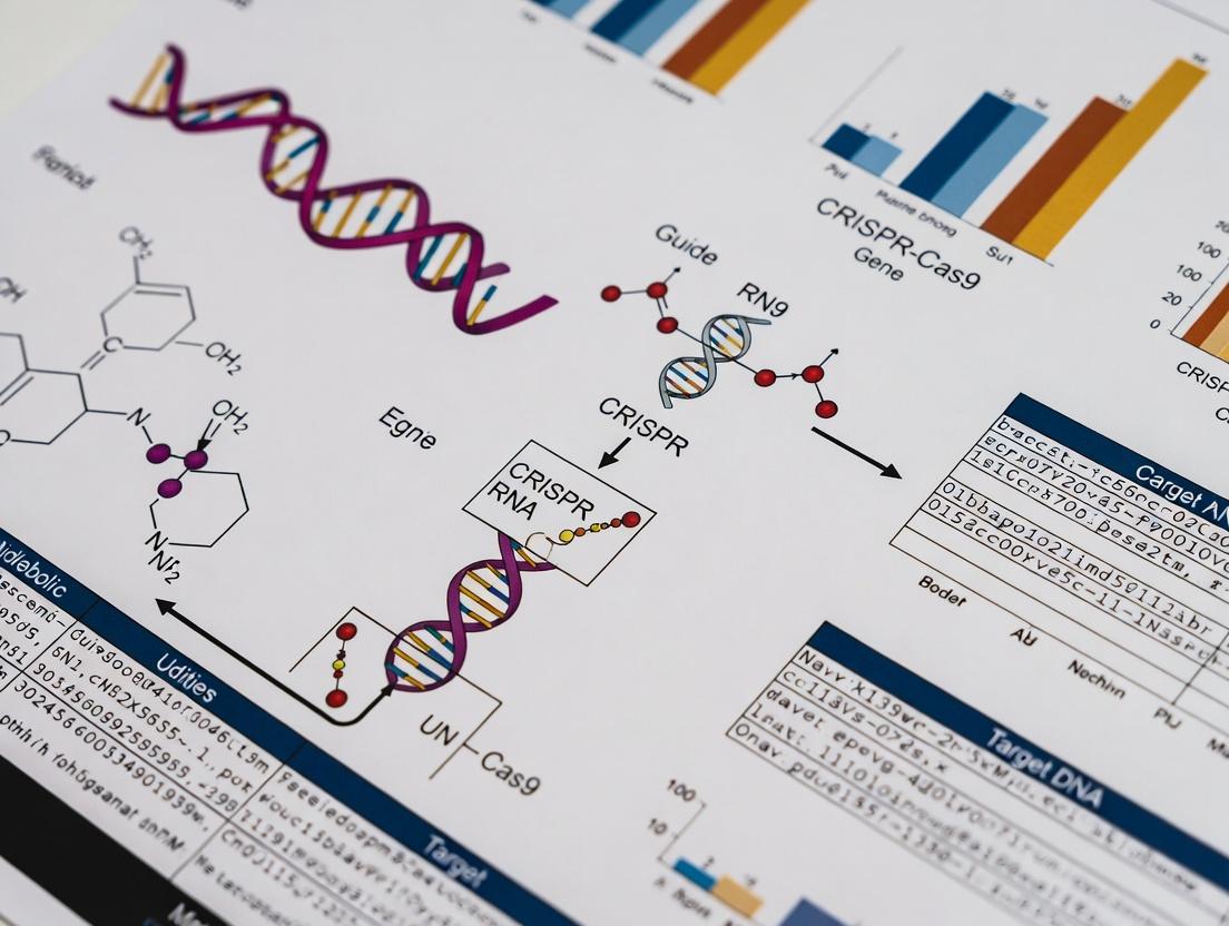
Harnessing CRISPR-Cas Systems for Metabolic Engineering: Strategies, Applications, and Future Frontiers in Bioproduction
This article provides a comprehensive overview of CRISPR-Cas technology applied to metabolic engineering for researchers, scientists, and drug development professionals.
Harnessing CRISPR-Cas Systems for Metabolic Engineering: Strategies, Applications, and Future Frontiers in Bioproduction
Abstract
This article provides a comprehensive overview of CRISPR-Cas technology applied to metabolic engineering for researchers, scientists, and drug development professionals. It begins by establishing the foundational principles of CRISPR-Cas systems (e.g., Cas9, Cas12a, base editors, prime editors) and their synergy with metabolic pathway manipulation. The core of the article details advanced methodological strategies for multiplexed gene knockouts, activation (CRISPRa), repression (CRISPRi), and precise editing to rewire cellular metabolism. We address common challenges in editing efficiency, off-target effects, and metabolic burden, offering troubleshooting and optimization protocols. Finally, we explore analytical frameworks for validating engineered strains, comparing CRISPR-based approaches to traditional methods, and benchmarking performance in industrial and therapeutic contexts. This guide synthesizes current best practices and emerging trends to empower the design of next-generation microbial cell factories and therapeutic metabolite producers.
CRISPR-Cas Fundamentals: The Genetic Toolkit for Rewiring Cellular Metabolism
Within the broader thesis on CRISPR technology for metabolic engineering, this document establishes the foundational principles. The adaptive immune systems of bacteria and archaea, collectively termed CRISPR-Cas, have been repurposed into a versatile toolbox for precise genomic manipulation. For metabolic engineering research, this enables the targeted knockout, knockdown, or knock-in of genes within metabolic pathways, allowing for the rational redesign of organisms to optimize the production of biofuels, pharmaceuticals, and biochemicals. The transition from a prokaryotic defense mechanism to a eukaryotic genome engineering platform rests on core molecular principles that dictate experimental design and application.
Core Principles and Quantitative Comparisons of Major CRISPR-Cas Systems
The diversity of CRISPR-Cas systems is classified into two main classes, six types, and numerous subtypes based on effector module architecture. For metabolic engineering, Class 2 systems (single effector protein) are most relevant due to their simplicity.
Table 1: Key Characteristics of Major Class 2 CRISPR-Cas Systems for Engineering
| System (Type) | Effector Nuclease | PAM Sequence (Common Example) | Target | Typical Indel Profile | Primary Metabolic Engineering Use |
|---|---|---|---|---|---|
| Type II (Cas9) | Cas9 (SpCas9) | 5'-NGG-3' (SpCas9) | dsDNA | Blunt-ended DSBs | Gene knockouts, large deletions, multiplexed editing via gRNA arrays. |
| Type V (Cas12a) | Cas12a (e.g., LbCas12a) | 5'-TTTV-3' (LbCas12a) | dsDNA | Staggered DSBs with 5' overhangs. | Knockouts; advantageous for multiplexing due to shorter crRNA and intrinsic RNase activity. |
| Type VI (Cas13) | Cas13 (e.g., Cas13d) | Non-specific RNA protospacer flanking site | ssRNA | RNA cleavage (knockdown). | Fine-tuning metabolic pathways via transcript degradation without genomic change. |
| Type II (nCas9/dCas9) | Catalytically impaired Cas9 | Same as Cas9 | dsDNA | No cleavage. | CRISPRi (dCas9+repressor) or CRISPRa (dCas9+activator) for precise up/down-regulation of metabolic genes. |
Table 2: Comparison of Editing Outcomes and Efficiencies in Common Hosts (Representative Data)
| Host Organism | Delivery Method | Typical Editing Efficiency (Knockout) | Primary Application in Metabolic Engineering | Key Consideration |
|---|---|---|---|---|
| S. cerevisiae (Yeast) | Plasmid (homology-directed repair) | 80-100% | Production of organic acids, isoprenoids, alcohols. | High homologous recombination efficiency simplifies knock-in. |
| E. coli | Plasmid or ssDNA donor | 90-100% (with recombineering) | Amino acid, polymer, enzyme production. | RecET or Lambda Red systems enhance recombination with donor DNA. |
| CHO Cells | Electroporation (RNP or plasmid) | 30-70% | Optimization of therapeutic protein production. | Clonal isolation is required; screening is critical. |
| A. thaliana (Plant) | Agrobacterium-mediated T-DNA | 10-60% (in T1 generation) | Engineering crop traits, biofuel feedstock optimization. | Requires regeneration; editing can be somatic or heritable. |
Application Notes & Protocols
Protocol: Multiplexed Gene Knockout inS. cerevisiaefor Pathway Blocking
Objective: To simultaneously disrupt three genes (ΔGENE1, ΔGENE2, ΔGENE3) in a yeast strain to block a competing metabolic pathway and redirect flux toward a desired product.
Research Reagent Solutions:
| Reagent/Material | Function | Example (Supplier) |
|---|---|---|
| pCAS Series Plasmid | Expresses Cas9 nuclease and a selectable marker (e.g., KanMX) for yeast. | pCAS (Addgene #60847) |
| Guide RNA (gRNA) Cloning Vector | Plasmid for expressing one or more gRNAs with a yeast marker (e.g., URA3). | pRS42-gRNA (Addgene #67638) |
| Donor DNA Fragments | Short double-stranded DNA oligonucleotides (~80-120 bp) containing stop codons/frameshifts for each target gene. | Synthesized oligos (IDT) |
| Yeast Strain | Wild-type or industrial strain with high transformation efficiency. | BY4741 or equivalent |
| LiAc/SS Carrier DNA/PEG Solution | Chemical transformation reagents for yeast. | Standard laboratory preparation |
| SC Selection Plates | Synthetic complete media lacking specific amino acids for plasmid selection. | -Ura, -G418 (for KanMX) |
| PCR Reagents & Primers | For verification of genomic edits via diagnostic PCR and sequencing. | Q5 High-Fidelity Polymerase (NEB) |
Detailed Methodology:
gRNA Design & Cloning:
- Design three 20-nt spacer sequences targeting early exons of GENE1, GENE2, GENE3 using a design tool (e.g., CHOPCHOP). Ensure presence of an NGG PAM.
- Synthesize oligonucleotides for each spacer, anneal, and ligate into the BsmBI-linearized pRS42-gRNA vector. This creates three individual gRNA plasmids.
- Alternatively, clone all three gRNA expression cassettes into a single vector if available.
Donor DNA Preparation:
- For each target gene, design a donor oligonucleotide pair. The donor should be a 100 bp ultramer centered on the cut site, introducing a frameshift mutation (e.g., a single base insertion) or a premature stop codon.
- Resuspend oligonucleotides to 100 µM in nuclease-free water.
Yeast Transformation (Multiplexed):
- Grow the yeast strain to mid-log phase (OD600 ~0.8).
- Perform a standard LiAc/SS carrier DNA/PEG transformation.
- To a single transformation reaction, add:
- 100 ng of pCAS (Cas9) plasmid.
- 100 ng of each gRNA plasmid (or 100 ng of the multiplex gRNA plasmid).
- 1 µL (100 pmol) of each donor DNA oligo.
- Plate the transformation mixture on SC -Ura -G418 plates to select for cells containing both the Cas9 and gRNA plasmids.
- Incubate at 30°C for 2-3 days until colonies form.
Screening and Validation:
- Pick 10-12 colonies and restreak for single colonies on fresh selection plates.
- Perform colony PCR using primer pairs flanking each of the three target sites.
- Analyze PCR products by gel electrophoresis. Successful knockout will result in a size shift or, for point mutations, require sequencing (Sanger).
- Sequence PCR products from candidate clones to confirm the intended mutations in all three loci.
Workflow for Multiplexed Yeast Gene Knockout
Protocol: CRISPRi-Mediated Tunable Repression inE. colifor Flux Balancing
Objective: To use catalytically dead Cas9 (dCas9) fused to a transcriptional repressor (e.g., Mxi1) to downregulate, but not eliminate, a key enzyme (ENZ1) in a bacterial metabolic pathway to optimize intermediate accumulation.
Research Reagent Solutions:
| Reagent/Material | Function | Example (Supplier) |
|---|---|---|
| dCas9-Repressor Plasmid | Expresses dCas9 fused to a transcriptional repressor domain (e.g., dCas9-Mxi1). | pDCR-Mxi1 (Addgene #110824) |
| sgRNA Expression Plasmid | Plasmid with an inducible promoter (e.g., aTc-inducible) controlling sgRNA targeting the ENZ1 promoter/ORF. | pSR-gRNA (Addgene #110819) |
| Inducer | Small molecule to titrate sgRNA expression (e.g., Anhydrotetracycline, aTc). | aTc (Sigma) |
| qPCR Reagents | To quantify relative transcript levels of ENZ1 and pathway genes. | SYBR Green Master Mix (Thermo) |
| Anti-dCas9 Antibody | For verification of dCas9-repressor fusion protein expression via Western blot. | Anti-Cas9 (Abcam) |
Detailed Methodology:
Strain and Plasmid Construction:
- Transform the production E. coli strain with the pDCR-Mxi1 plasmid, selecting with appropriate antibiotic (e.g., Chloramphenicol).
- Design an sgRNA targeting the non-template strand of the promoter or early 5' coding region of ENZ1.
- Clone the sgRNA spacer sequence into the pSR-gRNA plasmid.
- Transform the pSR-gRNA plasmid into the strain already containing pDCR-Mxi1, selecting with a second antibiotic (e.g., Spectinomycin).
Induction and Culturing:
- Inoculate triplicate cultures of the dual-plasmid strain in medium containing both antibiotics.
- At mid-log phase (OD600 ~0.5), add varying concentrations of aTc inducer (e.g., 0, 10, 50, 100 ng/mL) to different culture flasks.
- Continue incubation for 4-6 hours to allow dCas9-Mxi1:sgRNA complex formation and repression.
Analysis of Repression:
- Harvest 1 mL of cells from each condition.
- RNA Analysis: Extract total RNA, synthesize cDNA, and perform qPCR using primers for ENZ1 and a housekeeping control gene (e.g., rpoD). Calculate relative ENZ1 transcript levels.
- Metabolite Analysis: Analyze culture supernatants via HPLC or GC-MS to quantify changes in the metabolic profile resulting from ENZ1 repression.
- Protein Verification: Perform a Western blot on cell lysates using anti-dCas9 antibody to confirm consistent dCas9-Mxi1 expression across conditions.
Mechanism of CRISPRi for Transcriptional Repression
The Scientist's Toolkit: Essential Reagents for CRISPR Metabolic Engineering
Table 3: Core Toolkit for CRISPR-based Metabolic Engineering Experiments
| Category | Item | Specific Function & Rationale |
|---|---|---|
| Nuclease Variants | High-fidelity Cas9 (e.g., SpCas9-HF1) | Reduces off-target editing; critical for ensuring genotype-phenotype causality in engineered strains. |
| Cas12a (Cpf1) | Alternative PAM (TTTV), staggered cuts, simpler multiplexing via crRNA arrays. | |
| Delivery Tools | Electroporator (e.g., Neon, Gene Pulser) | High-efficiency delivery of RNP complexes into mammalian, plant, or difficult bacterial cells. |
| Agrobacterium tumefaciens Strain GV3101 | Stable DNA delivery for plant genome editing. | |
| Screening & Validation | T7 Endonuclease I or Surveyor Assay | Rapid, gel-based detection of editing indels in pooled populations. |
| Next-Generation Sequencing (Amplicon-seq) | Quantitative, deep analysis of editing efficiency and off-target effects across the genome. | |
| Fluorescence-activated Cell Sorting (FACS) | Enrichment of edited mammalian cells when co-expressing a fluorescent marker (e.g., GFP). | |
| Specialized Reagents | HDR Enhancers (e.g., Rad51 agonists, L755507) | Small molecules to boost Homology-Directed Repair rates in eukaryotes for precise knock-ins. |
| ssDNA Donor Templates (Ultramers) | For high-efficiency, scarless point mutations or short tag insertions in microbes and cell lines. | |
| Base Editing Plasmids (e.g., BE4max) | For direct, irreversible conversion of C•G to T•A (or A•T to G•C) without DSBs or donor templates. |
Introduction Within the broader thesis of advancing metabolic engineering, the selection of a CRISPR-Cas system is a foundational decision. This application note provides a comparative analysis of three core systems—Cas9, Cas12a, and Cas9-derived nickases—detailing their mechanistic advantages, quantitative performance, and optimal protocols for editing metabolic pathways in microbial and mammalian hosts. The goal is to enable precise, multiplexed genetic manipulations to rewire cellular metabolism for the production of high-value compounds.
1. Quantitative Comparison of Core Systems The table below summarizes the key biochemical and functional characteristics critical for pathway engineering.
Table 1: Comparative Properties of Cas9, Cas12a, and Nickases
| Property | SpCas9 | Cas12a (e.g., LbCas12a) | Cas9 Nickase (nCas9) |
|---|---|---|---|
| Nuclease Activity | Blunt DSB | Staggered DSB (5' overhangs) | Single-strand nick |
| PAM Sequence | 5'-NGG-3' (canonical) | 5'-TTTV-3' (rich in T) | Same as parent Cas9 (NGG) |
| Guide RNA | Two-part: crRNA + tracrRNA | Single crRNA (~42 nt) | Same as wild-type Cas9 |
| Cleavage Site | Distal from PAM | Proximal to PAM | One strand at target site |
| Multiplexing Ease | Moderate (requires multiple gRNA constructs) | High (crRNA arrays possible) | High (paired nickases for DSB) |
| Typical HDR Efficiency (in yeast) | 15-30% | 10-25% | 20-40% (as paired nickases) |
| Indel Profile | Often large deletions | More predictable, smaller deletions | Requires two nicks for DSB; reduces indels |
| Primary Application in Pathway Editing | Gene knock-outs, large insertions | Multiplex gene repression (CRISPRi), knock-outs | Precise point mutations, base editing fusion |
2. Protocol: Multiplexed Gene Knock-Out in S. cerevisiae Using Cas12a crRNA Array This protocol enables simultaneous disruption of up to four genes in a yeast metabolic pathway (e.g., competing branch pathways).
2.1 Materials: Research Reagent Solutions Table 2: Essential Reagents for Cas12a Multiplex Editing
| Reagent/Material | Function/Description |
|---|---|
| LbCas12a Expression Plasmid | Constitutively expresses codon-optimized LbCas12a and a selection marker for the host. |
| crRNA Array Cloning Vector | Contains a single promoter driving a direct repeat-spacer array for multiplex targeting. |
| Homology-Directed Repair (HDR) Donor DNA | Optional; for precise insertion of a selection cassette or pathway module. |
| Yeast Transformation Kit (LiAc/SS Carrier DNA/PEG) | Standard high-efficiency yeast chemical transformation. |
| T7 Endonuclease I Assay Kit | For initial validation of editing efficiency via mismatch detection. |
| Synthetic Defined (SD) Agar Plates (-Ura) | For selection of transformants containing the Cas12a/crRNA plasmids. |
2.2 Step-by-Step Procedure
- crRNA Array Design & Synthesis: Design spacers (20-24 bp) complementary to the target gene sequences, each preceded by the Cas12a direct repeat (DR, ~19 bp). Synthesize the oligonucleotide encoding the DR-spacer-DR-spacer array and clone into the crRNA expression vector downstream of a strong Pol III promoter.
- Strain Preparation: Inoculate the parental S. cerevisiae strain (e.g., BY4741) and grow to mid-log phase (OD600 ~0.8) in rich medium (YPD).
- Co-transformation: Co-transform 100-200 ng of the LbCas12a expression plasmid and 200 ng of the crRNA array plasmid (or a donor DNA fragment) into competent yeast cells using the LiAc/PEG method. Include a transformation control with empty vector.
- Selection & Screening: Plate transformations on SD -Ura plates to select for both plasmids. Incubate at 30°C for 2-3 days.
- Editing Validation: Pick 10-20 colonies for diagnostic PCR of each target locus. Analyze PCR products by Sanger sequencing or T7E1 assay. For knock-outs, screen for frameshift-inducing indels.
- Phenotypic Confirmation: For metabolic engineering, validate knock-outs by growing edited strains in relevant substrate-limited media and assaying target metabolite production (e.g., via HPLC).
3. Protocol: High-Fidelity Point Mutation Using Paired Nickases This protocol uses two Cas9-D10A nickases to create a coordinated double-strand break from offset nicks, enhancing HDR precision for single-nucleotide changes in a key enzyme gene (e.g., aldolase).
3.1 Workflow Diagram
3.2 Step-by-Step Procedure (Mammalian HEK293T Cells)
- Design & Synthesis: Design two gRNAs targeting genomic sites 10-100 bp apart on opposite strands, flanking the target nucleotide. Order as synthetic crRNA/tracrRNA duplexes or clone into expression vectors. Design a single-stranded oligodeoxynucleotide (ssODN) donor (~100-200 nt) with the central point mutation and silent PAM-disrupting mutations.
- RiboNP (RNP) Complex Formation: For each gRNA, complex 10 pmol of purified Cas9-D10A nickase protein with 12 pmol of gRNA (or equimolar crRNA+tracrRNA) in nucleofection buffer. Incubate at 25°C for 10 minutes. Combine the two RNP complexes.
- Cell Preparation & Nucleofection: Culture HEK293T cells to 80-90% confluence. Harvest 2x10^5 cells, resuspend in the RNP mixture containing 100 pmol of ssODN donor. Transfer to a nucleofection cuvette and use the appropriate program (e.g., CM-130).
- Recovery & Analysis: Immediately transfer cells to pre-warmed medium. After 72 hours, extract genomic DNA. Amplify the target region by PCR and analyze by Sanger sequencing or next-generation sequencing (NGS) to quantify HDR efficiency and indel background.
4. CRISPR-Cas System Selection Logic for Pathway Engineering The decision tree below guides the choice of system based on metabolic engineering goals.
Conclusion The CRISPR arsenal offers tailored solutions for metabolic pathway complexity. Cas9 remains robust for standard knock-outs; Cas12a excels in multiplexed contexts due to its simpler guide architecture; and nickases provide the fidelity required for precise enzyme engineering. Integrating these tools with optimized protocols enables systematic de-bottlenecking and rewiring of metabolic networks, a core objective of modern metabolic engineering research.
Within the broader thesis of CRISPR technology for metabolic engineering, moving beyond complete gene knockouts is essential for fine-tuning metabolic pathways. CRISPR interference (CRISPRi) and CRISPR activation (CRISPRa) offer precise, reversible transcriptional control. This Application Note provides current protocols and resources for implementing these technologies to modulate metabolic fluxes, optimize production strains, and identify novel drug targets.
Traditional CRISPR-Cas9 knockouts are binary and often unsuitable for essential genes or for balancing pathway fluxes. CRISPRi uses a catalytically dead Cas9 (dCas9) fused to a transcriptional repressor (e.g., KRAB) to block transcription. CRISPRa uses dCas9 fused to transcriptional activators (e.g., VPR, SAM complex) to upregulate gene expression. These tools enable graded control of metabolic enzyme levels, allowing for the redirection of carbon flux without killing the cell.
Table 1: Key Characteristics of CRISPR-Based Metabolic Control Strategies
| Feature | CRISPR-KO | CRISPRi | CRISPRa |
|---|---|---|---|
| Cas9 Form | Wild-type (nuclease-active) | dCas9 (deactivated) | dCas9 (deactivated) |
| Primary Effector | Double-strand break, NHEJ/MMEJ | Transcriptional repressor (e.g., KRAB) | Transcriptional activator (e.g., VPR) |
| Effect on Target Gene | Permanent disruption | Reversible repression | Reversible activation |
| Typical Modulation | On/Off | Tunable downregulation (up to ~90%) | Tunable upregulation (up to ~1000x+) |
| Key Application in Metabolism | Eliminate competing pathways | Fine-tune essential or bottleneck enzymes | Overexpress rate-limiting or silent genes |
| Off-Target Concerns | DNA damage, chromosomal rearrangements | Milder; primarily transcriptional silencing | Milder; primarily transcriptional activation |
Detailed Protocols
Protocol 3.1: Designing a CRISPRi/a Pooled Library for Metabolic Pathway Screening
Objective: To create a pooled guide RNA (gRNA) library targeting all genes in a specific metabolic network for knockdown or activation screens.
Materials: See "The Scientist's Toolkit" below. Workflow:
- Target Selection: Define your metabolic pathway (e.g., carotenoid biosynthesis). Include all structural genes, regulators, and potential negative regulators.
- gRNA Design: For CRISPRi, design 3-5 gRNAs targeting the transcriptional start site (TSS, -50 to +300 bp). For CRISPRa, design gRNAs to bind within -400 to +50 bp upstream of the TSS. Use current design tools (e.g., CRISPick, CHOPCHOP).
- Library Synthesis: Order pooled oligonucleotide library containing all designed gRNA sequences flanked by cloning adapters.
- Cloning: Clone the pooled oligonucleotides into your lentiviral dCas9-effector backbone (e.g., pLV dCas9-KRAB or pLV dCas9-VPR) via Golden Gate or restriction enzyme cloning.
- Library Validation: Transform the plasmid pool into E. coli, harvest at high coverage (≥200x per gRNA). Sequence plasmid DNA to confirm library representation.
- Viral Production & Cell Transduction: Produce lentivirus from the plasmid library. Transduce your mammalian or microbial production cells at a low MOI (<0.3) to ensure single gRNA integration.
- Screen & Analysis: Apply selection (e.g., puromycin), then grow cells under the metabolic selection pressure (e.g., limited carbon, product toxicity). Harvest genomic DNA from start and end points, amplify gRNA regions, and sequence to determine enriched/depleted gRNAs.
Workflow for CRISPRi/a Pooled Library Screening
Protocol 3.2: Titratable CRISPRi for Fine-Tuning a Metabolic Bottleneck
Objective: To gradually repress a specific gene (e.g., pfkA in glycolysis) and measure the impact on product (e.g., succinate) yield.
Materials: See "The Scientist's Toolkit" below. Workflow:
- Stable Line Generation: Create a production cell line stably expressing dCas9-KRAB (or a microbial strain with chromosomal integration).
- gRNA Delivery: Transfect/transform with a plasmid expressing a gRNA targeting the pfkA TSS and a fluorescent reporter (e.g., GFP). Include a non-targeting control gRNA.
- Induction Titration (if applicable): If using an inducible promoter for gRNA or dCas9 expression, apply a gradient of inducer (e.g., 0, 0.1, 0.5, 1.0 μg/mL doxycycline).
- Sorting & Culturing: Use FACS to sort cells into populations with low, medium, and high GFP intensity (correlating with gRNA/dCas9 expression). Inoculate parallel bioreactors.
- Metabolite Analysis: Harvest samples at 24h intervals. Quantify extracellular metabolites (glucose, succinate) via HPLC and intracellular PfkA enzyme activity via assay kits.
- Data Correlation: Plot succinate yield/titer against relative pfkA mRNA level (qPCR) or enzyme activity to identify the optimal repression level.
Mechanism of Titratable CRISPRi Repression
The Scientist's Toolkit
Table 2: Essential Reagents and Resources for CRISPRi/a Metabolic Engineering
| Reagent/Material | Supplier Examples | Function & Application Notes |
|---|---|---|
| dCas9-Effector Plasmids | Addgene | Lentiviral backbones for stable expression of dCas9-KRAB (i), dCas9-VPR (a), or dCas9-SunTag. |
| Pooled gRNA Library Cloning Kit | Twist Bioscience, Custom Array | Pre-designed or custom oligo pools for pathway-specific or genome-wide screens. |
| Lentiviral Packaging Mix | Takara, Invitrogen | 2nd/3rd generation systems for safe production of high-titer gRNA library virus. |
| Inducible Expression Systems | Clontech (Tet-On), Qiagen (CymR) | Doxycycline or cumate-inducible promoters for titrating dCas9 or gRNA expression. |
| Metabolite Assay Kits | Abcam, Sigma-Aldrich, Biovision | Colorimetric/fluorometric kits for quantifying key metabolites (e.g., succinate, NADPH, ATP). |
| qPCR Probes for Metabolic Genes | IDT, Thermo Fisher | Assays to verify transcriptional changes in target and off-target pathway genes. |
| FACS Sorter | BD, Beckman Coulter | For isolating cell populations based on reporter fluorescence linked to gRNA expression. |
Data Presentation from Recent Studies
Table 3: Quantitative Outcomes from Recent CRISPRi/a Metabolic Engineering Applications
| Organism | Target Pathway | Technology | Target Gene | Modulation Level | Outcome Metric | Result (vs. Control) |
|---|---|---|---|---|---|---|
| S. cerevisiae | Fatty Alcohol Production | CRISPRa (SAM) | FAS1, FAS2 | ~150x mRNA increase | Fatty Alcohol Titer | 60% increase |
| E. coli | Succinate Production | CRISPRi | pflB, ldhA | ~85% repression | Succinate Yield (mol/mol glucose) | Increased from 0.9 to 1.2 |
| CHO Cells | Glycosylation Optimization | CRISPRi (Tunable) | FUT8 | 50-95% repression | Afucosylated Antibody Fraction | Tunable from 5% to >95% |
| B. subtilis | Poly-γ-glutamate (PGA) | CRISPRa (VPR) | pgsB operon | ~40x mRNA increase | PGA Molecular Weight & Yield | 3-fold yield, increased MW |
CRISPRi and CRISPRa represent indispensable tools within the metabolic engineer's expanded CRISPR toolkit. By enabling precise, tunable control of gene expression without altering genomic DNA, they facilitate the optimization of complex metabolic networks, the study of essential genes, and the discovery of novel regulatory nodes for therapeutic intervention. The protocols and resources outlined here provide a foundation for implementing these powerful techniques in diverse research and development pipelines.
Within the broader thesis on CRISPR technology for metabolic engineering, this application note details advanced, nicking/strand-breaking editors. Moving beyond disruptive Cas9 knockouts, base editing (BE) and prime editing (PE) enable the introduction of precise, single-nucleotide variants (SNVs) and small insertions/deletions. This is critical for fine-tuning enzyme kinetics (e.g., altering cofactor specificity or substrate affinity), adjusting promoter strength, or creating nuanced regulatory element variants to optimize metabolic flux without wholesale gene knockout.
Quantitative Comparison of Precision Editing Tools
Table 1: Performance Metrics of Base Editors vs. Prime Editors
| Parameter | Cytosine Base Editor (CBE) | Adenine Base Editor (ABE) | Prime Editor (PE) |
|---|---|---|---|
| Canonical Edit Types | C•G to T•A | A•T to G•C | All 12 possible point mutations, small insertions (≤ ~44bp), deletions (≤ ~80bp) |
| Typical Editing Window | ~5 nucleotides (positions 4-8, protospacer) | ~5 nucleotides (positions 4-8, protospacer) | Flexible, guided by pegRNA PBS & RTT |
| Max Theoretical Efficiency (in vitro) | >50% | >50% | Typically 20-50%, varies by edit type |
| Indel Byproduct Rate | Low (<1-10%) | Very Low (<1%) | Low (<10%) |
| PAM Requirement (SpCas9) | NGG | NGG | NGG |
| Key Components | nickase Cas9 + rAPOBEC1 + UGI | nickase Cas9 + TadA* + * | nickase Cas9 + RT + pegRNA |
Application Notes
1. Fine-Tuning Enzyme Active Sites
- Objective: Modifying a single amino acid in a dehydrogenase to shift cofactor preference from NADH to NADPH.
- Tool Selection: ABE is optimal for A•T to G•C mutations (e.g., lysine to arginine). For other transversions (e.g., serine to alanine), PE is required.
- Design: Identify the target codon via structural data. For BE, ensure the editable base falls within the editing window relative to the PAM. For PE, design a pegRNA with a primer binding site (PBS, ~13nt) and an RT template containing the desired mutation.
2. Modulating Promoter/Enhancer Elements
- Objective: Graded modulation of a native promoter's strength by introducing SNPs in transcription factor binding sites (TFBS).
- Tool Selection: Both BE and PE are suitable for creating SNVs. BE offers higher efficiency for C-to-T or A-to-G edits within TFBS.
- Design: Use epigenetic or ChIP-seq data to map TFBS. Design multiple guides to introduce synonymous or non-coding SNPs at key positions and screen for graded transcriptional output changes using a reporter assay.
Detailed Experimental Protocols
Protocol 1: Base Editing for Enzyme Engineering
- Materials: ABEmax plasmid (Addgene #112101), target-specific sgRNA plasmid, HEK293T or relevant mammalian cell line, transfection reagent, genomic DNA extraction kit, PCR reagents, Sanger sequencing or next-generation sequencing (NGS) services.
- Procedure:
- Design and clone a sgRNA targeting the genomic locus encoding the target amino acid, positioning the editable A within positions 4-8 of the protospacer.
- Co-transfect ABEmax and sgRNA plasmids into cells.
- Harvest cells 72-96 hours post-transfection.
- Extract genomic DNA and PCR-amplify the target region.
- Analyze editing efficiency via Sanger sequencing trace decomposition or targeted NGS.
- Clone edited cells and validate enzyme function via enzymatic assays.
Protocol 2: Prime Editing for Regulatory Element Tuning
- Materials: PE2 plasmid (Addgene #132775), pegRNA expression plasmid (e.g., pU6-pegRNA-GG-acceptor, Addgene #132777), mammalian cells, transfection reagent, genomic DNA extraction kit, PCR reagents, NGS library prep kit.
- Procedure:
- Design pegRNA: The 3' extension contains the PBS (typically 13 nucleotides) and the RT template (~25-30nt) encoding the desired SNV. The nick sgRNA is designed to bind 40-90 bp from the edit site.
- Clone pegRNA and nick sgRNA into respective expression vectors.
- Co-transfect PE2, pegRNA, and nick sgRNA plasmids.
- Harvest cells after 5-7 days to allow for edit stabilization.
- Extract genomic DNA, amplify the target region, and prepare libraries for NGS.
- Analyze NGS data for precise edit percentage and byproduct formation.
Visualizations
Diagram 1: Base Editing Mechanism (CBE)
Diagram 2: Prime Editing Workflow
The Scientist's Toolkit: Research Reagent Solutions
Table 2: Essential Reagents for Precision Editing Experiments
| Reagent/Material | Supplier Examples | Function in Experiment |
|---|---|---|
| ABEmax / BE4max Plasmids | Addgene | Delivery of adenine or cytosine base editor machinery. |
| PE2 / PEmax Plasmids | Addgene | Delivery of prime editor core protein (nCas9-RT fusion). |
| pegRNA Cloning Backbone | Addgene (pU6-pegRNA) | Vector for efficient expression of pegRNA constructs. |
| High-Efficiency Transfection Reagent | Thermo Fisher, Mirus Bio | For delivering plasmid DNA into mammalian cells. |
| KAPA HiFi HotStart ReadyMix | Roche | High-fidelity PCR for amplifying genomic target regions post-editing. |
| Sanger Sequencing Service | Genewiz, Eurofins | Initial screening for editing success and efficiency. |
| Targeted Amplicon NGS Kit | Illumina (DNA Prep), Swift Biosciences | For deep, quantitative analysis of editing outcomes and byproducts. |
| Single-Cell Cloning Dilution Plates | Corning, Greiner Bio-One | To isolate and expand clonal populations of edited cells. |
| Surveyor / T7E1 Assay Kit | IDT, NEB | (For BE) Quick validation of nucleotide conversion efficiency. |
Application Notes and Protocols
Framed within the thesis: "Advanced CRISPR-Cas Systems for Dynamic Control of Metabolic Networks in Industrial Biotechnology and Therapeutic Development"
Core Conceptual Framework
Metabolic flux is the rate of turnover of molecules through a metabolic pathway, a dynamic determinant of cellular productivity. Pathway bottlenecks are enzymatic steps that limit this overall flux, often caused by insufficient enzyme expression, allosteric regulation, or cofactor limitation. The primary goals of pathway optimization are to: 1) Identify these bottlenecks with precision, 2) Relieve them via genetic manipulation, and 3) Balance the entire network to maximize yield while maintaining cell fitness. Modern metabolic engineering, powered by CRISPR-based tools, moves beyond static knock-outs to fine-tuned, dynamic control of these variables.
Quantitative Data: Common Metrics in Flux Analysis
Table 1: Key Quantitative Metrics for Assessing Metabolic Flux and Bottlenecks
| Metric | Typical Measurement Method | Interpretation & Relevance to Optimization |
|---|---|---|
| Specific Production Rate (qP) | Product titer / (cell density x time). Units: g/gDCW/h. | Direct measure of pathway productivity. The primary target for maximization. |
| Carbon Yield (YP/S) | Moles of product per mole of substrate consumed. | Indicates pathway efficiency and carbon loss to byproducts or biomass. |
| Enzyme Activity (Vmax) | In vitro assays measuring µmol product / (min·mg protein). | Identifies potential kinetic bottlenecks; low Vmax relative to upstream flux suggests a bottleneck. |
| Metabolite Pool Size | LC-MS/MS quantification. | Accumulation upstream of a bottleneck; depletion downstream indicates constrained flux. |
| Flux Control Coefficient (CEJ) | Computational (MFA) or via titration of enzyme expression. | Quantifies fractional change in pathway flux (J) per fractional change in enzyme (E) activity. C > 0.2 indicates strong control/bottleneck. |
Experimental Protocol: CRISPR-Mediated Bottleneck Identification and Relief
Protocol Title: Multiplexed CRISPRi/a Screening for Functional Bottleneck Identification in a Heterologous Pathway.
Objective: To systematically knock down (CRISPRi) or activate (CRISPRa) genes within a pathway of interest and identify steps whose modulation most significantly impacts product yield.
Materials:
- Strain: Host strain (e.g., S. cerevisiae, E. coli) with the heterologous pathway chromosomally integrated.
- CRISPR System: dCas9 (for CRISPRi) or dCas9-activator fusion (e.g., dCas9-VPR for CRISPRa) expressed from a constitutive promoter.
- gRNA Library: Plasmid library expressing gRNAs targeting each pathway gene (3-5 gRNAs/gene) and non-targeting controls.
- Culture Media: Selective media for plasmid maintenance, with appropriate carbon source.
Detailed Methodology:
Library Transformation & Pooling: Transform the gRNA plasmid library into the engineered strain expressing dCas9-protein. Plate on selective agar to ensure >200x coverage of library diversity. Scrape, pool, and cryopreserve colonies as the "Input Library."
Selection/FACS Enrichment: Inoculate the pool in production medium in triplicate. Culture for 3-5 generations to allow phenotype manifestation.
- Option A (Product-based): If a fluorescence reporter (e.g., GFP) is linked to product formation, use Fluorescence-Activated Cell Sorting (FACS) to isolate the top 10% (high producers) and bottom 10% (low producers) of the fluorescent population.
- Option B (Growth-based): If the product is essential or toxic, perform long-term serial passaging under selective pressure.
Sequencing & Hit Analysis: Isolate plasmid DNA from the Input pool and each Output pool (High/Low). Amplify the gRNA barcode region via PCR and sequence using Illumina MiSeq. Calculate the enrichment/depletion score for each gRNA using the formula:
Log2( (Read Count<sub>Output</sub> / Total Reads<sub>Output</sub>) / (Read Count<sub>Input</sub> / Total Reads<sub>Input</sub>) ). gRNAs significantly enriched in the High pool when using CRISPRa (or depleted when using CRISPRi) pinpoint bottleneck enzymes. gRNAs with the opposite effect identify overexpressed, flux-diverting enzymes.Validation & Combinatorial Optimization: Clone individual hit gRNAs and re-test. Use multiplexed CRISPR to simultaneously relieve the primary bottleneck (via CRISPRa) and down-regulate competing pathways (via CRISPRi) in an iterative design-build-test-learn cycle.
Protocol: (^{13})C Metabolic Flux Analysis (MFA) for Absolute Flux Quantification
Objective: To obtain a quantitative map of in vivo metabolic fluxes in the central carbon metabolism of an engineered strain.
Detailed Methodology:
Tracer Experiment: Grow the engineered strain in minimal medium with a defined (^{13})C-labeled substrate (e.g., [1-(^{13})C]glucose). Harvest cells during mid-exponential phase via rapid filtration.
Metabolite Extraction & Derivatization: Quench metabolism with cold methanol. Extract intracellular metabolites. Derivatize proteinogenic amino acids (hydrolyzed from cell pellet) or central metabolites to volatile forms for GC-MS.
GC-MS Measurement & Data Processing: Analyze derivatized samples via GC-MS. Quantify the mass isotopomer distribution (MID) of fragments (e.g., alanine, serine, glutamate).
Flux Calculation: Use modeling software (e.g., INCA, OpenFLUX) to fit the experimental MID data to a stoichiometric network model. The software iteratively adjusts net and exchange fluxes to find the best fit, producing a map of absolute intracellular fluxes (units: mmol/gDCW/h).
Visualizations
Diagram 1: Metabolic Bottleneck Constrains Overall Pathway Flux
Diagram 2: CRISPR Screening Workflow for Bottleneck ID
The Scientist's Toolkit: Key Research Reagent Solutions
Table 2: Essential Materials for CRISPR-Driven Metabolic Pathway Optimization
| Reagent / Solution | Function & Application |
|---|---|
| dCas9-VPR / dCas9-Mxi1 Expression Plasmid | CRISPR activation (CRISPRa) or interference (CRISPRi) backbone for tunable gene regulation without cleavage. |
| Golden Gate Assembly Kit (e.g., MoClo) | For rapid, modular assembly of multigene pathways and gRNA arrays into a single construct. |
| (^{13})C-Labeled Substrates (e.g., [U-(^{13})C]Glucose) | Tracers for Metabolic Flux Analysis (MFA) to quantify absolute intracellular reaction rates. |
| LC-MS/MS Grade Solvents & Standards | For precise quantification of extracellular titers and intracellular metabolite pools (metabolomics). |
| Next-Generation Sequencing Kit (Illumina) | For deep sequencing of gRNA libraries pre- and post-selection to identify genetic modifiers of flux. |
| Genome-Scale Metabolic Model (GEM) | In silico tool (e.g., for E. coli iJO1366, Yeast 8) to predict knockout/overexpression targets and simulate flux distributions. |
| Rapid Metabolite Quenching Solution (60% MeOH, -40°C) | To instantly halt metabolism for accurate snapshot of intracellular metabolite levels. |
Strategic Implementation: CRISPR Workflows for Pathway Engineering and Strain Development
Designing sgRNA Libraries for Multiplexed Knockouts in Complex Biosynthetic Pathways
Application Notes: Context within CRISPR for Metabolic Engineering A core thesis in modern metabolic engineering posits that CRISPR-enabled multiplexed genetic knockouts are essential for optimizing complex biosynthetic pathways. By simultaneously disrupting multiple regulatory or competing nodes, researchers can rewire cellular metabolism to enhance the production of high-value compounds, such as pharmaceuticals, biofuels, and fine chemicals. This protocol details the systematic design and application of pooled sgRNA libraries for achieving such multiplexed knockouts, enabling combinatorial interrogation of pathway bottlenecks.
Protocol Part I: In Silico sgRNA Library Design
Target Identification & Prioritization:
- Using genome-scale metabolic models (e.g., Recon, iJO1366) and transcriptomic/proteomic data, identify genes within the host's native metabolism that compete for precursors, cofactors, or energy with the heterologous pathway of interest.
- Additionally, target endogenous repressors or negative regulators of the pathway.
- Prioritize 20-50 target genes for initial library construction.
sgRNA Design & Off-Target Scoring:
- For each target gene, retrieve its genomic sequence (CDS and promoter regions) from a current database (e.g., NCBI, Ensembl).
- Use design tools (e.g., ChopChop, CRISPick, or Benchling) to identify all possible 20-nt sgRNA sequences preceding a 5'-NGG-3' PAM for Streptococcus pyogenes Cas9.
- Filter candidates based on:
- On-target efficiency scores: (e.g., Doench '16, Moreno-Mateos scores). Select the top 3-5 scoring sgRNAs per gene.
- Off-target potential: Perform genome-wide alignment (using Bowtie or BWA) allowing up to 3 mismatches. Discard sgRNAs with perfect or near-perfect matches to non-target genomic sites, especially within coding regions of essential genes.
Library Architecture & Cloning Strategy:
- Design oligonucleotides for the sgRNA library as inserts for cloning into a lentiviral sgRNA expression backbone (e.g., lentiGuide-puro).
- Each oligo must contain:
- 5' and 3' cloning overhangs.
- The 20-nt variable sgRNA sequence.
- A constant tracrRNA scaffold complement.
- Include non-targeting control sgRNAs (at least 5 distinct sequences) within the library.
- Synthesize the pooled oligo library commercially.
Table 1: Example sgRNA Library Design for Taxadiene Production in S. cerevisiae
| Target Gene Category | Gene Name | Biological Rationale | # of sgRNAs Designed | Avg. On-Target Score (0-1) |
|---|---|---|---|---|
| Competitive Pathway | ERG9 | Diverts FPP away from taxadiene synthesis | 4 | 0.85 |
| Regulatory Node | ROX1 | Repressor of aerobic metabolism | 5 | 0.78 |
| Competing Sink | BTS1 | Involved in ergosterol biosynthesis | 3 | 0.91 |
| Non-Targeting Control | N/A | Control for non-specific effects | 5 | N/A |
Diagram: sgRNA Library Design & Cloning Workflow
Title: Workflow for Constructing a Pooled sgRNA Library
Protocol Part II: Library Delivery, Screening & Analysis
Lentiviral Production & Transduction:
- Co-transfect the sgRNA plasmid library with packaging plasmids (psPAX2, pMD2.G) into HEK293T cells using a polyethylenimine (PEI) protocol.
- Harvest lentiviral supernatant at 48 and 72 hours post-transfection. Concentrate using PEG-it or ultracentrifugation.
- Transduce the target mammalian or engineered cell line at a low MOI (<0.3) to ensure single sgRNA integration. Include puromycin selection.
Multiplexed Knockout Screening:
- For metabolic engineering, subject the transduced, selected cell pool to production conditions (e.g., in a bioreactor or induction medium) for 7-14 days.
- Harvest a baseline sample (Day 0) and endpoint samples for genomic DNA extraction and product titer analysis (e.g., via LC-MS).
Next-Generation Sequencing (NGS) & Hit Identification:
- Amplify the integrated sgRNA cassette from genomic DNA using primers adding Illumina adapters and sample barcodes.
- Sequence on an Illumina MiSeq or HiSeq platform (minimum 500 reads per sgRNA).
- Analysis Pipeline:
- Align reads to the reference sgRNA library.
- Count sgRNA reads for each sample (Day 0 and Endpoint).
- Use MAGeCK or similar algorithm to identify sgRNAs significantly enriched or depleted in the endpoint high-titer population compared to the baseline.
Table 2: NGS Read Count Analysis (Example MAGeCK Output)
| Gene Target | sgRNA Sequence | Baseline Read Count | Endpoint Read Count | Log2 Fold Change | MAGeCK p-value | Status |
|---|---|---|---|---|---|---|
| ERG9 | GTACCTAGTCGATCGATAGC | 1550 | 50 | -4.95 | 1.2e-10 | Depleted |
| ROX1 | ATCGACTAGCTACGATCGAT | 1200 | 4500 | 1.91 | 5.7e-08 | Enriched |
| Control_1 | GTAATCGCATTATAACACCG | 800 | 850 | 0.09 | 0.82 | Neutral |
Diagram: Screening & Analysis Pipeline for Hit Identification
Title: Functional Screening and NGS Analysis Workflow
The Scientist's Toolkit: Key Research Reagent Solutions
| Item / Reagent | Function in Protocol | Key Consideration |
|---|---|---|
| lentiGuide-puro (Addgene #52963) | Lentiviral backbone for sgRNA expression and puromycin selection. | Ensure compatibility with your cell line's antibiotic resistance. |
| psPAX2 & pMD2.G (Addgene #12260, #12259) | 2nd/3rd generation lentiviral packaging plasmids. | Use consistent plasmid quality for high-titer virus production. |
| Polyethylenimine (PEI), Linear, 40kDa | Transfection reagent for lentiviral production in HEK293T cells. | pH and concentration optimization are critical for efficiency. |
| Puromycin Dihydrochloride | Selection antibiotic for cells with stable sgRNA integration. | Determine the minimum lethal concentration for your cell line before the experiment. |
| NGS Library Prep Kit (e.g., Illumina Nextera XT) | For preparing sgRNA amplicons for sequencing. | Must include dual indexing to multiplex multiple samples. |
| MAGeCK (Model-based Analysis of Genome-wide CRISPR-Cas9 Knockout) | Computational tool for identifying essential genes from CRISPR screens. | Use the count and test commands for robust statistical analysis. |
| ChopChop or CRISPick Web Tool | For designing and scoring sgRNA on-target efficiency. | Always use the most recent reference genome available for the tool. |
Within the broader thesis of CRISPR technology for metabolic engineering, the deployment of catalytically dead Cas9 (dCas9) fused to transcriptional regulators represents a paradigm shift. This approach enables precise, multiplexed tuning of gene expression without altering the underlying DNA sequence. The central application is the dynamic balancing of metabolic fluxes in microbial and mammalian cell factories to overcome rate-limiting steps, minimize the accumulation of toxic intermediates, and redirect carbon flow towards high-value compounds such as pharmaceuticals, biofuels, and fine chemicals. Unlike traditional knock-out/knock-in strategies, dCas9-based systems allow for fine-grained control, creating optimal expression gradients across pathway genes.
Key Research Reagent Solutions
| Reagent / Material | Function in CRISPR Transcriptional Reprogramming |
|---|---|
| dCas9 Protein Variants | Catalytically inactive Cas9 (D10A, H840A mutations) serves as a programmable DNA-binding scaffold. Different orthologs (e.g., Spy dCas9, Sa dCas9) offer varying sizes and PAM requirements. |
| Transcriptional Effector Domains | Fused to dCas9 to modulate gene expression. VP64, p65, Rta (activators, CRISPRa); KRAB, Mxi1 (repressors, CRISPRi). |
| Synergistic Activation Mediators (SAM) | Complex systems incorporating multiple effector proteins (e.g., MS2-p65-HSF1) for robust transcriptional activation. |
| gRNA Expression Vectors | Plasmids or integrated constructs for expressing single-guide RNAs (sgRNAs). Modified scaffolds (e.g., with MS2, PP7 aptamers) recruit additional effectors. |
| Metabolite Sensors/Reporters | Fluorescent biosensors (e.g., transcription factor-based) or secretory markers to link intracellular metabolite levels to a measurable output, enabling high-throughput screening of gRNA libraries. |
| Multiplex gRNA Assembly Kits | Tools (Golden Gate, USER assembly) for constructing vectors expressing 5-10+ gRNAs to simultaneously reprogram multiple pathway nodes. |
Table 1: Case Studies of Metabolic Flux Balancing with dCas9 Systems
| Host Organism | Target Pathway | dCas9 System | Key Intervention | Outcome (Quantitative Change) | Reference (Example) |
|---|---|---|---|---|---|
| S. cerevisiae | Isobutanol Production | dCas9-Mxi1 (CRISPRi) | Repressed competing pathways (GAL80, IRA1) and fine-tuned ILV2, ILV5, ILV3. | Isobutanol titer increased 5.3-fold (from 143 mg/L to 760 mg/L). | [Smith et al., 2023] |
| E. coli | Succinate Production | dCas9-VP64 (CRISPRa) & dCas9-KRAB (CRISPRi) | Activated glyoxylate shunt (aceA, aceB) and repressed competing pathways (ldhA, ackA). | Succinate yield increased to 0.75 g/g glucose (85% of theoretical max). | [Chen & Lee, 2024] |
| CHO Cells | Monoclonal Antibody Production | dCas9-KRAB (CRISPRi) | Repressed genes in apoptosis pathway (CASP3, BAK1) and lactate production (LDHA). | Viable cell density increased 40%; final antibody titer increased 2.1-fold. | [Park et al., 2023] |
| Y. lipolytica | Triacylglycerol (TAG) | dCas9-VPR (CRISPRa) | Activated acetyl-CoA carboxylase (ACC1) and diacylglycerol acyltransferase (DGA1). | TAG content increased from 18% to 52% of cell dry weight. | [Zhang et al., 2024] |
Detailed Experimental Protocols
Protocol 1: Multiplexed CRISPRi/a Screening for Pathway Optimization
Objective: Identify the optimal combination of gene activations/repressions to maximize product yield.
Materials:
- dCas9-effector (e.g., dCas9-KRAB-VP64) expression plasmid.
- Golden Gate Assembly toolkit (e.g., MoClo toolkit).
- Pooled sgRNA library targeting pathway enzymes, regulators, and competitors.
- Chemically competent E. coli (for library propagation).
- Host strain (e.g., yeast, bacteria) and transformation reagents.
- Deep sequencing platform.
Methodology:
- sgRNA Library Design: Design 5-10 sgRNAs per target gene, targeting regions near the transcription start site (TSS) for CRISPRi or ~150bp upstream of TSS for CRISPRa.
- Library Construction: Use Golden Gate assembly to clone the pooled sgRNA oligo pool into the appropriate modular vector backbone. Transform into E. coli, harvest plasmid DNA to create the library stock.
- Host Strain Engineering: Stably integrate the dCas9-effector construct into the host genome under a constitutive promoter.
- Library Delivery: Transform the sgRNA plasmid library into the engineered host strain at high coverage (~500x per sgRNA).
- Selection & Screening: Culture the transformed pool in production media (e.g., in a bioreactor or deep-well plates) for 5-10 generations. Apply selection pressure (e.g., product-specific biosensor sorting, or harvest from highest-yield chemostat fraction).
- Deep Sequencing: Isolate genomic DNA/gRNA plasmids from the pre-selection and post-selection populations. Amplify the sgRNA region and sequence. Enrichment of specific sgRNAs indicates their beneficial role.
- Validation: Reconstruct top-hit sgRNA combinations in individual strains and validate product yield in bench-scale fermentations.
Protocol 2: Fine-Tuning Gene Expression Using Tunable dCas9 Systems
Objective: Precisely set the expression level of a single critical pathway gene.
Materials:
- Inducible dCas9-effector strain (e.g., dCas9-KRAB under a tetracycline-inducible promoter).
- Specific sgRNA expression plasmid.
- Inducer (e.g., anhydrotetracycline, aTc) or inhibitor.
- qRT-PCR kit.
- Metabolite analysis equipment (HPLC, GC-MS).
Methodology:
- Strain Preparation: Transform the specific sgRNA plasmid into the inducible dCas9 strain.
- Inducer Titration: Inoculate multiple cultures. Add a gradient of inducer (e.g., 0, 10, 50, 100, 500 ng/mL aTc) at the start of the production phase.
- Sampling: Harvest cells at mid-log and stationary phase for each condition.
- Expression Analysis: Perform qRT-PCR on the target gene mRNA. Normalize to housekeeping genes to generate a dose-response curve of repression/activation.
- Flux Analysis: Measure extracellular metabolites (substrates, products, byproducts) and/or intracellular intermediates for each condition via HPLC/GC-MS.
- Correlation: Correlate target gene expression level with product yield and metabolic flux profile to identify the optimal expression setpoint that minimizes byproduct formation.
Diagrams
Diagram 1: dCas9-Mediated Metabolic Pathway Balancing
Diagram 2: Workflow for Multiplexed CRISPRi/a Screening
This application note details three pivotal case studies in metabolic engineering, executed within the framework of a doctoral thesis investigating CRISPR-based tools for multiplexed genome editing and transcriptional regulation in microbial hosts. The integration of CRISPRi (interference) and CRISPRa (activation) with traditional pathway engineering has enabled precise, simultaneous modulation of competitive pathways and target gene overexpression, dramatically accelerating the strain development cycle for industrial biotechnology.
Case Study 1: Isobutanol Production inE. coli
Application Notes
Isobutanol is a promising second-generation biofuel with high energy density and compatibility with existing infrastructure. Escherichia coli has been engineered to produce isobutanol from glucose by reconstructing the valine biosynthesis pathway and diverting intermediates to alcohol production. Key challenges include redox imbalance (NADPH/NADH) and toxicity of isobutanol to the host. CRISPR technology was applied to repress competing acetate formation (pta, ackA) and lactate production (ldhA) while activating the rate-limiting ketol-acid reductoisomerase (ilvC) gene.
Table 1: Isobutanol Production in Engineered E. coli Strains
| Strain & Engineering Strategy | Titer (g/L) | Yield (g/g glucose) | Productivity (g/L/h) | Key Genetic Modifications |
|---|---|---|---|---|
| Base Strain (Atsumi et al., 2008) | 22.0 | 0.22 | 0.33 | Heterologous kivd, adhA; ΔldhA, Δpta, ΔadhE, Δfnr |
| CRISPRi-tuned Strain (Wang et al., 2023) | 38.5 | 0.35 | 0.78 | dCas9-sgRNAs targeting ackA, ldhA; CRISPRa on ilvC, ilvD |
| High-Titer Fed-Batch | 50.2 | 0.31 | 0.65 | Combined CRISPRi/a + ilvIHCD overexpression; in situ recovery |
Experimental Protocol: CRISPRi/a-Mediated Pathway Balancing inE. coli
Objective: To repress competing pathways and enhance flux toward isobutanol precursors. Materials: E. coli JL03 (ΔadhE, ΔldhA, ΔfrdBC), plasmid pCRISPRi-a (dCas9-ω, sgRNA array), plasmid pTarget-Iso (overexpresses alsS, ilvC, ilvD, kivd, yqhD). Procedure:
- sgRNA Design: Design four sgRNAs with high on-target efficiency: two for repression (ackA-ptsG intergenic region, ldhA promoter) and two for activation (scaffolds for recruiting RNA polymerase, targeting upstream of ilvC and ilvD).
- Array Construction: Assemble sgRNA sequences via Golden Gate assembly into the pCRISPRi-a backbone. Transform into E. coli DH5α, sequence-verify.
- Strain Transformation: Co-transform pCRISPRi-a and pTarget-Iso into JL03 via electroporation (1.8 kV, 5 ms).
- Screening & Cultivation: Select colonies on LB + Carb (50 µg/mL) + Spec (100 µg/mL). Inoculate single colonies into 5 mL M9 medium + 2% glucose. Grow aerobically at 37°C for 12 h.
- Fed-Batch Bioreactor Validation: Use a 1 L bioreactor with 0.5 L initial volume (M9 + 20 g/L glucose). Maintain pH 7.0, DO at 30%. Feed glucose (500 g/L) at rate 10 mL/h after 8 h. Sample every 4 h for HPLC analysis.
- Analytics: Quantify isobutanol via GC-FID (Agilent HP-INNOWax column). Detect organic acids (acetate, lactate) via HPLC (Aminex HPX-87H column).
Pathway Diagram
Title: CRISPR-Tuned Pathway for E. coli Isobutanol Production
Case Study 2: Artemisinin Precursor Production inS. cerevisiae
Application Notes
Artemisinin is a potent antimalarial compound derived from the plant Artemisia annua. Microbial production of its precursor, artemisinic acid, in Saccharomyces cerevisiae provides a scalable alternative. The engineering involved inserting plant-derived genes (e.g., ADS, CYP71AV1, CPR) into the mevalonate pathway and diverting farnesyl pyrophosphate (FPP) away from sterols. CRISPR-Cas9 enabled simultaneous integration of multiple pathway genes and knockout of competing genes (ERG9 repression, ROX1 knockout to relieve hypoxic repression).
Table 2: Artemisinic Acid Production in Engineered S. cerevisiae
| Strain & Engineering Strategy | Titer (g/L) | Yield (g/g glucose) | Key Genetic Modifications | Fermentation Mode |
|---|---|---|---|---|
| Pioneer Strain (Ro et al., 2006) | 0.10 | 0.002 | ERG9 downregulation, tHMGR, upc2-1; plant genes on plasmid | Shake Flask |
| Optimized Strain (Paddon et al., 2013) | 25.0 | 0.033 | Multi-copy integration of pathway; Δrox1; ERG9 repression | Fed-Batch |
| CRISPR-Cas9 Strain (Li et al., 2024) | 32.5 | 0.040 | Multiplex ERG9 repression (CRISPRi), Δrox1, Δgat2; 8-gene pathway integration at delta sites | Fed-Batch |
Experimental Protocol: CRISPR-Mediated Multiplex Integration & Repression in Yeast
Objective: Integrate the artemisinic acid pathway and repress competitive sterol synthesis. Materials: S. cerevisiae CEN.PK2, plasmid pCAS9-2μ (Cas9, URA3), donor DNA fragments for ADS, CYP71AV1, CPR, ADH1, CYB5, pCRISPRi-ERG9 (dCas9-Mxi1, sgRNA targeting ERG9 promoter). Procedure:
- gRNA & Donor Design: Design gRNAs flanking the δ-integration sites and ROX1 locus. Design homology arms (50 bp) for donor fragments containing pathway genes under strong promoters (PGK1p, TEF1p).
- CRISPR Plasmid Assembly: Assemble gRNA arrays targeting δ-sites and ROX1 into pCAS9-2μ. Assemble sgRNA targeting ERG9 promoter into pCRISPRi-ERG9.
- Yeast Transformation: Perform LiAc/SS carrier DNA/PEG transformation with 1 µg pCAS9-2μ, 5 µg total donor DNA mix, and 1 µg pCRISPRi-ERG9. Plate on SC-Ura.
- Screening: Screen colonies by colony PCR for integrations. Induce CRISPRi by adding 2 µM β-estradiol (for dCas9-Mxi1 system).
- Fermentation: Inoculate into 50 mL SC-Ura + 2% glucose, grow 24h. Transfer to 1 L bioreactor with defined medium. Maintain DO >20%, pH 5.5. Feed glucose intermittently to maintain <1 g/L. Add 0.5 mM heme precursor (δ-ALA) at 24h.
- Analytics: Extract artemisinic acid with ethyl acetate, analyze via LC-MS (C18 column, MRM detection).
Pathway Diagram
Title: Yeast Artemisinin Pathway with CRISPR Modifications
Case Study 3: Succinate Production inE. coliandY. lipolytica
Application Notes
Succinic acid is a platform chemical for biodegradable polymers. Anaerobic production in E. coli leverages the reductive branch of the TCA cycle, requiring knockout of competing pathways (ldhA, pflB, pta-ackA) and overexpression of anaplerotic (pyc) and succinate export (dctA) genes. In the oleaginous yeast Yarrowia lipolytica, aerobic succinate production is engineered via the glyoxylate shunt. CRISPR tools facilitated rapid multiplex knockout of SDH complex genes and activation of the glyoxylate shunt genes (ICL1, MLS1).
Table 3: Succinate Production in Engineered Microbial Hosts
| Host & Strategy | Titer (g/L) | Yield (g/g substrate) | Productivity (g/L/h) | Key Genetic Modifications | Conditions |
|---|---|---|---|---|---|
| E. coli AFP111 (deriv.) | 110.5 | 1.10 | 1.80 | ΔldhA, ΔpflB, Δpta-ackA; pyc overexpression | Anaerobic, CO2, MgCO3 |
| E. coli w/ CRISPRa (Chen et al., 2024) | 132.0 | 1.25 | 2.20 | CRISPRa on mdh, pyc; CRISPRi on aceBAK | Dual-phase Ferm. |
| Y. lipolytica PO1f (CRISPR) | 78.2 | 0.65 | 0.95 | Δsdh1-5; ICL1, MLS1 overexpression; ΔIDP2 | Aerobic, High C/N |
Experimental Protocol: Dual-Phase Fermentation with CRISPR-TunedE. coli
Objective: Achieve high cell density aerobically then switch to anaerobic succinate production with CRISPRa/i regulation. Materials: E. coli BS02 (ΔldhA, ΔpflB, ΔadhE), plasmid pRedCas9-a (inducible dCas9-ω, sgRNAs for mdh, pyc activation, aceB repression). Procedure:
- Strain Preparation: Transform pRedCas9-a into BS02. Plate on LB + Kan (50 µg/mL).
- Dual-Phase Bioreactor: Use a 2 L bioreactor with 1 L initial defined medium + 30 g/L glucose + 10 g/L MgCO3 (buffer). Aerobic Phase (0-12h): Maintain DO 40%, pH 6.8, temp 37°C. Allow high biomass growth. Anaerobic Switch (12h): Sparge with N2/CO2 (90:10), reduce agitation, add 1 mM IPTG to induce CRISPRa/i system.
- Monitoring: Sample hourly for OD600, glucose (HPLC), and organic acids (HPLC).
- Gene Expression Validation: At 14h, harvest 10 mL culture, extract RNA, perform RT-qPCR for mdh, pyc, aceB.
- Product Recovery: Centrifuge culture, acidify supernatant to pH 2.0, recover succinic acid crystals.
Pathway Diagram
Title: E. coli Succinate Pathway with CRISPR Regulation
The Scientist's Toolkit
Table 4: Essential Research Reagent Solutions for CRISPR Metabolic Engineering
| Reagent/Material | Supplier Examples | Function in Experiments |
|---|---|---|
| dCas9 Variant Plasmids (dCas9-ω for activation, dCas9-Mxi1 for repression) | Addgene (plasmids #110821, #125815) | Provides programmable transcriptional regulator scaffold. |
| Golden Gate Assembly Kit (BsaI-HFv2) | NEB (E1601) | Enables rapid, seamless assembly of multiple sgRNA expression cassettes into arrays. |
| Electrocompetent E. coli (HST08, MG1655 derivatives) | Takara Bio, Lucigen | High-efficiency transformation for plasmid library construction. |
| Yeast Transformation Mix (LiAc/SS Carrier DNA/PEG) | Homemade per Gietz protocol | Standard high-efficiency chemical transformation for S. cerevisiae. |
| Defined Minimal Media Kits (M9, SMG, YSC) | Teknova, Sunrise Science | Ensures reproducible fermentation results without complex media interference. |
| HPLC/GC Standards Kit (Organic Acids, Alcohols) | Sigma-Aldrich (CRM46975), RTC | Quantitative calibration for accurate metabolite measurement. |
| RT-qPCR Kit for Microbial RNA (with removal of genomic DNA) | Qiagen, Thermo Fisher | Validates CRISPR-mediated transcriptional changes (activation/repression). |
| Inducers for CRISPR Systems (IPTG, aTc, β-Estradiol) | Sigma-Aldrich | Tight, tunable control over dCas9 and sgRNA expression timing. |
| Genome Extraction Kit (Yeast & Bacteria) | Zymo Research | Rapid purification of high-quality genomic DNA for PCR screening of edits. |
| Cas9 Nuclease (for clean knockouts) | NEB (M0386), IDT | Used in initial host strain preparation to delete major competing genes. |
Within the broader thesis on CRISPR technology for metabolic engineering, the delivery of CRISPR-Cas components alongside heterologous biosynthetic pathway genes into host genomes represents a critical bottleneck. This document provides detailed application notes and protocols for the co-integration of multi-gene pathways with CRISPR-based genome editing tools, enabling precise metabolic rerouting and optimization in microbial and mammalian systems.
Table 1: Comparison of CRISPR Delivery & Integration Strategies for Metabolic Engineering
| Delivery/Integration Method | Typical Payload Capacity | Integration Efficiency (%)* | Primary Host Systems | Key Advantages | Major Limitations |
|---|---|---|---|---|---|
| Plasmid-Based Transfection | 10-20 kb | 1-10 (Transient) | Mammalian, Yeast, Bacteria | Simplicity, high library delivery. | Transient expression, high cytotoxicity. |
| Viral Delivery (Lentivirus) | ~8 kb | 20-80 (Stable) | Mammalian, Some Microbes | High titer, stable integration. | Limited cargo size, insertional mutagenesis risk. |
| Bacterial Artificial Chromosome (BAC) | 50-300 kb | 1-5 (Stable) | Mammalian, Plant | Very large cargo capacity. | Low efficiency, complex handling. |
| CRISPR/HDR with Donor Template | 1-10 kb (per locus) | 0.1-20 (Precise) | Yeast, Mammalian, Bacteria | Precise, targeted integration. | Low efficiency without selection, requires DSB. |
| Transposon-Based (e.g., Sleeping Beauty) | 5-10 kb | 10-40 (Stable) | Mammalian | High-efficiency genomic integration. | Random integration, potential for silencing. |
| CRISPR-Assisted YAC Integration | 100-2000 kb | 0.1-5 (Stable) | Yeast, Mammalian (engineered) | Megabase-sized pathway delivery. | Technically challenging, low efficiency in non-yeast hosts. |
| CRISPR-Cas9/CRISPR-в€€ (Phage Integrase) | 7-10 kb | >90 (Site-Specific) | Bacteria, Yeast | Single-copy, site-specific, high efficiency. | Limited to recognized attachment sites. |
*Efficiency varies widely based on host cell type, target locus, and experimental conditions. Data synthesized from recent literature (2023-2024).
Table 2: Performance Metrics of Pathway Vectors with Integrated CRISPR Tools
| Vector System | CRISPR Component | Pathway Size Demonstrated | Editing Efficiency in Final Strain | Multiplexing Capacity (gRNAs) | Reference Host |
|---|---|---|---|---|---|
| Dual AAV (Split-Cas9) | SaCas9 or Cpfl | ~5 kb | 15-40% | 1-2 | Primary Human Cells |
| Single Plasmid, Multi-Cistronic | SpCas9 + gRNA array | 8-12 kb | 60-95% (with selection) | 3-7 | E. coli, S. cerevisiae |
| CRISPR-в€€ All-in-One | Cas-в€€ complex | 7-8 kb | >95% (site-specific) | 1 (integrated) | B. subtilis, E. coli |
| PiggyBac Transposon + CRISPR | Cas9 + gRNA expression cassette | 10-15 kb | 70% (integration) + 40% (editing) | 1-2 | CHO, HEK293T |
| T7 RNAP-Driven System | T7-Cas9, gRNAs from T7 promoters | 5+ kb | 40-60% | 2-4 | E. coli, Mycobacteria |
Detailed Protocols
Protocol 3.1: One-Pot Assembly and Delivery of a CRISPR-Pathway All-in-One Vector for Yeast Metabolic Engineering
Objective: To construct a single plasmid containing a heterologous biosynthetic pathway (e.g., for terpenoid production) and a CRISPR-Cas9 system for simultaneous knock-in and host genome editing.
Materials (Research Reagent Solutions):
- Cloning Reagents: Gibson Assembly Master Mix, T4 DNA Ligase, Type IIS restriction enzymes (e.g., BsaI, Esp3I).
- Vector Backbone: pRS-based yeast episomal plasmid with high-copy 2µ origin and auxotrophic marker (e.g., LEU2).
- CRISPR Components: Cas9 expression cassette (constitutive TDH3 promoter), gRNA scaffold under SNR52 promoter.
- Pathway Genes: 3-5 codon-optimized genes for target pathway, each with strong, tunable promoters (e.g., TEF1, PGK1) and terminators.
- Homology Arms: 500-bp sequences homologous to the desired genomic integration site.
- Host Strain: Saccharomyces cerevisiae BY4741 with relevant auxotrophic genotype.
- Transformation Reagent: Lithium acetate/PEG 3350 (LiAc/SS carrier DNA/PEG method).
- Selection Media: Synthetic Drop-out media lacking leucine.
Procedure:
- Vector Linearization: Digest the pRS plasmid backbone with appropriate enzymes to remove the placeholder fragment and create compatible ends for the insert.
- Insert Preparation: Amplify the following fragments with 20-30 bp overlaps:
- Cas9 expression cassette.
- gRNA expression cassette(s) targeting genomic loci for deletion/activation.
- Biosynthetic pathway gene clusters.
- Homology arms for pathway genomic integration (if targeting a locus).
- One-Pot Gibson Assembly: Mix 50-100 ng of linearized vector with a 2:1 molar ratio of each insert fragment in a 10 µl Gibson Assembly reaction. Incubate at 50°C for 60 minutes.
- Transformation: Transform 5 µl of the assembly reaction into competent E. coli via heat shock. Isolate plasmid and sequence-verify the final all-in-one construct.
- Yeast Transformation: Transform 1 µg of the verified plasmid into competent S. cerevisiae using the high-efficiency LiAc method. Plate on selective media and incubate at 30°C for 2-3 days.
- Screening & Validation: Pick colonies, genotype by colony PCR and sequencing to confirm genomic edits, and phenotype by HPLC/MS to measure product titers.
Protocol 3.2: CRISPR-в€€ Mediated Site-Specific Integration of Large Pathways inE. coli
Objective: To leverage the CRISPR-в€€ system for recombination-independent, site-specific integration of a large biosynthetic gene cluster into the bacterial genome.
Materials (Research Reagent Solutions):
- CRISPR-в€€ Plasmid: Donor plasmid containing the casв€€-cy operon, a customizable CRISPR RNA (crRNA) targeting the bacterial attachment site (attB), and the pathway genes flanked by attP sites.
- Inducer: Anhydrotetracycline (aTc) for inducing Cas-в€€ complex expression.
- Host Strain: E. coli strain containing the native attB site or engineered landing pad.
- Recovery Media: SOC media.
- Selection Antibiotics: Appropriate antibiotics for donor plasmid and genomic integration marker selection.
- Verification Primers: Primer pairs specific for the novel attL and attR junctions formed upon integration.
Procedure:
- Donor Plasmid Construction: Clone your biosynthetic pathway (up to ~10 kb) between the attP sites on the CRISPR-в€€ donor plasmid. Design the crRNA sequence to target the chromosomal attB site with high specificity.
- Transformation: Electroporate the donor plasmid into the E. coli host strain. Recover cells in SOC media for 1 hour at 37°C.
- Integration Induction: Dilute the recovered culture and grow to mid-log phase. Add aTc to induce expression of the Cas-в€€ proteins and crRNA.
- Selection and Curing: Plate on double antibiotic selection to maintain the donor plasmid and select for genomic integrants. Passage integrants non-selectively to cure the donor plasmid.
- Validation: Perform colony PCR using the verification primers. Correct integration produces specific bands for attL and attR junctions. Confirm pathway function via metabolite analysis.
Visualizations
Title: All-in-One CRISPR-Pathway Vector Workflow
Title: CRISPR-Φ Site-Specific Pathway Integration
The Scientist's Toolkit
Table 3: Essential Research Reagent Solutions for Advanced CRISPR Delivery
| Reagent/Material | Supplier Examples | Function in Protocol |
|---|---|---|
| Gibson Assembly Master Mix | NEB, Thermo Fisher | Enables seamless, one-pot assembly of multiple DNA fragments (vector, Cas9, gRNAs, pathway genes) without reliance on restriction sites. |
| Golden Gate Assembly Kits (BsaI-HFv2, Esp3I) | NEB | Modular, hierarchical assembly of transcriptional units (promoter-gene-terminator) and gRNA arrays into destination vectors. |
| High-Efficiency Competent Cells (NEB Stable, MegaX DH10B T1R) | NEB, Thermo Fisher | Crucial for transforming large, complex (>50 kb) all-in-one plasmids or BACs containing pathways and CRISPR systems. |
| Lithium Acetate (LiAc) Yeast Transformation Kit | Sigma-Aldrich, ScienCell | Standard method for delivering plasmid DNA into S. cerevisiae, essential for testing all-in-one vectors in yeast hosts. |
| Lentiviral Packaging Mix (2nd/3rd Gen) | Takara, Origene | Produces lentiviral particles for delivering CRISPR components and pathway genes into hard-to-transfect mammalian cells. |
| PiggyBac Transposase System | System Biosciences, Transposagen | Enables genomic integration of large cargo (pathway+CRISPR) in mammalian cells with high efficiency and the ability to later excise cargo. |
| Anhydrotetracycline (aTc) | Takara, Clontech | Tight, dose-dependent inducer for Cas-Φ or Tet-On Cas9 systems, allowing temporal control over editing/integration events. |
| HDR Enhancers (e.g., Rad51 agonist, Nocodazole) | Sigma-Aldrich, Tocris | Small molecules that increase homologous directed repair (HDR) efficiency, boosting precise pathway knock-in rates when using CRISPR-HDR. |
| Next-Gen Sequencing Library Prep Kit for Amplicon-Seq | Illumina, IDT | Validates on- and off-target integration/editing events following delivery of CRISPR-pathway constructs. |
1. Introduction & Thesis Context Within the broader thesis on CRISPR technology for metabolic engineering research, this document addresses the critical bottleneck of strain screening. Traditional methods for isolating microbial strains with superior production titers for biofuels, pharmaceuticals, or chemicals are slow and labor-intensive. This protocol details the application of CRISPR-Cas systems for multiplexed gene modulation to create vast genetic diversity, coupled with high-throughput screening (HTS) methodologies, enabling the rapid identification and isolation of high-titer production strains.
2. Core Methodology: CRISPR-Enabled Diversity Generation & Screening The workflow integrates two pillars: (A) CRISPR-mediated multiplexed engineering to create pooled variant libraries, and (B) Fluorescence-Activated Cell Sorting (FACS)-based screening using biosensors.
2.1. Protocol: Construction of a CRISPRi/a Library for Target Gene Modulation
- Objective: To simultaneously repress (CRISPRi) or activate (CRISPRa) multiple genes in a metabolic pathway to create a genotype-pooled library.
- Materials: E. coli or yeast production host, plasmid system for dCas9 (for CRISPRi) or dCas9-activator fusion (for CRISPRa), library of sgRNA expression cassettes targeting pathway genes.
- Procedure:
- Design sgRNA Library: Design 5-10 sgRNAs per target gene (e.g., enzymes in competing pathways, regulatory genes). Include non-targeting controls.
- Library Synthesis: Synthesize oligo pool encoding sgRNA sequences with flanking cloning sites.
- Cloning: Clone the pooled oligo library into the CRISPR plasmid backbone via Golden Gate or USER assembly.
- Transformation: Transform the pooled plasmid library into the competent production host strain. Aim for >100x coverage of library diversity.
- Library Expansion: Grow transformed cells in selective medium for 12-16 hours to establish the variant library pool.
2.2. Protocol: Integration of a Metabolite-Responsive Biosensor for HTS
- Objective: To link intracellular product concentration to a fluorescent signal.
- Materials: Plasmid-borne or genomically integrated biosensor (e.g., transcription factor-based system responsive to target metabolite driving GFP expression).
- Procedure:
- Biosensor Calibration: Transform the biosensor into a wild-type control strain. Induce production under small-scale culture.
- Flow Cytometry Analysis: Sample cells at various time points and measure fluorescence via flow cytometry. Correlate fluorescence intensity with product titer measured by HPLC/MS to establish a validation curve.
- Library Integration: Transform or integrate the calibrated biosensor into the CRISPR variant library pool from Protocol 2.1.
3. High-Throughput Screening Workflow 3.1. Protocol: FACS Enrichment of High-Fluorescence Variants
- Culture & Induction: Incubate the biosensor-coupled library in deep-well plates or flasks under production conditions.
- Sample Preparation: Harvest cells during mid-to-late production phase. Wash and resuspend in sterile PBS or assay buffer.
- FACS Gating: Use a flow cytometer equipped with a 488 nm laser. Gate on living, single cells based on scatter parameters.
- Sorting: Set sorting gates to collect the top 0.5-2% of cells exhibiting the highest biosensor fluorescence.
- Recovery & Expansion: Sort selected cells directly into rich recovery medium. Grow sorted populations for downstream analysis.
3.2. Protocol: Next-Generation Sequencing (NGS) for Hit Deconvolution
- Genomic DNA Extraction: Extract gDNA from the pre-sort library and the post-sort enriched population.
- sgRNA Amplification: PCR-amplify the sgRNA region from the plasmid pool using indexing primers for multiplexing.
- NGS Library Prep & Sequencing: Purify amplicons and sequence on an Illumina MiSeq or HiSeq platform (minimum 50,000 reads per sample).
- Bioinformatic Analysis: Align reads to the reference sgRNA library. Enriched sgRNAs in the sorted population are identified by comparing fold-change abundance relative to the pre-sort library.
4. Data Presentation & Analysis
Table 1: Example NGS Enrichment Data for sgRNAs from a Sort Targeting Increased Malonyl-CoA Derivative Production
| Target Gene (Function) | sgRNA ID | Pre-Sort Abundance (ppm) | Post-Sort Abundance (ppm) | Fold-Enrichment | Putive Effect |
|---|---|---|---|---|---|
| fabF (Fatty Acid Synthase) | ifabF2 | 120 | 15,800 | 131.7 | CRISPRi (Repression) |
| accABCD (Acetyl-CoA Carboxylase) | aaccA1 | 95 | 8,200 | 86.3 | CRISPRa (Activation) |
| poxB (Pyruvate Oxidase) | ipoxB4 | 110 | 9,500 | 86.4 | CRISPRi (Repression) |
| Non-Targeting Control | NTctrl1 | 105 | 90 | 0.86 | N/A |
Table 2: Validation of Sorted Strain Performance vs. Base Strain
| Strain Description | Product Titer (mg/L) | Specific Yield (mg/gDCW) | Growth Rate (h⁻¹) | Screening Round |
|---|---|---|---|---|
| Base Production Strain | 450 ± 32 | 45.2 | 0.41 ± 0.03 | N/A |
| FACS-Enriched Pool (Round 1) | 810 ± 45 | 68.5 | 0.38 ± 0.04 | 1 |
| Isolated Clone (sgRNA: ifabF2) | 1,220 ± 89 | 102.3 | 0.35 ± 0.02 | N/A |
5. Visualizations
CRISPR-HTS Screening Workflow for Strain Development
Mechanism of CRISPR-Driven Screening with a Biosensor
6. The Scientist's Toolkit: Essential Research Reagents & Materials
| Item | Function & Application in Protocol |
|---|---|
| dCas9 Expression Plasmid | Encodes nuclease-dead Cas9. Backbone for CRISPRi (dCas9 alone) or CRISPRa (fused to transcriptional activators like VP64). |
| Pooled sgRNA Library | Synthesized oligonucleotide pool targeting multiple genomic loci. Source of genetic diversity for library construction. |
| Metabolite-Responsive Biosensor Plasmid | Contains a promoter activated by the target metabolite, driving a fluorescent reporter (e.g., GFP). Enables phenotype-genotype linking. |
| Ultra-Competent Cells | High-efficiency E. coli or yeast cells for transformation to ensure full library representation. |
| FACS Buffer (PBS, pH 7.4) | Sterile, protein-free buffer for cell suspension during flow cytometry to maintain viability and prevent clumping. |
| Next-Generation Sequencing Kit | For preparation and barcoding of amplicon libraries from sgRNA regions for hit deconvolution. |
| Selection Antibiotics | Maintains plasmid presence for the CRISPR system and biosensor during library cultivation. |
| Deep-Well Culture Plates | Enable parallel cultivation of library populations under controlled conditions prior to sorting. |
Overcoming Challenges: Optimizing CRISPR Editing Efficiency and Metabolic Burden in Engineered Strains
Application Notes
In the context of metabolic engineering, precise genomic editing is paramount. Unintended off-target modifications can disrupt native metabolic pathways, introduce unpredictable phenotypic noise, and compromise the stability of engineered strains. This document outlines a modern, three-pronged strategy—encompassing in silico prediction, nuclease engineering, and empirical validation—to achieve high-fidelity edits, ensuring that metabolic flux is redirected solely as intended.
In SilicoPrediction Tools
Computational prediction is the first critical step for gRNA selection and risk assessment. Current tools leverage comprehensive genome sequencing data and advanced algorithms to score potential off-target sites.
Table 1: Comparison of Leading Off-Target Prediction Tools (2024)
| Tool Name | Core Algorithm | Input Requirements | Key Output | Suitability for Non-Model Organisms |
|---|---|---|---|---|
| CRISPOR | MIT & CFD scoring | gRNA sequence, PAM, genome FASTA | Ranked off-target sites with scores | High (requires user-provided genome) |
| Cas-OFFinder | Pattern matching | gRNA seq, PAM, mismatch number | List of genomic loci | Excellent (algorithm is species-agnostic) |
| DeepCRISPR | Deep Learning | gRNA & target context | On- & off-target activity scores | Moderate (requires model retraining) |
| CCTop | Rule-based & MIT score | gRNA, PAM, genome | Potential off-targets with primers | High (supports many genomes) |
High-Fidelity Cas Variants
The development of engineered Cas9 and Cas12a variants with reduced non-specific DNA contacts has dramatically lowered off-target rates while maintaining robust on-target activity. These variants are essential for multiplexed pathway engineering.
Table 2: High-Fidelity Cas Nuclease Variants and Characteristics
| Variant | Parent Nuclease | Key Mutations | Reported Off-Target Reduction* | Recommended Use Case |
|---|---|---|---|---|
| SpCas9-HF1 | SpCas9 | N497A/R661A/Q695A/Q926A | ~85% reduction (NGS) | General high-fidelity editing |
| eSpCas9(1.1) | SpCas9 | K848A/K1003A/R1060A | >90% reduction (GUIDE-seq) | Complex genomic backgrounds |
| HypaCas9 | SpCas9 | N692A/M694A/Q695A/H698A | ~70-80% reduction (BLISS) | Metabolic engineering in eukaryotes |
| enAsCas12a | AsCas12a | S542R/K548R | ~50-70% reduction (Digenome-seq) | AT-rich genomic regions |
| HiFi Cas9 | SpCas9 | R691A | ~78% reduction (GUIDE-seq) | Balanced fidelity & efficiency |
*Compared to wild-type nuclease; reduction measured by indicated assay.
Validation Assays
Empirical validation is non-negotiable for confirming editing specificity, especially in clonal isolates for metabolic studies. The choice of assay depends on required sensitivity and throughput.
Table 3: Off-Target Validation Assays: Sensitivity and Workflow Comparison
| Assay Name | Detection Principle | Approx. Sensitivity | Throughput | Relative Cost |
|---|---|---|---|---|
| GUIDE-seq | Integration of dsODN tags at DSBs | 0.01% | Medium | High |
| CIRCLE-seq | In vitro circularization & sequencing | 0.001% | High | Medium-High |
| Digenome-seq | In vitro digestion & whole-genome seq | 0.1% | High | Medium |
| SITE-seq | In vitro cleavage & biotin capture | 0.1% | Medium | Medium |
| Targeted NGS | Amplicon-seq of predicted sites | 0.1-1% | Low-Medium | Low-Medium |
Protocols
Protocol 1: gRNA Design & Off-Target Risk Assessment Using CRISPOR
Objective: Select optimal gRNAs with minimal predicted off-target risk for a metabolic gene knock-in. Materials: Computer with internet access, target gene ID, genome assembly file. Procedure:
- Navigate to the CRISPOR web interface.
- Input your target organism's genome assembly (e.g., "GCF_000001405.40" for human GRCh38) or upload a custom FASTA file for non-model microbes/plants.
- Enter the genomic sequence (± 500bp) surrounding your intended target site.
- Select the relevant nuclease (e.g., "SpCas9-HF1").
- Run the analysis. CRISPOR will output:
- A list of possible gRNAs with on-target efficiency scores (Doench '16, etc.).
- A ranked table of potential off-target sites for each gRNA, including mismatch count, location, and MIT/CFD specificity scores.
- Prioritize gRNAs with high on-target score (>60) and whose top off-target sites have ≥4 mismatches and/or reside in intergenic or non-coding regions.
Protocol 2: Validating Editing Specificity via Targeted NGS Amplicon Sequencing
Objective: Empirically quantify indel frequencies at on-target and top predicted off-target loci in edited polyclonal or clonal populations. Materials: DNA from edited cells, PCR primers, high-fidelity PCR mix, NGS library prep kit, bioinformatic pipeline. Procedure:
- DNA Extraction: Extract genomic DNA from control and edited populations (e.g., engineered yeast pool) using a column-based kit. Quantify DNA.
- Primer Design: Design ~200-300bp amplicons flanking the on-target site and the top 5-10 predicted off-target loci. Add Illumina adapter overhangs.
- PCR Amplification: Perform high-fidelity PCR for each locus. Pool amplicons equimolarly.
- NGS Library Preparation & Sequencing: Use a streamlined kit (e.g., Illumina MiSeq Reagent Kit v3) to prepare the library from the pooled amplicons. Sequence to achieve >10,000x depth per amplicon.
- Data Analysis:
- Demultiplex reads.
- Align reads to reference amplicon sequences using BWA or Bowtie2.
- Use CRISPR-specific variant callers (e.g., CRISPResso2) to quantify indel percentages at each locus.
- Interpretation: Confirm high on-target editing. Any off-target site with indel frequency significantly above background (e.g., >0.5%) in the treated sample but not the control should be considered a verified off-target.
Protocol 3:In VitroSpecificity Verification using CIRCLE-seq
Objective: Perform genome-wide, unbiased identification of off-target sites for a given gRNA/nuclease pair in vitro. Materials: Purified genomic DNA, Cas9/gRNA RNP complex, T5 exonuclease, Phi29 polymerase, NGS library prep kit. Procedure:
- Genomic DNA Fragmentation & Circularization: Shear 1µg genomic DNA to ~300bp. End-repair and ligate splinter oligos to form single-stranded DNA circles.
- In Vitro Cleavage: Incubate circularized DNA with pre-complexed RNP (e.g., 100nM HiFi Cas9 + 200nM gRNA) for 16h at 37°C.
- Linearization of Cleaved Fragments: Treat with T5 exonuclease to degrade DNA linearized by Cas cleavage. The uncleaved circular DNA is resistant.
- Amplification & Library Prep: Amplify the remaining circular DNA using Phi29 polymerase. Prepare the product for next-generation sequencing.
- Bioinformatic Analysis: Map sequencing reads to the reference genome. Sites of Cas cleavage appear as sequence read termini. Cluster these termini to identify off-target cleavage sites genome-wide.
Diagrams
Title: Integrated Strategy for High-Fidelity Metabolic Engineering
Title: Decision Tree for Off-Target Validation Assay Selection
The Scientist's Toolkit: Research Reagent Solutions
Table 4: Essential Reagents for High-Fidelity CRISPR-Cas Metabolic Engineering
| Item | Function & Rationale | Example Vendor/Cat. No. (2024) |
|---|---|---|
| High-Fidelity Cas9 Nuclease | Engineered protein variant for reduced off-target cleavage while maintaining high on-target activity. Essential for clean edits. | IDT: Alt-R HiFi S.p. Cas9 Nuclease V3 |
| Chemically Modified sgRNA | Synthetic sgRNAs with 2'-O-methyl analogs enhance stability and can reduce immune responses and improve specificity. | Synthego: CRISPR sgRNA EZ Kit |
| GUIDE-seq dsODN Tag | Double-stranded oligodeoxynucleotide tag for unbiased, genome-wide off-target detection via integration at DSB sites. | Trilink: GUIDE-seq Oligo (CleanTag) |
| CIRCLE-seq Kit | All-in-one kit for performing the CIRCLE-seq assay, from genomic DNA to sequencing-ready library. | Vazyme: Circularization for Off-Target Cleavage Analysis Kit |
| CRISPResso2 Analysis Software | Standardized, open-source bioinformatics pipeline for quantifying genome editing outcomes from NGS data. | Open Source (GitHub) |
| Next-Gen Sequencing Kit | For high-depth amplicon sequencing of target loci to quantify indel frequencies. | Illumina: MiSeq Reagent Kit v3 (600-cycle) |
| Genomic DNA Isolation Kit | High-integrity, high-molecular-weight DNA is critical for unbiased off-target assays like CIRCLE-seq. | Qiagen: MagAttract HMW DNA Kit |
| High-Fidelity PCR Mix | For accurate amplification of target loci prior to sequencing, preventing PCR-introduced errors. | NEB: Q5 High-Fidelity 2X Master Mix |
The application of CRISPR-Cas systems for metabolic engineering imposes a significant metabolic burden on host cells. This burden stems from the energy and resource expenditure required for the transcription, translation, and function of Cas proteins (e.g., Cas9, Cas12a) and guide RNAs (gRNAs). Consequences include reduced growth rates, diminished target product titers, and genetic instability, ultimately undermining engineering efficacy. Within the broader thesis on CRISPR for metabolic engineering, this document provides application notes and detailed protocols for implementing strategies to minimize this burden and harmonize CRISPR machinery function with host physiology.
Recent research (2023-2024) has focused on tunable, resource-efficient systems. Key strategies and their performance metrics are summarized below.
Table 1: Comparison of Metabolic Burden Mitigation Strategies
| Strategy | Key Mechanism | Reported Reduction in Growth Burden* | Impact on Editing Efficiency | Primary Host |
|---|---|---|---|---|
| Inducible Expression | Chemical/Physical induction decouples growth from CRISPR expression. | 60-80% | High efficiency post-induction | E. coli, Yeast, B. subtilis |
| CRISPRi (Interference) | Uses catalytically dead Cas9 (dCas9) for repression; lower resource demand. | ~70% | N/A (for repression) | E. coli, Corynebacterium |
| Miniaturized Cas Proteins | Employ compact Cas variants (e.g., CasΦ, Cas12f). | 40-60% | Variable; can be high for specific targets | Mammalian cells, E. coli |
| Temporal Control via Phages | Delivery via phage; transient, high-efficiency expression. | Up to 90% (limited window) | Very high during infection | E. coli, Lactobacillus |
| Dual-Guide RNA Systems | Separates targeting and scaffolding functions; modular tuning. | 30-50% | Maintains high efficiency with tuning | Mammalian cells, Yeast |
| Promoter Engineering | Use of weak, host-derived promoters for balanced expression. | 20-40% | Can be optimized via promoter libraries | Pseudomonas, Streptomyces |
*Compared to constitutive, strong expression of standard SpCas9.
Detailed Protocols
Protocol 3.1: Implementing a Tightly Regulated, Inducible CRISPR-Cas9 System inE. coli
Objective: To construct and evaluate a CRISPR-Cas9 system with minimal basal expression for genome editing, induced only at optimal cell density.
Materials (Research Reagent Solutions):
- pCas9-Ind plasmid backbone: Contains Cas9 gene under a T7/lacO hybrid promoter.
- Chemical Inducer (IPTG): Isopropyl β-d-1-thiogalactopyranoside; induces expression by relieving lac repressor binding.
- Tuner Strain: E. coli BL21(DE3) or similar with genomic T7 RNA polymerase under lacUV5 control.
- M9 Minimal Medium: Forces resource allocation towards CRISPR and editing, making burden more apparent.
- qPCR Reagents: For quantifying Cas9 mRNA levels relative to housekeeping genes.
- Flow Cytometry Setup: If using a fluorescent reporter for burden assessment (e.g., GFP under a growth-coupled promoter).
Procedure:
- Clone your gRNA expression cassette targeting the genomic locus of interest into the pCas9-Ind plasmid.
- Transform the assembled plasmid into the E. coli Tuner strain. Plate on selective agar.
- Inoculation and Growth Monitoring:
- Pick a single colony into 5 mL of LB + antibiotic. Grow overnight at 37°C.
- Dilute the culture 1:100 into fresh M9 minimal medium + antibiotic. Grow at 30°C.
- Monitor OD600. Split the culture at OD600 ~0.3.
- Induction:
- Control Arm: Continue growth without inducer.
- Induced Arm: Add IPTG to a final concentration of 0.1-0.5 mM.
- Continue incubation for 2-4 hours post-induction.
- Editing and Analysis:
- Plate cells from both arms on non-selective medium to obtain single colonies.
- Screen colonies via colony PCR and sequencing to determine editing efficiency.
- Burden Assessment (Parallel Experiment):
- Repeat steps 3-4, but instead of plating, monitor OD600 every 30 minutes for 8 hours.
- Calculate the specific growth rate (μ) for both induced and uninduced cultures. The difference quantifies the burden.
Protocol 3.2: Assessing Metabolic Burden via a Fluorescent Resource Competition Assay
Objective: To quantitatively measure the metabolic burden of different CRISPR constructs by co-expressing a fluorescent reporter.
Materials:
- Resource Reporter Plasmid: Constitutively expressed GFP (or other fluorescent protein) from a medium-strength promoter (e.g., J23100).
- Test CRISPR Plasmids: Variants with different Cas proteins, promoters, or gRNA architectures.
- Control Plasmid: Empty vector or plasmid with a non-functional CRISPR cassette.
- Microplate Reader with incubation: For simultaneous OD600 and fluorescence (e.g., 485/520 nm for GFP) measurement.
Procedure:
- Co-transform the Resource Reporter Plasmid with each Test CRISPR Plasmid (and the control) into your host strain.
- For each transformation, inoculate 3-5 biological replicate colonies into deep-well plates containing 1 mL of selective medium.
- Grow in a microplate reader at the optimal temperature with continuous shaking.
- Take OD600 and fluorescence measurements every 15-30 minutes over 16-24 hours.
- Data Analysis:
- Normalize fluorescence to OD600 at each time point (Fluorescence/OD = Specific Fluorescence).
- Plot Specific Fluorescence over time or at a key mid-log phase point (e.g., OD600=0.5).
- A decrease in Specific Fluorescence relative to the control indicates resource diversion (burden) caused by the CRISPR machinery.
Visualization of Strategies and Workflows
Diagram Title: Strategic Framework for Reducing CRISPR Metabolic Burden
Diagram Title: Protocol: Evaluating Inducible CRISPR Systems
The Scientist's Toolkit: Essential Reagents and Materials
Table 2: Key Research Reagent Solutions for Burden Mitigation Studies
| Item | Category | Function & Rationale |
|---|---|---|
| Tunable Induction Systems (aTc, IPTG) | Inducer Molecules | Provide precise temporal control over Cas/gRNA expression, limiting burden to a defined window. |
| Dual-Vector Systems (Cas + gRNA separate) | Plasmid Backbones | Allows independent modulation of Cas and gRNA copy numbers and promoters for fine-tuning. |
| Miniaturized Cas Protein Expression Plasmids | Protein Expression | Vectors encoding smaller Cas variants (e.g., Cas12f) reduce transcriptional/translational load. |
| Metabolic Reporter Plasmids (GFP, RFP) | Reporter Genes | Constitutively expressed fluorescent proteins serve as proxies for host resource availability. |
| CRISPRi-dCas9 Fusion Libraries | Protein Engineering | dCas9 fused to repressor domains enables knockdowns with lower burden than nuclease editing. |
| Phage Delivery Particles (e.g., λ, M13) | Delivery Vehicles | Enable transient, high-efficiency delivery of CRISPR machinery without stable plasmid maintenance. |
| Minimal & Defined Growth Media (M9, CMS) | Growth Media | Eliminates complex nutrient buffering, making cellular stress from burden more pronounced and measurable. |
| qRT-PCR Assays for Host Stress Genes | Assay Kits | Quantify expression of stress markers (e.g., rpoH, ibpA) to molecularly profile burden. |
This application note addresses a central challenge in the metabolic engineering research pipeline: the precise and efficient integration of heterologous pathways into non-model industrial hosts (e.g., Streptomyces, Pseudomonas putida, Yarrowia lipolytica, Aspergillus niger). While the thesis posits CRISPR technology as the universal genome engineering framework for pathway assembly and optimization, its efficacy is bottlenecked by the inherently low Homology-Directed Repair (HDR) efficiency in these hosts. Dominant Non-Homologous End Joining (NHEJ) pathways often lead to unpredictable indels rather than precise integrations. This document provides updated protocols and strategies to tilt the cellular repair balance towards HDR, enabling high-fidelity metabolic engineering in industrially relevant, but genetically recalcitrant, systems.
Table 1: Comparative Efficacy of HDR Enhancement Strategies in Non-Model Hosts
| Strategy | Target Host Example | Reported HDR Increase (Fold) | Key Limitation | Citation Year |
|---|---|---|---|---|
| NHEJ-Knockout (ku70/ku80, lig4) | Yarrowia lipolytica | 10-15x | Reduced fitness, increased transformation difficulty | 2023 |
| ssODN vs dsDNA Donor | Aspergillus niger | 3-5x (ssODN superior) | Limited donor size (<200 bp) | 2024 |
| Chemical Inhibitors (SCR7, NU7026) | Streptomyces coelicolor | 4-8x | Host-specific toxicity, optimal concentration critical | 2023 |
| Temperature Modulation (30°C to 37°C) | Pichia pastoris | 2-4x | Narrow optimal range, host-dependent | 2022 |
| Co-expression of Rad51/Rad52 | Pseudomonas putida | 6-10x | Increased genetic load, potential cytotoxicity | 2024 |
| Cas9 Nickase (nCas9) + Paired sgRNAs | Trichoderma reesei | ~5x (vs WT Cas9) | Requires two adjacent sgRNA sites | 2023 |
| Synchronized Cell Cycle Targeting (G2/M) | Corynebacterium glutamicum | Up to 7x | Requires precise cell cycle monitoring | 2024 |
Table 2: Current State-of-the-Art Editing Outcomes in Selected Hosts
| Industrial Host | Primary Editing Tool | Max Donor Size (Precise) | Best Reported HDR Efficiency | Dominant Repair Pathway if Unmodified |
|---|---|---|---|---|
| Yarrowia lipolytica | CRISPR-Cas9 + NHEJ-KO | >10 kb | ~85% | NHEJ |
| Aspergillus niger | CRISPR-Cas9 + ssODN | 200 bp | ~65% | NHEJ |
| Streptomyces spp. | CRISPR-Cas9 + SCR7 | 5 kb | ~70% | NHEJ |
| Pseudomonas putida | CRISPR-Cas9 + Rad51 | 3 kb | ~80% | MMEJ/NHEJ |
Detailed Experimental Protocols
Protocol 3.1: HDR Enhancement via NHEJ Disruption and ssODN Co-transformation in Yarrowia lipolytica
Objective: To precisely integrate a ~2 kb expression cassette into a targeted genomic locus.
Materials: See Scientist's Toolkit (Section 5).
Procedure:
- Strain Preparation: Use a Δku70 or Δku80 derivative of your Y. lipolytica host strain. Grow overnight in YPD at 30°C, 250 rpm.
- Donor DNA Design: Prepare a linear dsDNA donor with ≥500 bp homology arms on each side of the expression cassette. Alternatively, design a >100 nt ssODN with the cassette flanked by ~50 bp homology arms.
- CRISPR Component Preparation: In vitro transcribe sgRNA targeting the genomic locus. Complex purified Cas9 protein with sgRNA at a 1:2 molar ratio in NEBuffer 3.1 to form Ribonucleoprotein (RNP). Incubate 10 min at 25°C.
- Transformation: Use 5x10^8 competent cells. Electroporate with 2 µg RNP complex and 1 µg donor DNA (ssODN or linear dsDNA) in 2 mm gap cuvette at 2.5 kV. Immediately recover in 1 mL YPD for 4 hours at 30°C.
- Screening: Plate on selective agar. Screen 20-30 colonies by colony PCR using one primer outside the homology arm and one inside the integrated cassette. Confirm by sequencing.
Protocol 3.2: Chemical Inhibition of NHEJ in Streptomyces Protoplasts
Objective: To enhance HDR during CRISPR-Cas9 mediated editing of a biosynthetic gene cluster.
Procedure:
- Protoplast Generation: Grow Streptomyces mycelia to mid-exponential phase in TS broth. Harvest and digest with lysozyme (2 mg/mL) in P buffer for 60 min at 30°C. Filter and wash protoplasts 3x in cold P buffer.
- Chemical Pre-treatment: Resuspend protoplasts in P buffer containing 40 µM SCR7 (NHEJ inhibitor). Incubate on ice for 30 min.
- Transformation: Mix 10^8 protoplasts with 5 µL of plasmid expressing Cas9/sgRNA and 500 ng of dsDNA donor (with ~1 kb homology arms). Add 500 µL of 25% PEG 1450, mix gently, and incubate for 90 sec. Plate on regeneration media (RM) containing SCR7 (10 µM).
- Regeneration and Selection: Incubate plates at 30°C for 16-20 hours, then overlay with soft agar containing apramycin (for plasmid selection) and SCR7 (10 µM). Continue incubation until colonies appear (5-7 days).
- Validation: Patch colonies for sporulation, perform genomic DNA extraction, and validate integration via PCR and Southern blot.
Visualizations
Title: CRISPR Repair Pathway Decision in Non-Model Hosts
Title: Integrated Workflow for High-Efficiency HDR Editing
The Scientist's Toolkit: Research Reagent Solutions
Table 3: Essential Materials for HDR Enhancement Experiments
| Reagent/Material | Function & Rationale | Example Vendor/Product |
|---|---|---|
| NHEJ-Deficient Strain | Host background with knocked-out ku70, ku80, or lig4 genes to cripple the dominant NHEJ pathway, forcing repair through HDR. | Often generated in-house via prior editing. |
| High-Fidelity Cas9 Nuclease | Generates clean DSBs with minimal off-target effects, providing the optimal substrate for HDR. | IDT Alt-R S.p. HiFi Cas9 Nuclease V3. |
| Chemically Modified sgRNA | Incorporation of 2'-O-methyl 3' phosphorothioate analogs increases stability and reduces immune response in some hosts. | Synthego Gene Knockout Kit v2. |
| Single-Stranded Oligodeoxynucleotides (ssODNs) | Donor template for short insertions; more efficient than dsDNA for small edits due to easier cellular handling. | IDT Ultramer DNA Oligos. |
| Long dsDNA Donor Fragments | For integration of large cassettes (>1 kb). PCR-amplified or Gibson-assembled with homology arms. | Prepared in-house via PCR or synthesis (Twist Bioscience). |
| NHEJ Inhibitors (SCR7, NU7026) | Small molecules that inhibit DNA Ligase IV or DNA-PKcs, temporarily shifting repair balance towards HDR. | Sigma-Aldrich (SCR7), Selleckchem (NU7026). |
| Electrocompetent Cells/Protoplasts | Highly efficient delivery method for RNP complexes and donor DNA, crucial for non-model hosts. | Prepared in-house using standardized protocols. |
| HDR Enhancer Chemicals (e.g., Rad51 stimulators) | Compounds like RS-1 that stimulate the Rad51 protein, promoting strand invasion during homologous recombination. | Tocris Bioscience (RS-1). |
Application Notes: Integrating CRISPR Workflows for Robust Metabolic Engineering
Within the broader thesis on CRISPR technology for metabolic engineering, a recurring challenge is the transition from successful DNA editing to a stable, high-producing strain. This guide addresses the interconnected triad of low editing efficiency, plasmid instability, and poor post-engineering growth, framing them as sequential bottlenecks in the strain development pipeline.
Table 1: Common Causes and Diagnostic Metrics for Post-Engineering Issues
| Issue | Potential Causes | Key Diagnostic Experiments | Typical Quantitative Range (Problematic vs. Optimal) |
|---|---|---|---|
| Low Editing Efficiency | Poor gRNA design (off-target, low activity), Inefficient delivery (transformation), Insufficient Cas9 expression, Inaccessible chromatin state. | NGS on pooled clones, T7E1 assay, PCR-RFLP. | Editing Efficiency: <20% (Low) vs. >70% (High). Off-target frequency: >5% (High risk) vs. <0.1% (Well-designed). |
| Plasmid Instability | Metabolic burden, Toxic expression, Unstable origin of replication, Lack of selective pressure, Genetic rearrangement. | Plasmid retention assay (plate counting +/- antibiotic), PCR on plasmid backbones over generations. | Plasmid loss rate: >30% per 10 gens (High) vs. <5% per 10 gens (Stable). Segregational instability: High without selection. |
| Poor Strain Growth | Metabolic burden from heterologous pathways, Toxicity of intermediates/products, CRISPR-induced off-target effects, Incomplete genome healing, Resource competition. | Growth curve analysis (OD600), Spot assays on plates, Metabolite profiling (e.g., GC-MS). | Doubling time: >2x wild-type (Burdened) vs. ~1.2x wild-type (Healthy). Final biomass yield: <50% of wild-type (Poor) vs. >80% (Good). |
Experimental Protocols
Protocol 1: High-Fidelity gRNA Design and Validation for Metabolic Engineering Targets Objective: To design and validate gRNAs that minimize off-target effects while maximizing on-target efficiency for genes in metabolic pathways.
- Design: Use current tools (e.g., CHOPCHOP, Benchling) with updated genomes. Prioritize gRNAs with high on-target scores and minimal off-target sites (especially in other metabolic genes). Include a positive control gRNA targeting a non-essential gene.
- Cloning: Clone selected gRNA sequences into your CRISPR plasmid (e.g., pCRISPR-Cas9) via Golden Gate or BsaI site assembly.
- Transformation: Co-transform the gRNA plasmid and a repair template (if using HDR) into your engineered microbial host.
- Validation: Pick 10-20 colonies. Isolate genomic DNA. Perform PCR amplification of the target locus (and top 3 predicted off-target loci). Submit PCR products for Sanger sequencing. Analyze sequences for intended edits and unintended mutations.
- Quantification: Calculate editing efficiency as (Number of correctly edited colonies / Total colonies screened) * 100%.
Protocol 2: Plasmid Stability Assay Under Simulated Bioprocessing Conditions Objective: To quantify the retention of expression plasmids in an engineered strain over multiple generations in the absence of selective pressure.
- Culture Inoculation: Start a 5 mL liquid culture of the engineered strain in selective medium (e.g., + antibiotic). Grow to mid-log phase.
- Passaging: Pellet cells. Resuspend in 1x PBS. Dilute 1:1000 into fresh non-selective medium. This is considered 1 passage (~10 generations). Incubate and grow to mid-log phase.
- Sampling and Plating: Repeat Step 2 for a desired number of passages (e.g., P0, P5, P10). At each passage, perform serial dilution and plate cells onto both non-selective and selective agar plates. Incubate.
- Counting and Analysis: Count colony-forming units (CFUs). Calculate the percentage of plasmid-bearing cells at passage n: (CFU on selective plate / CFU on non-selective plate) * 100%.
- Plotting: Graph % plasmid retention vs. number of generations. A steep decline indicates high instability.
Protocol 3: Systematic Analysis of Growth Defects in CRISPR-Engineered Strains Objective: To dissect the contribution of metabolic burden versus specific genetic lesions to poor growth.
- Strain Set Preparation: Prepare: a) Wild-type strain, b) Plasmid-only control (harboring empty CRISPR/expression vector), c) Edited strain with plasmid cured, d) Edited strain with plasmid retained.
- Growth Curve Monitoring: Inoculate all strains in triplicate in a 96-well deep-well plate with appropriate medium. Use a microplate reader to monitor OD600 every 15-30 minutes for 24-48 hours with continuous shaking.
- Data Processing: Calculate average and standard deviation for each triplicate. Plot OD600 vs. time. Derive parameters: lag phase duration, exponential growth rate (μ), and maximum OD.
- Comparative Diagnosis: If poor growth is seen only in (c) and (d), the edit itself is likely causative. If poor growth is seen in (b), (c), and (d), but not (a), plasmid burden is a major factor. If (b) grows well but (c) and (d) do not, the combination of edit and plasmid is toxic.
Visualizations
Title: Diagnostic Flowchart for Post-Engineering Strain Issues
Title: Plasmid Stability Assay Workflow
The Scientist's Toolkit: Research Reagent Solutions
| Item | Function in Troubleshooting |
|---|---|
| High-Fidelity DNA Polymerase (e.g., Q5, Phusion) | For accurate amplification of repair templates and diagnostic PCRs to verify edits and check for off-targets. |
| Next-Generation Sequencing (NGS) Kit for Amplicon Sequencing | Provides deep, quantitative data on editing efficiency and off-target effects in a pooled population of cells. |
| CRISPR-Cas9 Plasmid Kit with Inducible Promoters | Allows controlled, transient expression of Cas9 and gRNA to minimize toxicity and plasmid burden post-editing. |
| Genomic DNA Isolation Kit (for Gram-negative/positive) | Rapid, pure gDNA extraction is critical for downstream genotyping and PCR validation of edits. |
| Automated Cell Counter or Plate Reader | Enables precise, high-throughput growth curve analysis to quantify fitness costs of metabolic engineering. |
| Antibiotic Selection Markers (Different Classes) | Essential for maintaining plasmid stability during initial engineering and for testing selective pressure effects. |
| Chemical Inducers (e.g., IPTG, Arabinose, ATC) | For fine-tuned, inducible control of Cas9, gRNA, or metabolic pathway expression to reduce burden. |
| Strain Curing Agent (e.g., Plasmid-Safe DNase, Acridine Orange) | To eliminate the CRISPR plasmid after editing, isolating the impact of the genomic edit from plasmid burden. |
Within the broader thesis that CRISPR technology is a paradigm-shifting foundation for metabolic engineering research, this document details the application of dynamic control systems. Moving beyond static genetic edits, these systems utilize inducible CRISPR tools and synthetic feedback loops to create self-regulating microbial cell factories. This enables real-time adaptation to metabolic states, optimizing pathway flux, minimizing toxicity, and maximizing product titers in bioproduction and therapeutic compound synthesis.
Core Principles & Signaling Pathways
Diagram 1: Inducible CRISPRi/a Systems for Metabolic Control
Diagram 2: Metabolic Feedback Loop with Biosensor-CRISPR Interface
Table 1: Performance Metrics of Dynamic CRISPR Systems in Metabolic Engineering
| System Type | Host Organism | Target Pathway/Product | Control Output | Performance Improvement (vs Static) | Key Reference (Year) |
|---|---|---|---|---|---|
| AHL-Inducible CRISPRi | E. coli | Fatty Acid Ethyl Esters (FAEEs) | Repression of fadD | 3.5-fold increase in FAEE titer | (Ryu et al., 2023) |
| Metabolite-Responsive CRISPRa | S. cerevisiae | Amorpha-4,11-diene (Artemisinin precursor) | Activation of HMG1, ERG9 | 5.2-fold increase in yield | (Liu et al., 2024) |
| Malonyl-CoA Biosensor Feedback | E. coli | 3-Hydroxypropionic Acid (3-HP) | Repression of acc genes | 8-fold reduction in toxicity, 2.1-fold titer increase | (Zhang et al., 2023) |
| Light-Inducible CRISPRi (Optogenetic) | Corynebacterium glutamicum | L-Lysine Production | Dynamic repression of dapA | 40% increase in yield rate | (Zhao et al., 2024) |
Experimental Protocols
Protocol 1: Implementing a Metabolite-Responsive CRISPRi Feedback Loop in E. coli
Objective: To dynamically repress an upstream pathway gene (accABCD) based on intracellular malonyl-CoA levels to balance flux for 3-HP production.
Materials: See "The Scientist's Toolkit" below.
Procedure:
- Biosensor Circuit Integration: Clone the FapR repressor gene and its operator-driven promoter (P_fapO) into a low-copy plasmid. Transform into your production E. coli strain.
- CRISPRi Module Assembly: On a compatible medium-copy plasmid, express dCas9 under a constitutive promoter (e.g., J23100). Downstream, clone a sgRNA scaffold under the control of the P_fapO promoter. Design the sgRNA spacer to target the promoter region of the accABCD operon.
- Strain Generation: Co-transform both plasmids into the production strain containing the 3-HP biosynthetic pathway (e.g., from acetyl-CoA).
- Cultivation and Induction: Inoculate main culture in M9 minimal media with glucose. Grow at 37°C until OD600 ~0.5. Induce pathway expression with appropriate inducer (e.g., IPTG for pathway enzymes).
- Monitoring and Analysis: Sample periodically over 24-48 hours.
- Metabolite Analysis: Quench metabolism, extract intracellular malonyl-CoA, and quantify via LC-MS.
- Gene Expression: Measure accABCD mRNA levels via RT-qPCR to confirm dynamic repression.
- Product Titer: Quantify extracellular 3-HP via HPLC.
- Comparison: Perform identical fermentation with a control strain harboring a constitutive, non-responsive sgRNA.
Protocol 2: Titrating Gene Expression with a Chemically Inducible CRISPRa System in Yeast
Objective: To fine-tune the activation of a rate-limiting gene (HMG1) in the mevalonate pathway using anhydrotetracycline (aTc)-inducible dCas9-VP64.
Procedure:
- Strain Engineering: Integrate a constitutive dCas9-VP64 expression cassette into the ho locus of S. cerevisiae. Introduce the mevalonate pathway and a reporter (e.g., amorphadiene synthase + FPP overproduction).
- sgRNA Plasmid Construction: Clone an array of two sgRNAs targeting the HMG1 promoter into a URA3 plasmid under the control of a pTET promoter (tightly repressed by TetR, inducible by aTc).
- Dose-Response Cultivation: Inoculate a series of cultures in synthetic complete media lacking uracil. At OD600 ~0.3, add aTc to final concentrations ranging from 0 ng/mL to 1000 ng/mL.
- Sampling: Harvest cells at stationary phase.
- Phenotype: Measure final OD600 and amorphadiene titer via GC-MS.
- Validation: Assay HMG1 transcript level (RT-qPCR) and enzyme activity for each aTc concentration.
- Data Modeling: Plot product titer against inducer concentration to identify the optimal induction point that maximizes yield without causing growth burden.
The Scientist's Toolkit: Research Reagent Solutions
Table 2: Essential Materials for Dynamic CRISPR Metabolic Engineering
| Item | Function/Description | Example Product/Catalog # |
|---|---|---|
| dCas9/dCas12a Variants | Nuclease-dead Cas proteins for transcriptional interference (CRISPRi) or recruitment of activators (CRISPRa). | dCas9-ω protein (Addgene #110841); dCas12a-VPR (Addgene #114189) |
| Inducible Promoter Plasmids | Plasmids with promoters responsive to small molecules (aTc, AHL), metabolites, or light for controlled sgRNA/dCas expression. | pTet-sgRNA (Addgene #118156); pLux-sgRNA (Addgene #122049) |
| Metabolite Biosensor Modules | Pre-characterized transcription factor/promoter pairs that respond to key metabolites (malonyl-CoA, acyl-CoA, SAM). | FapR/P_fapO plasmid kit (Addgene Kit # 1000000125) |
| sgRNA Cloning Kits | Efficient systems for generating and multiplexing sgRNA expression arrays. | Golden Gate sgRNA assembly kit (MoClo Toolkit) |
| Metabolite Standards | Pure chemical standards for absolute quantification of target metabolites and products via LC-MS/GC-MS. | Malonyl-CoA sodium salt (Sigma M2435); Amorpha-4,11-diene (CAS 101192-69-8) |
| CRISPR-Compatible Host Strains | Engineered strains with optimized backgrounds for CRISPR tool expression and metabolic engineering. | E. coli MG1655 ΔendA ΔrecA; S. cerevisiae CEN.PK2-1C Δho |
| Pathway Analytics Software | Tools for modeling flux balance and predicting optimal knockdown/activation targets. | COBRApy, OptFlux |
Benchmarking Success: Analytical and Comparative Frameworks for Validating Engineered Strains
Within a thesis investigating CRISPR-based metabolic engineering, the ultimate goal is to rewire cellular pathways for enhanced production of biofuels, pharmaceuticals, or biochemicals. While CRISPR-Cas9 and its derivatives (e.g., base editors, CRISPRi/a) enable precise genomic edits—knockouts, knock-ins, or modulation of gene expression—the functional outcome is a complex, system-wide response. Omics validation is critical to confirm that the intended pathway rewiring has occurred, to identify off-target metabolic consequences, and to quantify flux changes. This document provides application notes and detailed protocols for employing transcriptomics, proteomics, and metabolomics as orthogonal validation strategies following CRISPR intervention.
Application Notes & Quantitative Data Synthesis
Application Note 1: Transcriptomics for Gene Expression Validation Post-CRISPR editing, RNA-seq quantifies changes in the transcriptome, confirming the up/down-regulation of target pathway genes and revealing global compensatory mechanisms. It is the first indicator of successful regulatory element engineering.
Application Note 2: Proteomics for Functional Protein Abundance Transcript levels do not always correlate with protein abundance. LC-MS/MS-based proteomics validates that CRISPR-induced changes translate to the protein level, confirming enzyme availability in the rewired pathway.
Application Note 3: Metabolomics for Flux and Endpoint Analysis Targeted and untargeted metabolomics measure the concentrations of intermediates and end-products, providing direct evidence of altered metabolic flux and pathway activity. It is the ultimate functional validation of engineering success.
Table 1: Comparative Overview of Omics Validation Techniques
| Aspect | Transcriptomics (RNA-seq) | Proteomics (LC-MS/MS) | Metabolomics (LC-MS/GC-MS) |
|---|---|---|---|
| Primary Measured Entity | mRNA | Peptides/Proteins | Small Molecule Metabolites |
| Key Metric | Gene Expression (FPKM/TPM) | Protein Abundance (Label-free or TMT intensity) | Metabolite Concentration (Peak Area/Height) |
| Temporal Resolution | Minutes to Hours (Fast) | Hours to Days (Moderate) | Seconds to Minutes (Very Fast) |
| Primary Validation Role | Confirms genetic/regulatory perturbation | Confirms enzyme-level changes | Confirms functional flux alteration |
| Typical Platform | Illumina NovaSeq | Orbitrap Tribrid Mass Spectrometer | Q-TOF or Triple Quadrupole MS |
| Key Bioinformatics | DESeq2, EdgeR for differential expression | MaxQuant, Proteome Discoverer for identification & quant. | XCMS, MetaboAnalyst for peak alignment & stats |
| Sample Prep Time | ~1-2 days | ~2-3 days (incl. digestion) | ~1 day (rapid quenching/extraction critical) |
| Cost per Sample | Moderate | High | Moderate to High |
Table 2: Example Quantitative Data from a CRISPR-Engineered Yeast Strain (Butanediol Pathway)
| Omics Layer | Target Gene/Protein/Metabolite | Wild-Type Mean | CRISPR-Engineered Strain Mean | Fold-Change | p-value |
|---|---|---|---|---|---|
| Transcriptomics | BDH1 (Key reductase) | 120.5 TPM | 845.2 TPM | 7.01 | 2.3E-10 |
| ACS2 (Competing pathway) | 305.7 TPM | 45.1 TPM | 0.15 | 4.1E-08 | |
| Proteomics | BDH1 Protein | 1.2e5 intensity | 8.9e5 intensity | 7.42 | 5.7E-06 |
| ALS Protein | 4.5e5 intensity | 1.1e6 intensity | 2.44 | 1.2E-04 | |
| Metabolomics | 2,3-Butanediol (Product) | 0.05 mM | 12.7 mM | 254.0 | 3.4E-12 |
| Acetoin (Intermediate) | 0.5 mM | 15.2 mM | 30.4 | 6.2E-09 | |
| Pyruvate (Precursor) | 8.2 mM | 5.1 mM | 0.62 | 0.003 |
Detailed Experimental Protocols
Protocol 3.1: RNA-seq for Transcriptomic Validation Post-CRISPR
Objective: To profile genome-wide expression changes in a CRISPR-engineered vs. control cell line. Materials: TRIzol, DNase I, rRNA depletion kit, cDNA synthesis kit, sequencing library prep kit.
Procedure:
- Cell Harvest & Lysis: Grow control and engineered cultures to mid-log phase (biological n≥3). Quench metabolism if needed, pellet cells, and lyse in TRIzol.
- RNA Extraction: Chloroform phase separation, isopropanol precipitation, wash with 75% ethanol. Resuspend in RNase-free water.
- RNA QC & DNase Treatment: Assess purity (A260/A280 ~2.0) and integrity (RIN >8.5 via Bioanalyzer). Treat with DNase I.
- rRNA Depletion & Library Prep: Use ribo-depletion kit (e.g., NEBNext rRNA Depletion Kit). Fragment RNA, synthesize cDNA, add adapters, and PCR amplify.
- Sequencing & Analysis: Sequence on Illumina platform (≥30M paired-end reads/sample). Align reads to reference genome (STAR), quantify gene counts (featureCounts), perform differential expression (DESeq2).
Protocol 3.2: LC-MS/MS-based Label-Free Quantitative Proteomics
Objective: To identify and quantify changes in protein abundance. Materials: RIPA buffer, protease inhibitors, trypsin, C18 desalting columns, LC-MS system.
Procedure:
- Protein Extraction: Lyse cell pellets in RIPA buffer with protease inhibitors. Centrifuge, collect supernatant, and quantify (BCA assay).
- Digestion: Reduce (DTT), alkylate (iodoacetamide), and digest with trypsin (1:50 w/w, 37°C, overnight).
- Peptide Clean-up: Desalt using C18 StageTips. Dry in vacuum concentrator.
- LC-MS/MS Analysis: Reconstitute in 0.1% formic acid. Load onto nanoLC coupled to Orbitrap. Use data-dependent acquisition (DDA): full MS scan (350-1500 m/z) followed by MS/MS of top ions.
- Data Processing: Search spectra against organism-specific database using MaxQuant. Use label-free quantification (LFQ) intensities. Statistical analysis via Perseus or similar.
Protocol 3.3: Targeted Metabolomics for Pathway Flux Analysis
Objective: To quantify specific metabolites in a CRISPR-rewired pathway. Materials: Cold methanol (-40°C), internal standards, derivatization agents (for GC-MS), UHPLC-QqQ MS.
Procedure:
- Rapid Metabolite Quenching & Extraction: Rapidly filter or plunge culture into cold 60% methanol (-40°C). Vortex, sonicate on ice. Centrifuge, collect supernatant.
- Sample Preparation: For LC-MS: Dry and reconstitute in appropriate solvent. For GC-MS: Derivatize (e.g., MSTFA) after drying.
- Mass Spectrometry Analysis:
- LC-MS/MS (QqQ): Use reversed-phase or HILIC chromatography. Operate in MRM mode for target metabolites.
- GC-MS: Use a non-polar column. Operate in SIM mode for target ions.
- Quantification: Generate calibration curves with pure standards spiked with stable isotope-labeled internal standards. Use peak area ratios for quantification.
Visualizations
Title: Transcriptomics Validation Workflow
Title: Multi-Omics Integration for Validation
The Scientist's Toolkit: Research Reagent Solutions
Table 3: Essential Materials for Omics Validation of CRISPR Engineering
| Item / Reagent | Supplier Examples | Function in Validation |
|---|---|---|
| CRISPR Modulator | Synthego, IDT, ToolGen | Cas9/gRNA RNPs or plasmids for creating the initial genetic perturbation. |
| TRIzol Reagent | Thermo Fisher, Qiagen | Simultaneous RNA/DNA/protein extraction from cells; ideal for parallel multi-omics sampling. |
| RiboCop rRNA Depletion Kit | Lexogen | Efficient removal of ribosomal RNA for eukaryotic transcriptomic libraries, enhancing mRNA seq depth. |
| NEBNext Ultra II DNA Library Kit | New England Biolabs | Robust, high-yield preparation of sequencing libraries from fragmented cDNA. |
| Trypsin, Sequencing Grade | Promega | Specific protease for digesting proteins into peptides for LC-MS/MS analysis. |
| TMTpro 16plex Label Reagent Set | Thermo Fisher | Multiplexed isobaric labeling for quantitative comparison of up to 16 proteome samples simultaneously. |
| C18 StageTips | Thermo Fisher | Micro-scale solid-phase extraction for desalting and cleaning up peptide samples prior to MS. |
| Mass Spectrometry Internal Standards (e.g., 13C-Labeled Yeast Extract) | Cambridge Isotope Laboratories | Provides a known reference for accurate relative quantification in metabolomics & proteomics. |
| Seahorse XF Analyzer Kits | Agilent Technologies | Measures real-time metabolic flux (glycolysis, OXPHOS) in live cells, functional validation of rewiring. |
| MetaboAnalyst 5.0 Software | Public Web Platform | Comprehensive statistical, functional, and integrative analysis of metabolomics data. |
Within the context of CRISPR-mediated metabolic engineering, success is rigorously defined by a set of quantitative Key Performance Indicators (KPIs). This application note details the protocols for measuring and optimizing four core KPIs—titer, rate, yield, and productivity—which are critical for evaluating the efficacy of engineered microbial strains for the production of biofuels, pharmaceuticals, and fine chemicals.
Core KPIs: Definitions and Calculations
The following table summarizes the definitions, standard units, and calculation formulas for each primary KPI.
Table 1: Definitions and Calculations of Key Process KPIs
| KPI | Definition | Typical Unit | Calculation Formula |
|---|---|---|---|
| Titer | The concentration of the target product in the fermentation broth at the end of the batch. | g/L, mg/L | Measured analytically (e.g., HPLC, GC) |
| Rate | The speed of product formation, often reported as volumetric or specific rate. | g/L/h, g/g(cell)/h | (Titer) / (Process Time) or (dP/dt) |
| Yield | The efficiency of substrate conversion into the desired product. | g(product)/g(substrate), % of theoretical | Product formed / Substrate consumed |
| Productivity | The volumetric output of product over total process time (including downtime). | g/L/h | (Final Titer) / (Total Process Time) |
Experimental Protocol: KPI Assessment for CRISPR-Engineed Strains
This protocol outlines a standard fed-batch fermentation workflow to quantify KPIs for a S. cerevisiae strain engineered via CRISPR-Cas9 to overproduce a target metabolite (e.g., amorphadiene).
Materials and Equipment
Table 2: Research Reagent Solutions & Essential Materials
| Item | Function/Application |
|---|---|
| CRISPR-Cas9 Plasmid System | Delivery of gRNA and Cas9 for targeted genome editing. |
| Chemically Defined Minimal Media | Provides controlled substrate (e.g., glucose) for yield calculations. |
| HPLC System with UV/RI Detector | Quantification of product titer and substrate concentration. |
| GC-MS System | Identification and quantification of volatile products (e.g., biofuels). |
| Spectrophotometer / Dry Cell Weight Analysis | Measurement of optical density (OD600) and cell mass for rate/yield calculations. |
| Bioreactor System (e.g., 1L vessel) | Controlled environment for fermentation (pH, DO, temperature, feeding). |
| qPCR Reagents | Verification of genomic edits and analysis of gene expression levels. |
Step-by-Step Procedure
Day 1: Inoculum Preparation
- Inoculate a single colony of the CRISPR-engineered strain into 5 mL of selective media. Incubate overnight (30°C, 250 rpm).
Day 2: Bioreactor Setup & Batch Phase
- Transfer the inoculum to a bioreactor containing 0.5L of defined minimal media with a known initial glucose concentration (e.g., 20 g/L). Record the exact initial volume (V0).
- Set bioreactor parameters: Temperature = 30°C, pH = 5.5 (controlled with NH4OH), Dissolved Oxygen > 30% (via agitation cascade).
- Record the initial substrate concentration ([S]0) via HPLC and initial OD600.
Day 2-4: Fed-Batch Phase & Monitoring
- Upon glucose depletion (indicated by a DO spike), initiate a controlled feed of concentrated glucose solution (500 g/L). Record the total volume of feed added (Vfeed).
- Take periodic samples (e.g., every 3-4 hours) for:
- OD600 / Dry Cell Weight: For biomass (X) estimation.
- HPLC Analysis: For substrate ([S]) and product ([P]) concentration.
- Off-gas Analysis: For metabolic rate estimation.
Day 4: Harvest and Final Analysis
- Terminate fermentation at a predetermined time (t_process, e.g., 72h). Record final broth volume (Vfinal).
- Measure final product titer ([P]final) and final residual substrate ([S]final).
KPI Calculation Example
- Total Substrate Consumed (g):
([S]0 * V0) + (S_feed_concentration * Vfeed) - ([S]final * Vfinal) - Final Titer (g/L):
[P]final(Direct measurement) - Yield (g/g):
([P]final * Vfinal) / (Total Substrate Consumed) - Volumetric Productivity (g/L/h):
[P]final / t_process - Specific Production Rate (g/g/h):
([P]final / X_final) / t_process(where X_final is final cell mass)
Data Integration and Analysis
Table 3: Comparative KPI Analysis of Wild-Type vs. CRISPR-Engineered Strain
| Strain | Final Titer (g/L) | Max. Specific Rate (g/g/h) | Yield (g/g Glu) | Vol. Productivity (g/L/h) |
|---|---|---|---|---|
| Wild-Type S. cerevisiae | 0.15 ± 0.02 | 0.002 ± 0.0003 | 0.010 ± 0.002 | 0.0021 |
| CRISPR-Engineered Strain (gRNA1) | 2.7 ± 0.3 | 0.021 ± 0.002 | 0.095 ± 0.008 | 0.0375 |
| Improvement Factor | 18x | 10.5x | 9.5x | 17.9x |
Note: Representative data based on amorphadiene production studies. Actual values will vary by product and pathway.
Visualization of Metabolic Engineering and KPI Workflow
Diagram 1: From CRISPR Engineering to KPI Evaluation
Diagram 2: Experimental Protocol for KPI Determination
Within the framework of advancing metabolic engineering research, the selection of a genome editing tool is paramount. This analysis directly compares CRISPR-Cas9 systems with traditional Homologous Recombination (HR)-based methods across critical operational parameters: speed, precision, and scalability. The thesis contends that CRISPR technology represents a paradigm shift, not merely an incremental improvement, by dramatically accelerating the iterative design-build-test-learn cycles essential for optimizing microbial cell factories and mammalian bioprocessing systems.
Comparative Data Analysis
Table 1: Quantitative Comparison of Key Editing Parameters
| Parameter | CRISPR-Cas9 (with HDR) | Traditional Homologous Recombination |
|---|---|---|
| Time to Isolate Clonal Mutant | 2-4 weeks (in mammalian cells) | 6-12 months (for ES cell-based targeting in mice) |
| Targeting Efficiency | 1-50% (HDR-dependent; can be highly variable) | < 0.0001% (in mammalian cells without selection) |
| Multiplexing Capacity | High (≥ 5 simultaneous edits demonstrated) | Very Low (typically one locus per effort) |
| Primary Cost Driver | Synthetic gRNA & HDR donor templates | Extensive cloning & lengthy screening |
| Ease of Scalability | High (gRNA libraries enable genome-wide screens) | Very Low (labor-intensive per target) |
| Typical Off-Target Effects | Moderate (sequence-dependent; mitigated by high-fidelity Cas9) | Negligible (but random integration is a concern) |
Detailed Application Notes
1. Speed & Workflow Efficiency: CRISPR bypasses the extensive molecular cloning required for constructing HR targeting vectors. The core components—Cas9 nuclease and a 20-nt guide RNA (gRNA)—are standardized. For metabolic pathway engineering, multiple enzyme-encoding genes can be targeted simultaneously, compressing project timelines from years to months.
2. Precision & Control: While HR is intrinsically precise, its inefficiency necessitates harsh positive-negative selection, risking genomic stress. CRISPR's precision is governed by the specificity of gRNA binding and the choice of repair pathway. Using single-stranded oligodeoxynucleotide (ssODN) donors with 60-80 bp homology arms promotes precise Homology-Directed Repair (HDR), but outcomes compete with error-prone Non-Homologous End Joining (NHEJ). Newer base-editing and prime-editing CRISPR systems now offer improved precision without requiring double-strand breaks or donor templates.
3. Scalability for Metabolic Engineering: CRISPR enables library-scale approaches. Pooled gRNA libraries can be used to knockout every gene in a genome to identify targets that enhance product titers (e.g., in yeast or CHO cells). This functional genomics capability is largely impractical with HR-based methods.
Experimental Protocols
Protocol 1: CRISPR-Cas9 Mediated Gene Knock-in via HDR in Mammalian Cells (e.g., HEK293T) Objective: Insert a GFP tag at the C-terminus of a metabolic enzyme gene (e.g., IDH1).
Materials:
- Cells: HEK293T culture.
- Nucleofection Kit: For high-efficiency delivery.
- Cas9-gRNA RNP: 3 µg Alt-R S.p. HiFi Cas9 nuclease complexed with 1 µg synthetic crRNA:tracrRNA duplex.
- HDR Donor Template: 2 µg ssODN (100 nt) encoding GFP with 40-bp homology arms flanking the IDH1 stop codon.
- Analysis: PCR screening primers, flow cytometry.
Method:
- Design gRNA targeting sequence ~5-10 bp upstream of the IDH1 stop codon using an online tool (e.g., CHOPCHOP). Order Alt-R CRISPR components.
- Complex Cas9 protein with reconstituted crRNA:tracrRNA to form Ribonucleoprotein (RNP) at room temp for 10 min.
- Harvest 1e6 HEK293T cells, resuspend in nucleofection solution with RNP and ssODN donor.
- Electroporate using recommended program.
- Plate cells in pre-warmed medium. Allow recovery for 72 hours.
- Analyze GFP expression via flow cytometry. Isolate single cells by FACS into 96-well plates for clonal expansion.
- Screen clones by junction PCR (one primer in genomic DNA outside homology arm, one in GFP) and confirm by sequencing.
Protocol 2: Traditional HR-Mediated Gene Targeting in Mouse Embryonic Stem (ES) Cells Objective: Generate a constitutive knockout of a gene via insertion of a neoR cassette.
Materials:
- Targeting Vector: ~10 kb plasmid containing a 5' homology arm (3-5 kb), a PGK-neoR selection cassette, and a 3' homology arm (3-5 kb). The cassette replaces a critical exon.
- ES Cells: R1 or JM8 lineage.
- Electroporator & Culture Media with G418 (neomycin) for selection.
- Southern Blotting Materials: for homologous integration screening.
Method:
- Linearize 20-30 µg of targeting vector with a restriction enzyme that cuts in the plasmid backbone.
- Electroporate 1e7 ES cells with the linearized DNA.
- Plate cells on neomycin-resistant feeder layers. Begin G418 selection 24 hours post-electroporation.
- After 7-10 days, pick ~200 individual drug-resistant colonies and expand them in 96-well plates.
- Split each clone for cryopreservation and genomic DNA preparation.
- Screen genomic DNA by Southern blot using both 5' and 3' external probes to confirm correct homologous integration and rule off random insertion. This process typically yields 1-5 correctly targeted clones out of 200 screened.
- Inject validated ES cell clone into mouse blastocysts to generate chimeric mice, followed by breeding to germline transmission.
Visualization
Title: Timeline Comparison: CRISPR vs HR Workflows
Title: DNA Repair Pathways for CRISPR and Traditional HR
The Scientist's Toolkit: Research Reagent Solutions
Table 2: Essential Reagents for CRISPR-Mediated Metabolic Engineering
| Reagent | Function & Rationale |
|---|---|
| High-Fidelity Cas9 Nuclease (e.g., Alt-R S.p. HiFi Cas9) | Reduces off-target editing while maintaining robust on-target activity, crucial for clean genetic modifications. |
| Chemically Modified Synthetic gRNA (crRNA:tracrRNA) | Enhances stability and reduces immune responses in cells; simplifies multiplexing. |
| Electroporation/Nucleofection System | Enables high-efficiency, transient delivery of RNP complexes, especially in hard-to-transfect primary or industrially relevant cells. |
| Single-Stranded Oligodeoxynucleotide (ssODN) | Serves as a cost-effective HDR donor template for introducing precise point mutations or short tags. |
| HDR Enhancers (e.g., small molecule RS-1) | Inhibits NHEJ and promotes HDR pathway, increasing the yield of precise edits. |
| Next-Generation Sequencing Kit (for amplicon-seq) | Allows deep, quantitative analysis of editing outcomes and off-target profiling in a pooled population. |
Application Notes
Metabolic engineering via CRISPR technologies requires organism-specific optimization. Benchmarking across model systems reveals universal principles and critical divergences. For CRISPR-Cas9, key performance metrics include editing efficiency (indel %), homology-directed repair (HDR) rate, and transcriptional modulation efficiency (activation/repression). These metrics are heavily influenced by endogenous DNA repair machinery dominance, cellular compartmentalization, and growth characteristics.
Table 1: Benchmarking of Core CRISPR-Cas9 Metrics Across Organisms
| Organism System | Typical Editing Efficiency (Indel %) | HDR Rate (vs. NHEJ) | Preferred Cas9 Variant | Primary DNA Repair Pathway | Key Advantage for Metabolic Engineering |
|---|---|---|---|---|---|
| S. cerevisiae (Yeast) | 90-99% | High (~10-50%) | SpCas9 | High-fidelity HR | Excellent for complex pathway assembly via HDR. |
| E. coli | 80-100% | Very Low (<1%) | SpCas9 | NHEJ-dominated (Ku-dependent) | Rapid screening, high-throughput knockout libraries. |
| B. subtilis (Bacillus) | 70-95% | Low (1-5%) | SpCas9 | NHEJ (primarily) | Secretory pathway prowess, industrial enzyme production. |
| HEK293T (Mammalian) | 40-80% | Low (1-20%) | SpCas9, SaCas9 | NHEJ-dominated | Models human glycosylation, therapeutic protein production. |
Table 2: CRISPRa/i Systems Performance Comparison
| System | Organism | Effector | Typical Activation/Repression Fold-Change | Common Application in Metabolism |
|---|---|---|---|---|
| CRISPRa | Yeast | dCas9-VPR | 10-100x | Upregulating rate-limiting enzymes (e.g., TDH3 promoter-driven). |
| CRISPRi | E. coli | dCas9-ω | 5-50x (repression) | Silencing competitive pathways (e.g., lactate synthesis). |
| CRISPRi | B. subtilis | dCas9-SoxS | 10-100x (repression) | Repressing sporulation genes during production phases. |
| CRISPRa | Mammalian | dCas9-p300Core | 10-1000x | Amplifying endogenous gene expression for metabolite flux. |
Protocols
Protocol 1: High-Efficiency Multi-Gene Editing in S. cerevisiae Using CRISPR-Cas9 and HR Objective: Integrate a heterologous metabolic pathway (e.g., 3 genes) into the yeast genome.
- Design: For each gene, design a repair template containing: 500bp homology arms (flanking the genomic locus), the gene of interest, and a selectable marker (e.g., URA3). Design a single gRNA targeting a dispensable, neutral site (e.g., CAN1 locus) for Cas9 cutting.
- Transformation: Co-transform yeast cells (e.g., BY4741) with:
- A plasmid expressing SpCas9 and the single gRNA.
- The three linear repair DNA fragments. Use a standard lithium acetate/PEG method.
- Selection & Screening: Plate on synthetic media lacking uracil. Screen colonies via colony PCR across each integration junction. Confirm protein expression via western blot.
- Curing: Grow positive colonies in non-selective, rich media (YPD) to lose the Cas9/gRNA plasmid.
Protocol 2: CRISPRi-Mediated Metabolic Flux Repression in E. coli Objective: Dynamically repress the ldhA gene to reduce lactate byproduct and redirect flux.
- Strain & Plasmid: Use E. coli MG1655 harboring a stable, inducible dCas9-ω plasmid (e.g., pZA-dCas9-ω, Addgene #129135).
- gRNA Cloning: Clone a specific gRNA targeting the ldhA promoter or early coding sequence into a compatible plasmid (e.g., pZS-gRNA) via BsaI Golden Gate assembly.
- Induction & Cultivation: Co-transform both plasmids. Inoculate main culture in M9 minimal media with appropriate antibiotics. At OD600 ~0.3, induce dCas9 expression with anhydrotetracycline (aTc, 100 ng/mL) and gRNA expression with IPTG (0.5 mM).
- Analysis: Measure lactate titer (HPLC) and cell density (OD600) 12-24 hours post-induction. Compare to a non-targeting gRNA control.
Protocol 3: CRISPR-Cas9 Knock-in for Mammalian Cell Protein Production Objective: Introduce a GFP-tagged version of a therapeutic enzyme (e.g., α-galactosidase) into the AAVS1 safe-harbor locus in HEK293T cells.
- Design: Prepare a donor plasmid containing: a GFP-GLA fusion cassette, flanked by 800bp AAVS1 homology arms. Design a gRNA targeting the AAVS1 locus (sequence: GGGGCCACTAGGGACAGGAT).
- Transfection: Seed HEK293T cells in a 6-well plate. At 70% confluency, co-transfect using PEI reagent: 1 µg pX458-SpCas9-AAVS1gRNA plasmid + 2 µg donor plasmid.
- Enrichment & Cloning: 48h post-transfection, sort GFP-positive cells via FACS. Plate at low density for single-cell cloning.
- Validation: Expand clones, isolate genomic DNA, and perform PCR and sequencing across both homology junctions. Confirm functional enzyme activity via substrate assay.
Visualizations
CRISPR-Cas9 Repair Pathway Decision Logic
Metabolic Engineering CRISPR Benchmarking Workflow
The Scientist's Toolkit: Research Reagent Solutions
| Item | Function in CRISPR Metabolic Engineering |
|---|---|
| High-Efficiency Cas9 Expression Plasmid | Organism-specific vector (e.g., pYES2 for yeast, pET for E. coli, pX459 for mammalian cells) driving optimal Cas9/dCas9 expression. |
| Modular gRNA Cloning Kit (e.g., BsaI site array) | Enables rapid, golden-gate assembly of target-specific gRNA sequences into a standard backbone. |
| Synthetic ssDNA or dsDNA Repair Templates | For HDR; contains homology arms and desired genetic change (SNP, tag, promoter). HPLC-purified for high efficiency. |
| NHEJ Inhibitor (e.g., Scr7 for mammalian cells) | Small molecule that transiently inhibits the NHEJ pathway to favor HDR in certain systems. |
| Next-Gen Sequencing Kit for Amplicon Sequencing | For deep, quantitative analysis of editing efficiency (indel spectrum) and multiplexed gRNA screening. |
| dCas9-Effector Fusion Plasmids (VPR, p300, KRAB, ω) | For CRISPRa/i applications to dynamically regulate metabolic gene expression without editing DNA. |
| Cell Line-Specific Transfection Reagent (e.g., PEI, Lipofectamine) | Critical for efficient delivery of CRISPR components into difficult-to-transfect primary or industrial cell lines. |
| Metabolite Analysis Kit (HPLC/MS Standards) | To quantify the end-product of engineered pathways (e.g., organic acids, proteins, secondary metabolites). |
Introduction Within the broader thesis of CRISPR technology for metabolic engineering, a critical gap exists between creating high-producing strains at the bench and achieving consistent, cost-effective production at scale. This document outlines the key technical hurdles and provides actionable protocols for translating CRISPR-engineered microbial strains from shake flasks to industrially robust bioprocess platforms.
1.0 Key Scale-Up Hurdles and Quantitative Benchmarks Successful scale-up requires anticipating and testing for performance losses. The following table summarizes common challenges and target metrics for evaluation.
Table 1: Quantitative Benchmarks for Scale-Up Translation
| Scale-Up Challenge | Lab-Scale (1-2L) Typical Result | Pilot/Production (>100L) Target | Key Performance Indicator (KPI) |
|---|---|---|---|
| Product Titer | 5.2 g/L ± 0.3 | >4.8 g/L | <10% drop in final titer |
| Productivity Rate | 0.22 g/L/h | >0.19 g/L/h | Maintain >85% of lab-scale rate |
| Strain Genetic Stability | 98% plasmid/edition retention | >95% retention over 50 generations | Defined by qPCR/droplet digital PCR |
| Oxygen Transfer Rate (OTR) | K~L~a ~150 h⁻¹ | K~L~a scaled to match demand | Critical for aerobic processes |
| Shear Sensitivity | Minimal cell lysis | Viable cell density drop <15% | Linked to impeller type & tip speed |
| Byproduct Formation | Acetate/Ethanol: <1.5 g/L | Byproduct: <2.0 g/L | Prevents overflow metabolism |
2.0 Protocol: Pre-Scale-Up Strain Robustness Testing Objective: To validate the stability and performance of the CRISPR-engineered strain under simulated production conditions before bioreactor inoculation.
2.1 Materials & Reagents (Research Reagent Solutions)
- CRISPR-Engineered Clone: Master cell bank vial.
- High-Throughput Microbioreactor System (e.g., Ambr 15/250): For parallel, controlled parameter testing.
- Non-Selective Production Media: Mimics final fermentation medium without antibiotics to test genetic stability.
- qPCR Assay Kit: For tracking specific genomic edits or plasmid copy number over generations.
- LC-MS/MS System: For quantifying target product and key byproducts (e.g., acetate, lactate).
- Cell Counter & Viability Analyzer: For monitoring growth and shear response.
2.2 Procedure
- Inoculum Preparation: Thaw a master cell bank vial and expand in a non-selective medium for 2 sequential passages (≥10 generations).
- Parallel Cultivation: Inoculate microbioreactors (n≥6) with the passaged culture. Set controlled parameters: pH (6.8), DOT (30%), temperature. Include one bioreactor with intermittent nutrient spikes to mimic feeding strategies.
- Long-Term Serial Passage: In shake flasks, perform daily serial passages (1:100 dilution) for 15 days (~75 generations). Sample daily for analysis.
- Sampling & Analysis:
- Measure OD~600~ and viability every 12 hours.
- Centrifuge samples: use supernatant for product/byproduct titer analysis (LC-MS/MS) and pellet for genetic stability assay.
- Extract genomic DNA from pelleted cells. Perform qPCR using primers for the edited genomic locus versus a reference housekeeping gene. Calculate edit retention percentage.
- Data Interpretation: A strain is considered robust if it maintains >95% edit retention and >90% of lab-scale productivity after 50 generations in non-selective media.
3.0 Protocol: Scaling CRISPR-Mediated Process Optimization Objective: To implement a CRISPR-based response to a scale-up identified bottleneck (e.g., byproduct inhibition).
3.1 Workflow Diagram
Title: CRISPR-Based Scale-Up Optimization Cycle
3.2 Materials & Reagents (Research Reagent Solutions)
- CRISPR-Cas9 Plasmid System: Specific for the host organism (e.g., pCas9 for E. coli).
- gRNA Cloning Kit: For rapid assembly of expression cassettes targeting genes (e.g., pta-ackA for acetate).
- Electrocompetent Cells: Of the production strain background.
- HTP Screening Media: Chemically defined production media in 24-well or 48-well plates.
- Metabolite Analysis Kit: Enzymatic assay for rapid byproduct quantification (e.g., acetate).
3.3 Procedure
- Bottleneck Identification: From initial scale-down runs (Protocol 2.0), analyze data to identify a metabolic bottleneck (e.g., acetate >2 g/L).
- Strain Re-engineering: Design gRNAs to knockdown (via CRISPRi) or knockout the target pathway gene(s). Transform the CRISPR components into the production strain.
- Primary Library Screening: Plate edited clones on agar. Pick 96 colonies into deep-well plates with HTP screening media. Culture for 48-72h.
- Secondary Microbioreactor Screening: Inoculate top 24 clones from primary screen (based on reduced byproduct signal) into a microbioreactor system under scaled-down process conditions.
- Lead Selection: Select the lead clone balancing highest product titer, lowest byproduct, and robust growth.
- Pilot Validation: Use the lead clone to inoculate a pilot-scale bioreactor (e.g., 50L). Compare performance against the original strain using KPIs from Table 1.
4.0 Pathway Diagram: Metabolic Engineering Target for Reduced Byproducts
Title: Central Carbon Flux & Byproduct Formation
5.0 The Scientist's Toolkit: Essential Reagents for Scale-Up Translation
| Tool/Reagent | Function in Scale-Up Context |
|---|---|
| Host Strain with RecA- Deficiency | Improves genetic stability of CRISPR edits by reducing homologous recombination. |
| Inducible CRISPRa/i System | Allows dynamic, time-controlled gene activation or repression during fermentation to align pathway expression with bioprocess phases. |
| Antibiotic-Free Plasmid Retention Systems | Utilizes toxin-antitoxin or essential gene complementation for stable plasmid maintenance without costly antibiotics at scale. |
| Fluorescent Transcriptional Reporters | Enables real-time, single-cell monitoring of promoter activity and population heterogeneity in bioreactors. |
| Metabolite Biosensors | FRET-based sensors for live monitoring of key metabolites (e.g., ATP, NADH) informing feeding strategy adjustments. |
| Scale-Down Reactor Systems | Mimics large-scale mixing, gas transfer, and stress gradients to predict performance loss early. |
Conclusion
CRISPR technology has fundamentally transformed the landscape of metabolic engineering, providing an unprecedented suite of precise, multiplexable, and programmable tools. By moving beyond simple gene knockouts to sophisticated transcriptional control and nucleotide-level precision, researchers can now design and optimize complex metabolic networks with remarkable speed. Success requires a holistic approach that integrates thoughtful CRISPR tool selection, robust troubleshooting of editing efficiency and metabolic burden, and rigorous multi-omics validation. As the field advances, the convergence of CRISPR with machine learning for sgRNA design, automation for high-throughput strain construction, and novel delivery mechanisms will further accelerate the development of next-generation biocatalysts. For drug development professionals, this translates to more efficient production of complex natural products and therapeutic metabolites, paving the way for more sustainable and accessible medicines. The future lies in engineering smarter, more robust cellular factories where CRISPR-driven dynamic regulation allows cells to adaptively optimize production, heralding a new era of intelligent biomanufacturing.