DPPH vs. ABTS+ Assays: A Complete Guide for Accurate Antioxidant Screening in Research & Drug Development
This comprehensive guide provides researchers, scientists, and drug development professionals with an in-depth analysis of the DPPH and ABTS+ radical scavenging assays, the cornerstone methods for *in vitro* antioxidant capacity...
DPPH vs. ABTS+ Assays: A Complete Guide for Accurate Antioxidant Screening in Research & Drug Development
Abstract
This comprehensive guide provides researchers, scientists, and drug development professionals with an in-depth analysis of the DPPH and ABTS+ radical scavenging assays, the cornerstone methods for *in vitro* antioxidant capacity screening. Covering foundational principles, detailed protocols, common pitfalls, and critical comparative validation, the article equips readers with the knowledge to select, optimize, and interpret these assays effectively for characterizing natural products, pharmaceuticals, and novel compounds. It addresses current methodological debates and offers practical insights for generating robust, reproducible data in preclinical research.
Understanding the Core Science: DPPH and ABTS+ Radicals in Antioxidant Screening
The Fundamental Role of Antioxidant Screening in Modern Biomedical Research
Antioxidant screening is a cornerstone of modern biomedical research, enabling the rapid identification and quantification of compounds capable of mitigating oxidative stress—a pathological hallmark of numerous diseases. Within this paradigm, chemical assays using stable radicals like DPPH (2,2-diphenyl-1-picrylhydrazyl) and ABTS⁺ (2,2′-azino-bis(3-ethylbenzothiazoline-6-sulfonic acid)) are foundational. They provide high-throughput, reproducible methods for initial antioxidant capacity (AOC) screening, guiding subsequent complex cellular and in vivo studies. This application note details the critical protocols, data interpretation, and translational context of these essential assays.
Research Reagent Solutions Toolkit
| Reagent/Material | Function in DPPH/ABTS⁺ Assays |
|---|---|
| DPPH Radical | Stable nitrogen-centered radical; its purple color (λ~517 nm) decolorizes upon reduction by an antioxidant. |
| ABTS Salt | Precursor for generating the blue-green ABTS⁺ radical cation (λ~734 nm) via oxidation with potassium persulfate. |
| Trolox (6-hydroxy-2,5,7,8-tetramethylchroman-2-carboxylic acid) | Water-soluble vitamin E analog used as a standard reference compound for quantifying AOC (Trolox Equivalents). |
| Potassium Persulfate (K₂S₂O₈) | Strong oxidizing agent used to generate the ABTS⁺ radical cation from ABTS salt. |
| UV-Visible Microplate Reader | Instrument for high-throughput measurement of absorbance decrease in DPPH/ABTS⁺ assays. |
| Methanol / Ethanol (for DPPH) | Common solvents for dissolving DPPH radical and lipophilic antioxidant samples. |
| Phosphate Buffered Saline (PBS) pH 7.4 (for ABTS⁺) | Standard aqueous buffer for ABTS⁺ assay, compatible with hydrophilic antioxidants. |
Table 1: Key Comparative Parameters of Standardized DPPH and ABTS⁺ Assay Protocols.
| Parameter | DPH Assay | ABTS⁺ Assay |
|---|---|---|
| Primary Radical | DPPH• (organic, neutral) | ABTS⁺• (cationic, soluble in aqueous/organic) |
| Working Wavelength | 515 - 517 nm | 734 nm |
| Typical Reaction Time | 30 min - 1 hr (kinetic or endpoint) | 4 - 6 min (endpoint) |
| Standard Curve Compound | Trolox (or other appropriate standard) | Trolox |
| Reported AOC Units | µmol Trolox Equivalents (TE) per g or L | µmol TE per g or L |
| Key Interferences | Colored samples, direct UV absorbers | None at 734 nm |
| Applicable Solvent Systems | Predominantly methanol, ethanol | PBS, ethanol, methanol - versatile |
Detailed Experimental Protocols
Protocol 1: DPPH Radical Scavenging Assay (Microplate)
Principle: The antioxidant reduces the DPPH radical, causing a decrease in absorbance at 517nm proportional to its concentration/activity.
Reagents:
- DPPH stock solution (0.1 mM): Dissolve 3.94 mg DPPH in 100 mL methanol. Store in amber vial at 4°C.
- Trolox standard (100 µM): Dilute in methanol from a 1 mM stock.
- Antioxidant samples: Dissolve/extract in methanol or compatible solvent.
Procedure:
- Sample Preparation: Prepare serial dilutions of Trolox standard and test samples in methanol.
- Reaction Setup: In a 96-well microplate, add 100 µL of DPPH working solution to 100 µL of each standard/sample dilution. For blank (control), add 100 µL methanol to 100 µL DPPH.
- Incubation: Cover plate, incubate in the dark at room temperature for 30 minutes.
- Measurement: Measure absorbance at 517 nm using a microplate reader.
- Calculation: Calculate % Scavenging = [(Acontrol - Asample) / A_control] x 100. Plot % scavenging vs. Trolox concentration for standard curve. Express sample AOC as µM TE/g.
Protocol 2: ABTS⁺ Radical Cation Scavenging Assay (Microplate)
Principle: Pre-formed ABTS⁺ radical is reduced by antioxidants, decreasing its intense blue-green color measured at 734 nm.
Reagents:
- ABTS⁺ stock solution: Mix equal volumes of 7.4 mM ABTS salt and 2.6 mM potassium persulfate. Incubate in the dark at RT for 12-16 hours. The solution becomes dark blue.
- Working solution: Dilute the stock with PBS (pH 7.4) to an absorbance of 0.70 (±0.02) at 734 nm.
- Trolox standard & samples: Prepare in PBS or ethanol (<50% final concentration).
Procedure:
- Radical Working Solution: Prepare fresh ABTS⁺ working solution.
- Reaction Setup: In a microplate, combine 20 µL of standard/sample with 180 µL of ABTS⁺ working solution. For blank, use PBS/solvent.
- Incubation & Measurement: Incubate at 30°C for exactly 6 minutes. Measure absorbance at 734 nm immediately.
- Calculation: Calculate % Inhibition as in DPPH protocol. Generate Trolox standard curve and report results as µM TE/g.
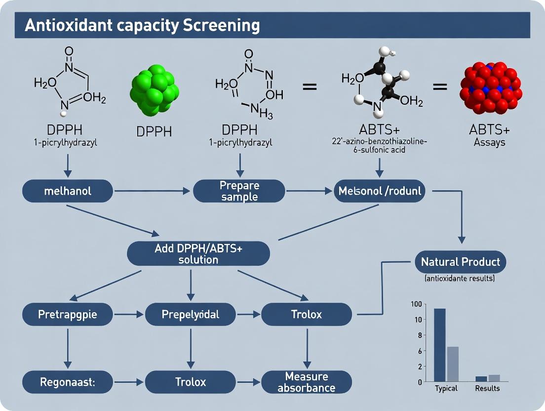
Visualization of Experimental Workflow and Biological Context
Diagram 1: High-Throughput Antioxidant Screening Pipeline.
Diagram 2: Oxidative Stress Pathway & Screening Intervention.
Within the context of a thesis focused on in vitro antioxidant capacity screening, the DPPH (2,2-diphenyl-1-picrylhydrazyl) radical assay stands as a foundational, rapid, and widely employed method. It serves as a critical first-pass screening tool for researchers, scientists, and drug development professionals to evaluate the free radical scavenging potential of pure compounds, plant extracts, and functional foods. This application note details the chemical underpinnings, practical protocols, and data interpretation for the DPPH assay, frequently referenced alongside the ABTS+ assay for comprehensive antioxidant profiling.
Chemical Structure and Properties
DPPH is a stable, organic nitrogen-centered radical. Its stability allows for convenient handling and storage, unlike highly reactive oxygen or hydroxyl radicals.
- Chemical Formula: C₁₈H₁₂N₅O₆
- IUPAC Name: 2,2-Diphenyl-1-(2,4,6-trinitrophenyl)hydrazyl.
- Structure: The molecule features a central hydrazyl group (>N–N•) bearing a phenyl group and a picryl (2,4,6-trinitrophenyl) group. The delocalization of the unpaired electron across the entire molecule, particularly into the picryl ring's π-system, confers remarkable stability.
- Key Physical Properties:
Table 1: Key Physicochemical Properties of DPPH
| Property | Value / Description | Relevance to Assay |
|---|---|---|
| Molecular Weight | 394.33 g/mol | Required for preparing molar solutions. |
| Appearance | Dark purple/black crystalline powder | Visual indicator of radical presence. |
| λmax (in methanol) | ~517 nm | Analytical wavelength for spectrophotometry. |
| Solubility | Soluble in organic solvents (methanol, ethanol, acetone). Low solubility in water. | Assay is typically performed in methanolic or ethanolic solutions. |
| Molar Absorptivity (ε) | ~12,000 M⁻¹cm⁻¹ (in methanol) | Used for quantitative radical concentration calculations. |
Reaction Mechanism
The DPPH assay is based on a single electron transfer (SET) mechanism. An antioxidant (AH) capable of donating a hydrogen atom reduces the DPPH• radical to its corresponding hydrazine form (DPPH-H), accompanied by a characteristic color change from purple to yellow.
Primary Reaction: DPPH• (Purple) + AH (Antioxidant) → DPPH-H (Yellow) + A• (Oxidized Antioxidant)
The extent of discoloration, measured by the decrease in absorbance at 517 nm, is proportional to the antioxidant's scavenging capacity and concentration.
Diagram 1: DPPH Radical Scavenging Reaction Mechanism (96 chars)
Application Notes & Protocols
Standard DPPH Radical Scavenging Assay Protocol
This protocol is optimized for a microplate or cuvette-based spectrophotometric readout.
The Scientist's Toolkit: Key Research Reagent Solutions
Table 2: Essential Materials for DPPH Assay
| Reagent / Material | Function & Specification |
|---|---|
| DPPH Powder | Source of the stable radical. High purity (>95%) recommended. Store desiccated at -20°C, protected from light. |
| Anhydrous Methanol or Ethanol | Primary solvent. Must be UV-spectrophotometric grade to avoid interfering absorbance. |
| DPPH Stock Solution (0.1-0.2 mM) | Prepared fresh daily or aliquoted and stored at -20°C for short-term use. Concentration must be verified via absorbance. |
| Antioxidant Stock Solutions | Test compounds/extracts dissolved in same solvent as DPPH solution. Serial dilutions prepared for dose-response. |
| Microplate Reader or UV-Vis Spectrophotometer | Equipped to measure absorbance at 515-517 nm. |
| 96-Well Microplates or Quartz/Glass Cuvettes | Must be compatible with the organic solvent used. |
Step-by-Step Workflow:
Diagram 2: DPPH Assay Experimental Workflow (64 chars)
Detailed Methodology:
- DPPH Working Solution: Accurately weigh ~1-2 mg DPPH and dissolve in 50-100 mL methanol to prepare a 0.1 mM solution. Verify concentration by measuring absorbance against a methanol blank (A ~1.0 ± 0.02 at 517 nm for 0.1 mM in a 1 cm pathlength).
- Sample Preparation: Prepare serial dilutions of test antioxidants in methanol.
- Reaction Mixture:
- Test: Mix 100 µL of antioxidant solution with 100 µL of DPPH working solution.
- Control: Mix 100 µL of methanol with 100 µL of DPPH working solution.
- Blank: Mix 100 µL of antioxidant solution with 100 µL of methanol (corrects for sample color).
- Incubation: Shake gently and incubate the mixture in the dark at room temperature for 30 minutes (time must be standardized).
- Measurement: Measure the absorbance of all wells/cuvettes at 517 nm.
- Calculation:
- Percent Radical Scavenging Activity (% RSA) = [ (Acontrol - Atest) / A_control ] x 100.
- Plot % RSA vs. sample concentration to determine IC₅₀ (concentration causing 50% inhibition).
Data Interpretation and Comparative Analysis
Table 3: Example DPPH Assay Data for Standard Antioxidants (Hypothetical Data)
| Antioxidant Standard | IC₅₀ (µM) * | Comments / Typical Range |
|---|---|---|
| Trolox (Water-soluble Vitamin E analog) | 15.2 | Common positive control. IC₅₀ often 10-20 µM. |
| Ascorbic Acid (Vitamin C) | 18.7 | Potent standard. Activity can vary with solvent pH. |
| Quercetin | 8.5 | Strong flavonoid antioxidant. Often < 10 µM. |
| BHA (Butylated Hydroxyanisole) | 22.1 | Synthetic reference. |
| Crude Plant Extract (e.g., Ginkgo biloba) | 45.0 µg/mL | Reported in µg/mL for extracts. |
Note: IC₅₀ values are highly dependent on protocol specifics (DPPH concentration, incubation time). Internal standardization is critical.
Critical Considerations in Broader Research Context
- Solvent Effects: Solvent polarity and hydrogen-bonding capacity significantly influence antioxidant reactivity and DPPH solubility. Consistency is key.
- Reaction Kinetics: Different antioxidants exhibit varying reaction rates with DPPH. End-point measurements (e.g., 30 min) may not reflect true capacity for fast or slow-reacting compounds. Kinetic monitoring is advised for robust thesis work.
- Interference: Colored samples can interfere with absorbance readings. The sample blank correction is essential but not always perfect.
- Complementarity with ABTS+ Assay: DPPH is largely selective for lipophilic antioxidants due to its solvent requirements. The hydrophilic ABTS+ assay complements it, providing a broader spectrum assessment of antioxidant capacity. A strong correlation between assays suggests general radical scavenging ability, while discrepancies reveal specificity related to antioxidant polarity or radical type.
The ABTS+ radical cation assay is a cornerstone spectrophotometric method for determining the antioxidant capacity of pure compounds, plant extracts, and biological fluids. This application note, framed within a broader thesis comparing DPPH and ABTS+ assays, details the generation, stability, and kinetic analysis of the ABTS+ radical. While the DPPH assay is non-ionic and best suited for organic-soluble antioxidants, the ABTS+ assay, operating in both organic and aqueous media at physiologically relevant pHs (7.4), offers broader applicability for screening hydrophilic and lipophilic antioxidants in drug development research.
Generation & Chemical Stability
The ABTS+ radical is generated by the oxidation of ABTS salt. Its stability is crucial for assay reproducibility. The following table summarizes standard generation methods and their impact on stability.
Table 1: Methods for ABTS+ Radical Generation and Stability Profile
| Generation Method | Oxidizing Agent | Incubation Conditions | Stable Concentration (λ=734 nm) | Storage Stability (at 4°C, in dark) | Key Considerations |
|---|---|---|---|---|---|
| Chemical Oxidation | Potassium Persulfate (K₂S₂O₈) | 12-16 hours, room temp, dark | A~0.70 ± 0.02 | 2-3 days | Most common; generates stock solution. |
| Chemical Oxidation | Manganese Dioxide (MnO₂) | 5-30 min, filtration required | A~0.70 ± 0.02 | 1-2 days | Fast but requires filtration; Mn²+ contamination risk. |
| Enzymatic Oxidation | Hydrogen Peroxide / Horseradish Peroxidase (HRP) | Minutes, controlled kinetics | Adjustable | Hours | Used for real-time kinetic studies; mimics biological systems. |
Protocol: Generation of ABTS+Stock Solution (Persulfate Method)
This protocol yields a stable stock solution suitable for high-throughput antioxidant screening.
Research Reagent Solutions & Essential Materials:
| Item | Function/Specification |
|---|---|
| ABTS diammonium salt | Substrate for radical generation. Purity >98%. |
| Potassium persulfate (K₂S₂O₈) | Strong oxidizing agent to generate ABTS+. |
| Phosphate Buffered Saline (PBS), 0.1 M, pH 7.4 | Assay buffer for physiological relevance. |
| Ethanol or Methanol (absolute) | Solvent for lipophilic antioxidants. |
| Deionized water (≥18 MΩ·cm) | Preparation of all aqueous solutions. |
| Amber volumetric flask or bottle | Protects radical stock from light degradation. |
| UV-Vis Spectrophotometer / Plate Reader | For measuring absorbance at 734 nm. |
| 0.2 μm syringe filter (optional) | For sterilizing/filtering final stock. |
Procedure:
- Dissolve ABTS diammonium salt in PBS (pH 7.4) or water to a final concentration of 7 mM.
- Dissolve potassium persulfate in water to a final concentration of 2.45 mM.
- Mix the two solutions at a 1:1 (v/v) ratio. Example: 5 mL ABTS solution + 5 mL persulfate solution.
- Incubate the mixture in the dark at room temperature for 12-16 hours to allow complete radical formation.
- The resulting solution is your ABTS+ stock solution. It should have a deep blue-green color.
- Before use, dilute the stock with PBS (pH 7.4) or ethanol (based on assay requirements) to an absorbance of 0.70 ± 0.02 at 734 nm (1 cm pathlength). This is the working solution.
- Store the stock solution in an amber bottle at 4°C for up to 3 days. Monitor absorbance before each use.
Reaction Kinetics & Antioxidant Screening Protocol
The scavenging reaction follows the general scheme: ABTS+ + AH (Antioxidant) → ABTS + A+ + H⁺. The kinetics can be monitored to distinguish between fast and slow-reacting antioxidants.
Protocol: Kinetic Mode Antioxidant Capacity Assay
- Preparation: Generate and dilute ABTS+ working solution as per Section 3. Prepare serial dilutions of antioxidant standards (e.g., Trolox) and samples in appropriate solvents.
- Reaction Setup: In a cuvette or microplate well, mix:
- x μL of antioxidant sample/standard (or blank solvent)
- (1000 - x) μL of ABTS+ working solution.
- Typical reaction volume: 1 mL (cuvette) or 200 μL (microplate).
- Kinetic Measurement: Immediately start recording absorbance at 734 nm.
- For cuvettes: Record every 5-10 seconds for 6-10 minutes.
- For microplate readers: Use kinetic mode, shaking before reading, with cycles every 15-30 seconds for 6-10 minutes.
- Data Analysis:
- Plot Absorbance (734 nm) vs. Time.
- Calculate percent inhibition at a fixed endpoint (e.g., 6 min): % Inhibition = [(A₀ - Aₜ)/A₀] x 100, where A₀ is initial absorbance of control and Aₜ is absorbance with antioxidant at time t.
- For IC₅₀: Determine antioxidant concentration causing 50% inhibition at the chosen endpoint.
- For Kinetics: Analyze the decay curve. Fast antioxidants (e.g., Trolox, Vitamin C) cause immediate decay. Slow antioxidants (e.g., some phenolics) show gradual decay.
Table 2: Kinetic Parameters for Reference Antioxidants in ABTS+ Assay (pH 7.4)
| Antioxidant | Reaction Type | Typical IC₅₀ Range (μM)* | Time to Reach Plateau | Notes for Drug Development Screening |
|---|---|---|---|---|
| Trolox (Water-soluble analog of Vitamin E) | Fast, single-step | 1.5 - 2.5 | < 2 minutes | Primary standard for TEAC (Trolox Equivalent Antioxidant Capacity). |
| Ascorbic Acid (Vitamin C) | Fast, single-step | 1.0 - 2.0 | < 1 minute | May show pro-oxidant effects at high concentrations. |
| Gallic Acid | Fast, single-step | 0.8 - 1.5 | < 3 minutes | High reactivity; can overestimate antioxidant potential. |
| Quercetin | Moderate, multi-step | 3.0 - 6.0 | 4-7 minutes | Flavonoid behavior can be complex; kinetic analysis is informative. |
| Glutathione (Reduced) | Moderate | 8.0 - 15.0 | 5-8 minutes | Important biological antioxidant; reactivity is pH-dependent. |
*IC₅₀ values are method-dependent and should be determined empirically in each lab.
Experimental Workflow and Logical Relationships
Title: ABTS+ Assay Workflow for Antioxidant Screening
Title: ABTS+ Stability Factors and Scavenging Reaction
Within antioxidant capacity screening research, the DPPH (2,2-diphenyl-1-picrylhydrazyl) and ABTS+ (2,2’-azino-bis(3-ethylbenzothiazoline-6-sulfonic acid) radical cation decolorization assays are two of the most established and widely employed methods. This article, framed within a broader thesis on comparative antioxidant assessment, provides detailed application notes and protocols. It underscores that while these assays share conceptual similarities, their inherent chemical differences necessitate complementary use for a comprehensive evaluation of antioxidant potential in drug development and phytochemical research.
Core Principles and Comparative Mechanism
Both assays are based on the principle of electron/hydrogen atom transfer from an antioxidant to a stable, colored radical, resulting in a measurable decolorization proportional to antioxidant capacity.
Table 1: Fundamental Comparison of DPPH• and ABTS•+ Assays
| Parameter | DPPH• Assay | ABTS•+ Assay |
|---|---|---|
| Active Radical Species | DPPH• (organic, nitrogen-centered) | ABTS•+ (organic, nitrogen-centered radical cation) |
| Generation Method | Commercially available, dissolved in organic solvent (e.g., methanol, ethanol). | Requires chemical or enzymatic generation (e.g., with K₂S₂O₈ or MnO₂). |
| Primary Solvent Compatibility | Predominantly organic (methanol, ethanol). Aqueous samples cause precipitation. | Both aqueous and organic solvents. Highly versatile. |
| Reaction pH | Not pH-sensitive; operates at ambient pH. | Can be performed at various pHs (e.g., pH 7.4 for physiological relevance). |
| λ max (Absorbance) | ~515-517 nm | ~734 nm (also 414, 645, 815 nm) |
| Typical Reaction Time | Slower (30 min - 2 hours to reach endpoint). | Faster (4-10 min to reach endpoint). |
| Sensitivity to Antioxidants | Less sensitive to certain antioxidants (e.g., thiols, proteins). | More sensitive; reacts with a broader range (phenolics, thiols, Vit C, proteins). |
| Lipophilic vs. Hydrophilic | Better for assessing lipophilic antioxidants. | Can assess both hydrophilic and lipophilic antioxidants. |
Diagram 1: Core Mechanism of Radical Decolorization Assays (91 chars)
Detailed Experimental Protocols
Protocol 3.1: DPPH Radical Scavenging Assay
Research Reagent Solutions:
- DPPH Stock Solution (0.1 mM): Accurately weigh 3.94 mg of DPPH• and dissolve in 100 mL of pure methanol. Store in amber glass at 4°C, stable for 48 hours.
- Antioxidant Standard (Trolox, 1 mM): Dissolve 2.5 mg of Trolox in 10 mL methanol. Dilute serially for a calibration curve (e.g., 50-500 µM).
- Sample Solutions: Prepare test compounds/extracts in methanol at appropriate concentrations. For aqueous samples, ensure final reaction mixture has <10% water.
Methodology:
- Prepare a series of test tubes with varying concentrations of the antioxidant sample or Trolox standard (e.g., 0-100 µL).
- Adjust the volume in each tube to 100 µL with methanol.
- Add 900 µL of the 0.1 mM DPPH• stock solution to each tube. Vortex thoroughly.
- Prepare a control tube with 100 µL methanol + 900 µL DPPH• solution.
- Prepare a blank with 1 mL methanol only.
- Incubate the reaction mixtures in the dark at room temperature for 30 minutes.
- Measure the absorbance of each mixture at 517 nm against the methanol blank.
- Calculate the radical scavenging activity (RSA) as a percentage:
- % RSA = [(Acontrol - Asample) / Acontrol] x 100
- Where Acontrol is the absorbance of the DPPH• + methanol control.
- Determine IC₅₀ (concentration causing 50% scavenging) from the dose-response curve. Express results as Trolox Equivalent Antioxidant Capacity (TEAC) if calibrated.
Protocol 3.2: ABTS Radical Cation Scavenging Assay
Research Reagent Solutions:
- ABTS Stock Solution (7 mM): Dissolve 38.4 mg ABTS diammonium salt in 10 mL deionized water.
- Potassium Persulfate Solution (2.45 mM): Dissolve 6.6 mg K₂S₂O₈ in 10 mL water.
- ABTS•+ Working Solution: Mix equal volumes of the two stock solutions (e.g., 5 mL each). Allow to react in the dark at room temperature for 12-16 hours to generate the radical cation. Critical: This solution must be diluted with phosphate-buffered saline (PBS, pH 7.4) or ethanol to an absorbance of 0.70 (±0.02) at 734 nm before use. Prepare fresh daily.
- Trolox Standard (1 mM): As in Protocol 3.1, but prepare in PBS or ethanol based on solvent system.
Methodology:
- Prepare sample/standard solutions in PBS or ethanol (30 µL volume typical).
- Add 1 mL of the pre-diluted ABTS•+ working solution to each. Vortex immediately.
- Incubate at 30°C for exactly 6 minutes (or as validated, typically 4-10 min).
- Measure absorbance at 734 nm against a PBS/ethanol blank.
- Calculate % Inhibition using the same formula as for DPPH.
- Construct a Trolox standard curve and express results as TEAC (µmol Trolox equivalents per gram or liter).
Diagram 2: Comparative Workflow of DPPH and ABTS Assays (78 chars)
The Scientist's Toolkit: Essential Reagents & Materials
Table 2: Key Research Reagent Solutions
| Item | Function & Critical Note |
|---|---|
| DPPH• (Free Radical) | Stable organic radical source. Purity is critical. Store desiccated at -20°C. Methanol dissolution creates the reactive purple species. |
| ABTS Diammonium Salt | Precursor for generating the radical cation (ABTS•+). Must be oxidized prior to assay. More stable in its salt form than the generated radical. |
| Potassium Persulfate (K₂S₂O₈) | Common oxidizing agent for generating ABTS•+. Alternative: Manganese dioxide (MnO₂) or enzymatic generation. |
| Trolox (6-hydroxy-2,5,7,8-tetramethylchroman-2-carboxylic acid) | Water-soluble vitamin E analog. The standard reference antioxidant for quantifying results (TEAC). |
| Methanol (HPLC Grade) | Preferred solvent for DPPH assay. Minimizes water-induced radical precipitation. |
| Phosphate Buffered Saline (PBS), pH 7.4 | Used to dilute and buffer the ABTS•+ working solution for physiologically relevant pH assessment. |
| Ethanol (Absolute) | Alternative solvent for both assays, especially for samples incompatible with methanol or for mixed aqueous-organic systems in ABTS. |
| Microplate Reader or UV-Vis Spectrophotometer | For high-throughput (96-well plate) or cuvette-based absorbance measurement at 517 nm (DPPH) or 734 nm (ABTS). |
Data Interpretation and Complementary Use
Table 3: Quantitative Data Interpretation and Reporting
| Output Metric | DPPH Assay Typical Range | ABTS Assay Typical Range | Interpretation & Note |
|---|---|---|---|
| IC₅₀ Value | Varies widely (e.g., 1-500 µg/mL for plant extracts). Lower IC₅₀ = higher potency. | Often lower than DPPH IC₅₀ for the same sample due to faster/broader reactivity. | Direct comparison is invalid due to different radicals, kinetics, and mechanisms. Use for rank-ordering within the same assay. |
| TEAC Value | Reported as µmol Trolox/g extract or µM Trolox eq. | Reported in same units. Typically yields higher values than DPPH for same sample. | Allows for relative comparison across studies using the same assay. Highlights the differential reactivity of sample components. |
| Reaction Kinetics | Monitored over 30-120 min. May show slow-reacting antioxidants. | Monitored over 4-10 min. Captures fast-reacting antioxidants. | Kinetic profiles differ. A full time-course can reveal antioxidant behavior (e.g., rapid vs. slow scavengers). |
The DPPH assay serves as a robust, simple model for lipophilic antioxidant activity in non-polar environments. In contrast, the ABTS assay offers versatility, speed, and the ability to probe activity at physiological pH and across solubility spectra. Their key similarity—the electron-transfer mechanism—makes them broadly applicable. Their inherent differences—in radical chemistry, solvent compatibility, pH sensitivity, and kinetic profile—mean that data from one assay cannot predict results from the other. Discrepancies between DPPH and ABTS results for the same sample are not errors but informative insights into the antioxidant's nature. Therefore, employing both assays in tandem is essential for comprehensive screening, providing a more holistic and mechanistically informative profile crucial for informed decision-making in nutraceutical and pharmaceutical development.
Within antioxidant screening research utilizing DPPH and ABTS⁺ assays, the interpretation of key metrics—% Inhibition, IC₅₀, and TEAC—is fundamental. These parameters allow researchers to quantify and compare the radical scavenging efficacy of novel compounds, natural extracts, and synthetic drugs. This application note details their interpretation and provides standardized protocols for generating robust, comparable data in a drug development context.
Core Metrics Interpretation Table
| Metric | Definition | Interpretation in DPPH/ABTS⁺ Assays | Ideal Value Range (General Guide) | Key Consideration |
|---|---|---|---|---|
| % Inhibition | Immediate measure of antioxidant activity at a fixed concentration/time. | The percentage of DPPH• or ABTS⁺• radicals quenched by the sample. | Higher % indicates stronger activity. Often reported at 50-100 µg/mL for extracts. | Concentration-dependent. Single-point data; does not reflect potency or efficiency. |
| IC₅₀ | Half Maximal Inhibitory Concentration. | The concentration of antioxidant required to scavenge 50% of the radicals. Lower IC₅₀ = higher potency. | Typically µM for pure compounds; µg/mL for extracts. A lower value indicates greater potency. | Derived from a dose-response curve. Critical for comparing potency independent of maximum effect. |
| TEAC | Trolox Equivalent Antioxidant Capacity. | The concentration of Trolox (water-soluble vitamin E analog) that produces the same % inhibition as the sample. Reported as µM Trolox Equivalents/g or /mL. | Allows direct comparison across different assay types. Higher TEAC = higher antioxidant capacity. | Standardizes results to a common reference. TEAC value can vary with assay duration and protocol. |
Key Experimental Protocols
Protocol A: DPPH Radical Scavenging Assay
Objective: To determine % Inhibition, IC₅₀, and TEAC for test samples. Principle: Antioxidants reduce the stable purple DPPH• radical to yellow non-radical DPPH-H, measurable by absorbance loss at 517 nm.
Procedure:
- DPPH Solution: Prepare 0.1 mM DPPH in methanol (or ethanol). Protect from light.
- Sample Preparation: Prepare serial dilutions of test compound/extract in the same solvent. Prepare a Trolox standard curve (e.g., 0-500 µM).
- Reaction: Mix 100 µL of each sample dilution (or standard) with 100 µL of DPPH solution in a 96-well microplate. Include solvent blank (sample + methanol) and negative control (solvent + DPPH).
- Incubation: Incubate in the dark at room temperature for 30 minutes.
- Measurement: Measure absorbance at 517 nm using a plate reader.
- Calculation:
- % Inhibition = [(Acontrol - Asample) / A_control] x 100.
- Plot % Inhibition vs. sample concentration to derive IC₅₀ via nonlinear regression.
- TEAC: From the Trolox standard curve (Absorbance vs. [Trolox]), determine the equivalent Trolox concentration that gives the same % inhibition as the sample.
Protocol B: ABTS⁺ Radical Cation Scavenging Assay
Objective: To generate the long-lived ABTS⁺• radical for antioxidant capacity measurement. Principle: Pre-formed ABTS⁺• (blue-green) is reduced to colorless ABTS by antioxidants, measured by absorbance decay at 734 nm.
Procedure:
- ABTS⁺• Stock: React 7 mM ABTS with 2.45 mM potassium persulfate (final concentration). Incubate in the dark at room temperature for 12-16 hours. The solution turns dark blue.
- Working Solution: Dilute the stock with PBS (pH 7.4) or ethanol to an absorbance of 0.70 (±0.02) at 734 nm.
- Reaction: Mix 20 µL of sample/Trolox standard with 200 µL of ABTS⁺• working solution in a microplate.
- Incubation & Measurement: Incubate for exactly 6 minutes in the dark. Measure absorbance at 734 nm.
- Calculation: Calculate % Inhibition as in DPPH. TEAC is directly determined by comparing sample inhibition to the Trolox standard curve run concurrently.
Visualization of Workflow and Data Relationship
Diagram Title: From Assay to Metrics: Deriving IC50 and TEAC
The Scientist's Toolkit: Key Research Reagent Solutions
| Item | Function in DPPH/ABTS+ Assays |
|---|---|
| DPPH (1,1-Diphenyl-2-picrylhydrazyl) | Stable organic radical. Source of DPPH• radical; absorbance at 517 nm decreases upon reduction by antioxidants. |
| ABTS (2,2'-Azino-bis(3-ethylbenzothiazoline-6-sulfonic acid)) | Chromogenic substrate. Oxidized to the stable blue-green ABTS⁺• radical cation, measured at 734 nm. |
| Trolox (6-hydroxy-2,5,7,8-tetramethylchroman-2-carboxylic acid) | Water-soluble vitamin E analog. Primary standard for constructing calibration curves to calculate TEAC values. |
| Potassium Persulfate (K₂S₂O₈) | Oxidizing agent. Used to generate the ABTS⁺• radical cation from ABTS stock. |
| Microplate Reader (with 517nm & 734nm filters) | Detection instrument. Enables high-throughput measurement of absorbance changes in 96-well or 384-well formats. |
| Methanol / Ethanol (HPLC grade) | Common solvents. Used to dissolve DPPH, antioxidants, and extracts while maintaining assay compatibility. |
| PBS Buffer (pH 7.4) | Physiological pH buffer. Used for diluting ABTS⁺• working solution to mimic biological conditions. |
Step-by-Step Protocols: Executing DPPH and ABTS+ Assays in the Lab
Within the framework of antioxidant capacity screening research utilizing DPPH (2,2-diphenyl-1-picrylhydrazyl) and ABTS+ (2,2'-azino-bis(3-ethylbenzothiazoline-6-sulfonic acid)) assays, the integrity of results is fundamentally dependent on the quality of materials and reagents. This document provides detailed application notes and protocols for sourcing, preparing, and validating key reagents, ensuring reproducibility and reliability in high-throughput screening environments for drug development.
Sourcing and Specifications
Critical reagents must be sourced from reputable suppliers with certificates of analysis (CoA). Key specifications for primary reagents are summarized below.
Table 1: Critical Reagent Specifications for Antioxidant Assays
| Reagent | CAS Number | Purity (Minimum) | Recommended Supplier(s) | Key Quality Indicator |
|---|---|---|---|---|
| DPPH Radical | 1898-66-4 | 95% | Sigma-Aldrich, TCI Chemicals | Absorbance at 517 nm in ethanol: ≥1.00 (0.1 mM) |
| ABTS Diammonium Salt | 30931-67-0 | 98% (HPLC) | Sigma-Aldrich, Cayman Chemical | Peroxidase activity negligible |
| Potassium Persulfate | 7727-21-1 | 99%+ | Fisher Scientific, Merck | Low heavy metal content |
| Trolox (6-hydroxy-2,5,7,8-tetramethylchroman-2-carboxylic acid) | 53188-07-1 | 97% | Sigma-Aldrich, Alfa Aesar | Primary standard for calibration |
| Ethanol (Absolute) | 64-17-5 | 99.8%, ACS grade | Various | Absence of stabilizers (e.g., benzene) |
| Methanol (HPLC grade) | 67-56-1 | 99.9% | Various | UV cut-off <205 nm |
| Phosphate Buffered Saline (PBS) | N/A | pH 7.4 ± 0.05 | Various | Sterile, endotoxin-free for cell-based studies |
Preparation and Standardization Protocols
Protocol 1: Preparation of Stable DPPH Radical Stock Solution
Objective: To prepare a standardized, stable DPPH working solution for radical scavenging assays.
- Weighing: Precisely weigh 3.94 mg of DPPH powder using an analytical balance (readability 0.01 mg).
- Dissolution: Transfer quantitatively to a 100 mL volumetric flask. Dissolve in and make up to volume with absolute ethanol. This yields a 0.1 mM stock solution.
- Standardization: Measure the absorbance of this solution at 517 nm using a UV-Vis spectrophotometer against an ethanol blank. The absorbance should be 1.00 ± 0.02 (1 cm path length). Adjust concentration if necessary.
- Storage: Aliquot into amber vials under inert gas (N₂) if available. Store at -20°C for up to 4 weeks. Discard if absorbance decreases by >10%.
Protocol 2: Generation and Standardization of ABTS⁺ Radical Cation
Objective: To generate a consistent batch of ABTS⁺ radical cation with defined kinetic stability.
- Stock Solution: Dissolve 38.4 mg of ABTS diammonium salt in 10 mL of distilled water to yield a 7 mM stock.
- Radical Generation: Add 176 µL of a 140 mM potassium persulfate solution (in water) to the ABTS stock. Mix thoroughly.
- Incubation: Allow the mixture to stand in the dark at room temperature (23±2°C) for 12-16 hours to ensure complete radical generation.
- Working Solution Dilution: Dilute the incubated solution with ethanol or PBS (depending on assay protocol) to an absorbance of 0.70 ± 0.02 at 734 nm. This working solution must be used within 4 hours of preparation for consistent activity.
Protocol 3: Preparation of Trolox Calibration Standard
Objective: To prepare a serial dilution of Trolox for quantification of antioxidant capacity (TEAC).
- Primary Stock: Dissolve 25.0 mg of Trolox in 100 mL of ethanol or buffer to make a 1.0 mM stock. Store at -80°C for up to 3 months.
- Working Standards: Prepare a dilution series in the same matrix as the assay (e.g., ethanol for DPPH, PBS for ABTS). A typical range is 0.0 (blank), 0.1, 0.2, 0.4, 0.6, and 0.8 mM.
- Calibration Curve: Run each standard in triplicate using the relevant assay protocol (see below). Plot mean absorbance against concentration. The R² value should be ≥0.995.
Critical Quality Control Checks
Table 2: Mandatory Pre-Experiment Quality Checks
| Check Parameter | Method | Acceptance Criterion | Frequency |
|---|---|---|---|
| DPPH Radical Purity | Absorbance Scan (400-600 nm) | Single peak λ_max at 517 nm in ethanol | Per new batch |
| ABTS⁺ Working Solution Stability | Kinetic Read at 734 nm | Absorbance decay <5% over 30 min | Before each assay run |
| Trolox Standard Curve Linearit | Linear Regression | R² ≥ 0.995, slope within 5% of historical data | With each assay plate |
| Solvent Interference | Blank Measurement | Absorbance < 0.02 at assay wavelength | Per solvent batch |
| Microplate Reader Performance | Absorbance Standard Test | Reads NIST-traceable standard within ±2% | Quarterly |
Experimental Protocols for Antioxidant Screening
Protocol 4: Microplate-Based DPPH Radical Scavenging Assay
- Materials: 96-well clear flat-bottom microplates, multichannel pipettes, plate reader capable of reading 517 nm.
- Procedure: a. Pipette 180 µL of standardized DPPH working solution (A517 ≈ 1.0) into sample wells. b. Add 20 µL of test compound (in ethanol or DMSO <1%) or Trolox standard to respective wells. For control, add 20 µL of solvent. c. Mix gently on an orbital shaker for 10 seconds. d. Incubate in the dark at room temperature for 30 minutes. e. Measure absorbance at 517 nm.
- Calculation: % Scavenging = [(Acontrol - Asample) / A_control] * 100. Calculate IC₅₀ from dose-response curve.
Protocol 5: Kinetic ABTS⁺ Decay Assay
- Materials: 96-well plates, plate reader capable of kinetic reads at 734 nm.
- Procedure: a. Add 20 µL of sample or standard to the well. b. Initiate reaction by rapidly adding 180 µL of freshly diluted ABTS⁺ working solution (A734 = 0.70 ± 0.02). c. Immediately place plate in reader and record absorbance at 734 nm every minute for 10 minutes. d. Use the absorbance at the 6-minute endpoint or calculate the area under the decay curve (AUC) for analysis.
- Calculation: Express results as Trolox Equivalents (TEAC) using the Trolox standard curve from the same plate.
The Scientist's Toolkit
Table 3: Essential Research Reagent Solutions for Antioxidant Assays
| Item | Function in DPPH/ABTS+ Assays | Critical Consideration |
|---|---|---|
| Analytical Balance (0.01 mg) | Precise weighing of reagents and standards | Regular calibration with certified weights is mandatory. |
| UV-Vis Spectrophotometer/Plate Reader | Quantification of radical absorbance decay | Wavelength accuracy and photometric linearity must be validated. |
| pH Meter (with temperature probe) | Preparation of PBS and buffer solutions | Daily calibration with 3-point buffers (pH 4, 7, 10). |
| Ultrasonic Bath | For degassing solvents and dissolving stubborn compounds | Prevents bubble formation in microplate wells. |
| Amber Volumetric Flasks & Vials | Protection of light-sensitive radicals (DPPH, ABTS⁺) from photodegradation. | Must be used for all stock solution storage. |
| Inert Atmosphere (N₂) Glove Bag/Box | Preparation of radical stocks under oxygen-free conditions | Extends shelf-life of DPPH stock solutions significantly. |
| Single-Channel and Multichannel Micropipettes | Accurate liquid handling for high-throughput microplate setups | Regular maintenance and volume accuracy checks required. |
| Laboratory Information Management System (LIMS) | Tracking reagent lot numbers, preparation dates, and QC data. | Essential for audit trails and troubleshooting reproducibility issues. |
Visualizations
Reagent QC and Preparation Workflow
ABTS+ Radical Generation and Scavenging Reaction
Within the broader thesis investigating standardized protocols for in vitro antioxidant capacity screening, the 2,2-diphenyl-1-picrylhydrazyl (DPPH•) radical scavenging assay remains a cornerstone. This protocol details the steps for the quantitative spectrophotometric DPPH assay, providing a foundational method for researchers and drug development professionals to screen natural compounds, synthetic molecules, and biological extracts for primary antioxidant activity. Its simplicity, speed, and reproducibility make it a critical first-tier screening tool, complementary to more complex assays like ABTS⁺, FRAP, and ORAC within a comprehensive antioxidant research framework.
The Scientist's Toolkit: Research Reagent Solutions
| Reagent/Material | Function & Specification |
|---|---|
| DPPH (2,2-Diphenyl-1-picrylhydrazyl) | The stable free radical compound. Source of the absorbance signal. Purity ≥ 95%. Store desiccated at -20°C, protected from light. |
| Methanol (HPLC Grade) | Preferred solvent for DPPH dissolution. Ensures clear solutions and minimizes solvent interference. Low water content is critical. |
| Ethanol (Absolute, 99.8%) | Common alternative solvent. Must be free from stabilizing antioxidants. |
| Trolox (6-hydroxy-2,5,7,8-tetramethylchroman-2-carboxylic acid) | Water-soluble vitamin E analog. Standard antioxidant used for calibration and expressing results as Trolox Equivalents (TEAC). |
| Ascorbic Acid (Vitamin C) | Reference standard for antioxidant activity validation. |
| Quercetin or Gallic Acid | Reference standard for plant polyphenol antioxidants. |
| Microplate Reader or UV-Vis Spectrophotometer | For absorbance measurement at 515-517 nm. Must be capable of kinetic or endpoint measurement. |
| 96-well Microplates (Flat-bottom, UV-compatible) or Quartz Cuvettes | Reaction vessels compatible with the detection system. |
Detailed Application Notes and Protocols
Stock and Working Solution Preparation
- DPPH Stock Solution (1 mM): Accurately weigh 3.94 mg of DPPH powder. Transfer to a 10 mL volumetric flask and dilute to the mark with HPLC-grade methanol (or ethanol). Vortex vigorously for 1-2 minutes until fully dissolved. This solution is deep violet and must be prepared fresh daily or stored in an amber vial at -20°C for up to 3 days. Note: Protect from light at all stages.
- DPPH Working Solution (100 µM): Dilute the 1 mM stock solution 1:10 with the same solvent (e.g., 1 mL stock + 9 mL methanol). The absorbance of this working solution at 517 nm should be between 0.9 and 1.2 AU (± 0.02) for optimal assay linearity.
- Antioxidant Standard (Trolox) Stock (1 mM): Weigh 2.50 mg of Trolox. Dissolve in methanol (or a methanol:water mix if needed) in a 10 mL volumetric flask. This can be aliquoted and stored at -20°C for up to one month.
- Sample Preparation: Test samples (compounds or extracts) should be dissolved in the same solvent as the DPPH solution. Prepare a series of concentrations (typically 5-7) to generate a dose-response curve. Include a solvent-only control.
Experimental Protocol: Microplate Method
Principle: The antioxidant donates a hydrogen atom to the violet-colored DPPH•, reducing it to yellow-colored DPPH-H, causing a decrease in absorbance at 517 nm.
Procedure:
- Blank Well: Add 150 µL of pure solvent (methanol) to designated wells.
- Control Well: Add 100 µL of solvent + 50 µL of DPPH working solution. This represents 0% scavenging (Abs_control).
- Sample Well: Add 100 µL of antioxidant sample at varying concentrations + 50 µL of DPPH working solution.
- Immediately seal the microplate with a transparent film, mix briefly on a plate shaker, and place in the microplate reader.
- Incubate in the dark at room temperature for 30 minutes (kinetic monitoring up to 60 minutes is recommended for reaction completion analysis).
- Measure the absorbance at 517 nm (Abs_sample). Use the blank well for baseline correction.
Data Analysis & Reporting
Calculate the radical scavenging activity (% RSA) for each sample concentration:
% RSA = [(Abs_control – Abs_sample) / Abs_control] × 100
Key Parameters:
- IC₅₀ (Half Maximal Inhibitory Concentration): The concentration of antioxidant required to scavenge 50% of DPPH radicals. Determine by plotting % RSA vs. log[sample concentration] and performing non-linear regression.
- Trolox Equivalent Antioxidant Capacity (TEAC): Express activity relative to the Trolox standard curve (µmol Trolox equivalents per gram or mL of sample).
Table 1: Example Data Set for Trolox Standard
| Trolox Concentration (µM) | Mean Absorbance (517 nm) | % RSA |
|---|---|---|
| 0 (Control) | 1.000 | 0 |
| 5 | 0.850 | 15.0 |
| 10 | 0.700 | 30.0 |
| 25 | 0.450 | 55.0 |
| 50 | 0.250 | 75.0 |
| 100 | 0.100 | 90.0 |
Calculated IC₅₀ for Trolox: ~15 µM (assay-dependent)
Workflow and Reaction Pathway Visualization
Diagram Title: DPPH Assay Experimental Workflow
Diagram Title: DPPH Radical Scavenging Reaction Mechanism
Within the broader investigation of antioxidant screening methods, the ABTS⁺ (2,2'-azino-bis(3-ethylbenzothiazoline-6-sulfonic acid)) radical cation decolorization assay stands alongside the DPPH assay as a cornerstone technique. This protocol details two primary methods for generating the stable ABTS⁺ chromophore—chemical oxidation and enzymatic generation—and emphasizes the importance of kinetic monitoring over single-time-point measurements for accurate assessment of antioxidant capacity. Kinetic analysis provides crucial data on reaction rates and mechanisms, distinguishing between rapid and slow-reacting antioxidants.
ABTS⁺ Radical Generation: Methods and Protocols
Chemical Generation using Potassium Persulfate
This is the most common method, producing a long-lived stock solution of the blue-green ABTS⁺ radical.
Protocol:
- Prepare a 7 mM ABTS stock solution in distilled water (e.g., 38.4 mg in 10 mL water).
- Prepare a 2.45 mM potassium persulfate (K₂S₂O₈) solution in water (e.g., 6.6 mg in 10 mL water).
- Mix equal volumes of the two solutions (e.g., 5 mL ABTS + 5 mL K₂S₂O₈).
- Allow the mixture to react in the dark at room temperature for 12-16 hours to ensure complete radical generation. The solution will turn an intense dark blue-green.
- Dilute the stock ABTS⁺ solution with an appropriate buffer (commonly phosphate-buffered saline, PBS, pH 7.4) to an absorbance of 0.70 (±0.02) at 734 nm. This working solution is stable for several days when stored in the dark at 4°C.
Table 1: Typical Composition for Chemical Generation
| Component | Final Concentration in Reaction Mixture | Volume for 10 mL Stock | Function |
|---|---|---|---|
| ABTS | 7 mM | 38.4 mg | Chromogen, forms radical cation |
| Potassium Persulfate (K₂S₂O₈) | 2.45 mM | 6.6 mg | Oxidizing agent |
Enzymatic Generation using Horseradish Peroxidase (HRP)/Hydrogen Peroxide
This method generates ABTS⁺ in situ and is useful for studying antioxidant activity under physiological-like conditions or for real-time kinetic studies.
Protocol:
- Prepare a reaction buffer (e.g., 50 mM phosphate buffer, pH 7.5).
- To a cuvette or microplate well, add:
- Buffer to the final desired volume.
- ABTS to a final concentration of 500 µM.
- Hydrogen peroxide (H₂O₂) to a final concentration of 50 µM.
- Initiate the reaction by adding Horseradish Peroxidase (HRP) to a final activity of 0.25–1.0 U/mL.
- Immediately mix and begin kinetic measurement at 734 nm. The formation of ABTS⁺ occurs rapidly (within minutes).
Table 2: Typical Composition for Enzymatic Generation
| Component | Final Concentration | Function |
|---|---|---|
| ABTS | 500 µM | Chromogen substrate |
| Hydrogen Peroxide (H₂O₂) | 50 µM | Oxidizing substrate for HRP |
| Horseradish Peroxidase (HRP) | 0.25 – 1.0 U/mL | Enzyme catalyst |
Table 3: Chemical vs. Enzymatic ABTS⁺ Generation
| Parameter | Chemical Oxidation (K₂S₂O₈) | Enzymatic Generation (HRP/H₂O₂) |
|---|---|---|
| Radical Solution | Stable pre-formed stock | Generated in situ, transient |
| Reaction Time | Slow (12-16 hrs for generation) | Fast (seconds to minutes) |
| Buffer Compatibility | Can be diluted in various buffers (PBS, acetate) | Requires optimal pH buffer for enzyme activity |
| Primary Use | High-throughput single-point & endpoint screening | Real-time kinetic studies, mechanistic analysis |
| Pro-oxidant Interference | Residual persulfate may oxidize some antioxidants | Possible interactions with H₂O₂-scavenging antioxidants |
| Physiological Relevance | Low | Higher (enzymatic oxidation) |
Kinetic Monitoring Protocol for Antioxidant Assessment
A single endpoint measurement can be misleading. Kinetic monitoring captures the entire reaction profile.
Protocol for Kinetic Measurement:
- Instrument Setup: Use a UV-Vis spectrophotometer or plate reader capable of kinetic measurement at 734 nm. Set the temperature (typically 25°C or 30°C).
- Baseline: Add appropriate buffer to a cuvette/well. Record baseline absorbance.
- Radical Addition: Add the pre-formed ABTS⁺ working solution (A734 ≈ 0.70) or the complete enzymatic generation mix without antioxidant. Monitor for 1-2 minutes to confirm radical stability.
- Reaction Initiation: Add a small volume (e.g., 10-50 µL) of the antioxidant sample (standard or unknown) to the ABTS⁺ solution. Mix rapidly.
- Data Acquisition: Immediately begin recording absorbance at 734 nm at regular intervals (e.g., every 5-30 seconds) for a period of 6-30 minutes, until the reaction reaches a plateau.
- Data Analysis: Plot absorbance (A) or percentage inhibition (%I) versus time (t). Calculate parameters such as:
- Initial Rate of Decolorization: Slope of the linear portion of the curve (ΔA/Δt).
- Time to Reach 50% Inhibition (T₅₀).
- Final Percentage Inhibition at Plateau.
The Scientist's Toolkit: Key Research Reagent Solutions
Table 4: Essential Materials for ABTS⁺ Assay
| Reagent / Material | Function & Notes |
|---|---|
| ABTS Diammonium Salt | The core chromogen. High-purity grade (>98%) is essential for reproducibility. |
| Potassium Persulfate (K₂S₂O₈) | Chemical oxidant for stable radical stock generation. Store desiccated. |
| Horseradish Peroxidase (HRP) | Enzyme for physiological radical generation. Use a defined activity (U/mg). |
| Hydrogen Peroxide (H₂O₂) 30% Solution | Substrate for enzymatic generation. Standardize concentration before use. |
| Phosphate Buffered Saline (PBS), pH 7.4 | Common dilution buffer for pre-formed ABTS⁺, simulates physiological ionic strength. |
| Trolox (6-hydroxy-2,5,7,8-tetramethylchroman-2-carboxylic acid) | Water-soluble vitamin E analog; the standard antioxidant for generating calibration curves (expressed as Trolox Equivalents). |
| Microplate Reader (with 734 nm filter) | Enables high-throughput kinetic analysis of multiple samples simultaneously. |
| Quartz or Polystyrene Cuvettes/Plates | Must be transparent at 734 nm. Verify plate compatibility for low-volume assays. |
Visualization of ABTS⁺ Assay Workflows and Context
Title: Chemical ABTS+ Radical Generation Protocol
Title: Enzymatic ABTS+ Generation & Kinetic Assay
Title: ABTS+ and DPPH in Antioxidant Screening Research
1. Introduction Within a thesis investigating DPPH and ABTS⁺ assays for antioxidant capacity screening, robust sample preparation is paramount. The efficacy of these assays is directly contingent on the compatibility of the prepared sample with the assay chemistry. This document details validated protocols for preparing complex biological and synthetic matrices to ensure accurate, reproducible quantification of antioxidant activity.
2. Sample Preparation Protocols
2.1. Plant and Food Extracts Objective: To solubilize and stabilize antioxidants from solid or semi-solid matrices while removing interfering compounds. Detailed Protocol:
- Homogenization: Weigh 1.0 g of fresh or dried sample. Add 10 mL of extraction solvent (e.g., 80% methanol, 70% ethanol, or aqueous acetone) to a 15 mL conical tube.
- Sonication: Sonicate the mixture in an ice-water bath for 15 minutes at 40 kHz.
- Centrifugation: Centrifuge at 12,000 × g for 15 minutes at 4°C.
- Filtration: Carefully decant the supernatant and filter through a 0.45 μm PTFE or nylon membrane filter.
- Concentration (Optional): Evaporate the filtrate under a gentle stream of nitrogen at 40°C. Reconstitute the dried extract in a known volume of the assay-compatible solvent (typically methanol or ethanol for DPPH, phosphate-buffered saline or ethanol for ABTS⁺).
- Storage: Store prepared extracts at -80°C for long-term use. Avoid repeated freeze-thaw cycles.
2.2. Blood Serum/Plasma Objective: To deproteinize samples, releasing protein-bound antioxidants and preventing turbidity in spectrophotometric assays. Detailed Protocol:
- Deproteinization: Mix 100 μL of serum/plasma with 300 μL of ice-cold methanol (or acetonitrile) in a 1.5 mL microcentrifuge tube. Vortex vigorously for 1 minute.
- Precipitation: Incubate the mixture at -20°C for 20 minutes to complete protein precipitation.
- Centrifugation: Centrifuge at 15,000 × g for 20 minutes at 4°C.
- Collection: Collect the clear supernatant. If using ABTS⁺ assay in aqueous buffer, ensure the final supernatant is compatible. For methanolic DPPH, the supernatant can often be used directly after a 1:5 or 1:10 dilution in methanol.
- Critical Note: For ABTS⁺ assay, a parallel blank must be prepared where the assay buffer replaces the radical cation solution to correct for inherent sample color.
2.3. Pharmaceutical Formulations (Tablets/Capsules) Objective: To completely extract active pharmaceutical ingredients (APIs) and excipients with antioxidant properties. Detailed Protocol:
- Grinding: Crush five tablets or empty the contents of five capsules into a fine powder using a mortar and pestle.
- Solvent Extraction: Weigh an amount of powder equivalent to one dose. Transfer to a volumetric flask (e.g., 100 mL). Add 50 mL of appropriate solvent (simulated gastric fluid, phosphate buffer pH 7.4, or methanol/water mix based on API solubility).
- Agitation: Sonicate for 30 minutes, then place on an orbital shaker for 2 hours at room temperature.
- Dilution: Bring to volume with the same solvent. Mix thoroughly.
- Clarification: Centrifuge an aliquot at 10,000 × g for 10 minutes. Filter the supernatant through a 0.2 μm syringe filter prior to analysis. Perform serial dilutions to fall within the assay's linear range.
3. Quantitative Data Summary
Table 1: Impact of Sample Preparation on Assay Recovery and Interference
| Matrix | Prep Method | Avg. Antioxidant Recovery (%) | Key Interference Mitigated | Optimal Assay |
|---|---|---|---|---|
| Berry Extract | 80% Methanol Sonication | 98.5 ± 2.1 | Chlorophyll, Tannins | DPPH |
| Human Serum | Methanol Deproteinization | 95.2 ± 3.5 | Protein Turbidity | ABTS⁺ |
| Vitamin C Tablet | Phosphate Buffer (pH 7.4) | 99.1 ± 1.8 | Starch, Fillers | Both (DPPH & ABTS⁺) |
| Herbal Oil | Direct Dissolution in Hexane, then MeOH | 89.7 ± 4.2 | Lipid Peroxides | DPPH (with correction) |
Table 2: Recommended Solvent Systems for Different Matrices
| Matrix Type | Primary Solvent | Alternative Solvent | Compatibility Note |
|---|---|---|---|
| Polyphenol-rich Plants | 70% Aqueous Acetone | 80% Aqueous Methanol | Excellent for flavonoids. Acetone must be evaporated for ABTS⁺. |
| Serum/Plasma | Cold Methanol | Cold Acetonitrile | Methanol gives higher recovery for hydrophilic antioxidants. |
| Lipid-rich Foods | Chloroform-Methanol (2:1) | Hexane followed by MeOH | Requires phase separation. Lipid-free extract must be used. |
| Aqueous Formulations | Phosphate Buffer Saline (PBS) | Deionized Water | Direct analysis possible; check for buffer-radical interaction. |
4. The Scientist's Toolkit: Essential Research Reagent Solutions
Table 3: Key Reagents and Materials for Sample Preparation
| Item | Function & Rationale |
|---|---|
| Solid Phase Extraction (SPE) Cartridges (C18) | For clean-up of complex extracts; removes sugars, organic acids, and polar pigments that can interfere. |
| Protein Precipitation Plates (96-well) | Enables high-throughput deproteinization of serum/plasma samples for antioxidant screening. |
| PTFE (0.45 μm & 0.2 μm) Syringe Filters | Clarifies samples post-extraction; PTFE is chemically inert and suitable for organic solvents. |
| Nitrogen Evaporation System | For gentle, rapid concentration of volatile solvent extracts without heat degradation of antioxidants. |
| Ultrasonic Bath (with temp control) | Ensures efficient, reproducible cell lysis and compound extraction from solid matrices. |
| Antioxidant Spike Standards (e.g., Trolox, Ascorbic Acid) | Used in standard addition protocols to validate recovery rates and identify matrix effects. |
| Oasis HLB SPE Sorbent | A hydrophilic-lipophilic balanced sorbent ideal for simultaneous extraction of acidic, basic, and neutral antioxidants from complex fluids. |
5. Workflow and Context Diagrams
Title: Overall Sample Preparation Workflow for Antioxidant Assays
Title: Sample Prep Role in a Broader Antioxidant Research Thesis
Application Notes & Protocols
The accurate quantification of antioxidant capacity via DPPH (2,2-diphenyl-1-picrylhydrazyl) and ABTS⁺ (2,2'-azino-bis(3-ethylbenzothiazoline-6-sulfonic acid)) assays is a cornerstone of phytochemical and nutraceutical screening. This thesis posits that instrument-specific variables—spectrophotometer configuration and microplate reader parameters—are critical, yet often under-optimized, sources of variance in reported IC₅₀ values. These application notes provide standardized protocols to minimize this methodological divergence.
Spectrophotometer Setup for Single-Cuvette DPPH/ABTS⁺ Assays
Core Principle: Ensure consistent pathlength, mixing, and temperature control for kinetic or endpoint measurements.
Detailed Protocol: DPPH Radical Scavenging Assay (Endpoint)
- Instrument Warm-up & Baseline: Power on UV-Vis spectrophotometer 30 minutes prior. Perform a baseline correction with the solvent used (commonly methanol for DPPH, PBS or ethanol for ABTS⁺) across 500-800 nm.
- Wavelength Selection: Set analytical wavelength: 517 nm for DPPH, 734 nm for ABTS⁺. Confirm peak absorbance of the fresh radical solution falls within ±2 nm of these values.
- Cuvette & Volume Standardization: Use matched quartz or optical glass cuvettes. For a 1 cm pathlength, a minimum working volume of 1.0 mL is recommended to ensure the light beam passes fully through the sample.
- Mixing Protocol: After adding antioxidant sample/extract to the radical solution, cap cuvette and invert 3x gently. Do not vortex to avoid introducing bubbles. Place in cuvette holder and start timer.
- Incubation & Measurement: Allow reaction to proceed in the dark for 30 minutes (DPPH) or 6 minutes (ABTS⁺) at controlled room temperature (25±1°C). Measure absorbance against a blank of radical solution + solvent.
- Calculation: Percent Inhibition = [(Acontrol - Asample) / A_control] × 100.
Table 1: Critical Spectrophotometer Parameters for Antioxidant Assays
| Parameter | DPPH Assay Setting | ABTS⁺ Assay Setting | Justification |
|---|---|---|---|
| Analytical Wavelength | 517 nm | 734 nm | Maximum absorbance of stable radical. |
| Bandwidth | 2 nm | 2 nm | Balances spectral purity with light throughput. |
| Scan Speed | Medium (480 nm/min) | Medium (480 nm/min) | Adequate for precise single-point reads. |
| Integration Time | 0.1 sec | 0.1 sec | Ensures stable signal capture. |
| Temperature Control | 25°C (if available) | 25°C (if available) | Kinetics are temperature-sensitive. |
Microplate Reader Optimization for High-Throughput Screening
Core Principle: Translate cuvette-based chemistry reliably to a microplate format, accounting for pathlength correction, evaporation, and edge effects.
Detailed Protocol: ABTS⁺ Scavenging Assay in 96-Well Plates
- Plate & Reader Pre-configuration:
- Use clear, flat-bottom 96-well plates. Pre-read plate at 734 nm to check for manufacturing irregularities.
- Configure reader: Set temperature to 25°C, enable orbital shaking (3 sec, 1 mm amplitude) before reading.
- Set kinetic mode: Read every minute for 10 minutes post-mixing.
- Pathlength Correction: For endpoint assays, include a water blank in unused wells. Use the reader's pathlength correction function if available, or apply the formula: Corrected Abs = (Measured Abs) × (Reference Pathlength / Actual Pathlength). For approximate correction in clear flat-bottom plates, a factor of ~0.3 cm is typical.
- Reaction Assembly (200 µL total volume):
- Well A1-H1: Blank (Solvent + ABTS⁺ working solution).
- Well A2-H2: Control (Solvent + ABTS⁺ working solution).
- Sample Wells: 20 µL of sample/standard + 180 µL of ABTS⁺ working solution.
- Use a multichannel pipette for simultaneous addition of ABTS⁺ to initiate reaction.
- Sealing & Incubation: Immediately seal plate with optically clear film. Incubate in the plate reader for exactly 6 minutes with shaking.
- Data Acquisition: Read absorbance at 734 nm. Use software to subtract blank and calculate % inhibition relative to the control column average.
Table 2: Microplate Reader Configuration for Antioxidant Assays
| Configuration | Recommended Setting | Impact on Data Quality |
|---|---|---|
| Read Mode | Absorbance, Endpoint or Kinetic | Suits assay design (single point vs. kinetic). |
| Z-Height/Calibration | Optimized for plate type | Ensures consistent focal point for all wells. |
| Number of Flashes | 10-25 per well | Improves signal-to-noise ratio. |
| Read Area/Well Diameter | 70-80% of well diameter | Avoids edge artifacts. |
| Shaking Before Read | Enabled (low intensity) | Ensures homogeneous mixing, crucial for kinetics. |
| Plate Layout | Randomization of samples | Mitigates positional (edge evaporation) bias. |
The Scientist's Toolkit: Key Research Reagent Solutions
Table 3: Essential Materials for DPPH & ABTS⁺ Assays
| Item | Function & Specification |
|---|---|
| DPPH Radical | Stable free radical powder. Store desiccated at -20°C. Prepare fresh 0.1 mM solution in methanol. |
| ABTS Diammonium Salt | Precursor for generating ABTS⁺ radical cation. Store at 4°C. |
| Potassium Persulfate | Oxidizing agent to generate ABTS⁺ (final 2.45 mM). Prepare fresh solution. |
| Trolox (6-hydroxy-2,5,7,8-tetramethylchroman-2-carboxylic acid) | Water-soluble Vitamin E analog; the standard reference antioxidant for both assays. |
| Optical Glass Cuvettes (1 cm path) | For spectrophotometer use; ensure matched set for sample vs. reference. |
| Clear Flat-Bottom 96-Well Plates | For microplate reader; ensure low UV absorbance and minimal meniscus formation. |
| Optically Clear Plate Sealing Film | Prevents evaporation and cross-contamination during incubation. |
| Multichannel Pipette (8 or 12 channel) | Enables simultaneous reagent dispensing for consistent reaction start times. |
Experimental Workflow and Data Analysis Pathway
Diagram Title: Workflow for Antioxidant Capacity Assays
Diagram Title: Data Analysis Path for IC50 Determination
This protocol details the systematic workflow for generating robust, high-quality data in the screening of antioxidant capacity using DPPH (2,2-diphenyl-1-picrylhydrazyl) and ABTS⁺ (2,2’-azino-bis(3-ethylbenzothiazoline-6-sulfonic acid)) assays. Framed within a broader thesis on comparative antioxidant screening, this document emphasizes the critical importance of experimental timing, appropriate replication, and the implementation of essential controls (Blank, Positive, Negative) to ensure accuracy, precision, and biological relevance. Adherence to this standardized workflow is paramount for producing reliable data suitable for publication and further drug development.
The Scientist's Toolkit: Research Reagent Solutions
| Item | Function in DPPH/ABTS⁺ Assays |
|---|---|
| DPPH Radical (in methanol/ethanol) | The stable free radical; its purple color reduction by antioxidants is measured at 517 nm. |
| ABTS⁺ Radical Cation (in PBS or ethanol) | The pre-formed radical cation; its blue-green color reduction is measured at 734 nm. |
| Trolox (6-hydroxy-2,5,7,8-tetramethylchroman-2-carboxylic acid) | A water-soluble vitamin E analog used as a standard to create a calibration curve (Positive Control reference). |
| Ascorbic Acid (Vitamin C) | A common natural antioxidant used as an additional Positive Control for assay validation. |
| Methanol/Absolute Ethanol | Solvents for dissolving DPPH and many lipophilic antioxidant samples. |
| Phosphate Buffered Saline (PBS), pH 7.4 | Aqueous reaction medium for ABTS⁺ assay, simulating physiological pH. |
| Potassium Persulfate | Used to oxidize ABTS to the stable radical cation (ABTS⁺) prior to the assay. |
| Quercetin/Rutin/Gallic Acid | Reference phenolic antioxidants for comparing sample activity. |
| Microplate Reader (with 517 nm & 734 nm filters) | Instrument for high-throughput measurement of absorbance changes. |
Core Experimental Protocols
Protocol 3.1: ABTS⁺ Radical Cation Stock Solution Generation
Principle: ABTS is oxidized by potassium persulfate to form the stable, blue-green ABTS⁺ radical. Procedure:
- Prepare 7 mM ABTS aqueous solution.
- Prepare 2.45 mM potassium persulfate aqueous solution.
- Mix equal volumes of both solutions (e.g., 1:1 v/v).
- Allow the mixture to react in the dark at room temperature for 12-16 hours before use.
- The resulting ABTS⁺ stock solution is stable for 2-3 days when stored in the dark at 4°C.
- Prior to assay, dilute the stock solution with PBS (pH 7.4) or ethanol to an absorbance of 0.70 (±0.02) at 734 nm.
Protocol 3.2: DPPH Assay Procedure (Microplate Format)
Principle: Antioxidants donate a hydrogen atom to the purple DPPH radical, reducing it to a yellow-colored diphenylpicrylhydrazine. Reagents: DPPH in methanol (0.1 mM, freshly prepared), Antioxidant samples/Trolox standard in suitable solvent, Methanol (for blank). Procedure:
- Preparation: Prepare a serial dilution of the Trolox standard (e.g., 0-500 µM) and antioxidant samples.
- Plate Setup: In a 96-well microplate, add:
- Test Wells: 100 µL DPPH solution + 100 µL sample/standard.
- Negative Control: 100 µL DPPH + 100 µL solvent (measures initial DPPH absorbance).
- Blank: 100 µL methanol + 100 µL sample/standard (corrects for sample color).
- Positive Control: 100 µL DPPH + 100 µL known antioxidant (e.g., Ascorbic acid).
- Reaction & Measurement: Mix gently. Incubate the plate in the dark at room temperature for 30 minutes. Measure absorbance at 517 nm.
- Calculation: % Inhibition = [(ANegative – ATest) / A_Negative] x 100. Plot % Inhibition vs. Trolox concentration for standard curve.
Protocol 3.3: ABTS⁺ Assay Procedure (Microplate Format)
Principle: Antioxidants decolorize the ABTS⁺ radical cation by electron/hydrogen atom donation. Reagents: Diluted ABTS⁺ working solution (A734nm = 0.70 ±0.02), Antioxidant samples/Trolox standard, PBS or ethanol (for blank). Procedure:
- Preparation: Prepare Trolox standard and sample dilutions.
- Plate Setup: In a 96-well microplate, add:
- Test Wells: 20 µL sample/standard + 180 µL ABTS⁺ working solution.
- Negative Control: 20 µL solvent (PBS/EtOH) + 180 µL ABTS⁺.
- Blank: 20 µL sample/standard + 180 µL PBS/EtOH.
- Positive Control: 20 µL Trolox standard + 180 µL ABTS⁺.
- Reaction & Measurement: Mix immediately after adding ABTS⁺. Incubate in the dark at 30°C for exactly 6 minutes. Measure absorbance at 734 nm.
- Calculation: % Inhibition calculated as in DPPH. Express results as Trolox Equivalent Antioxidant Capacity (TEAC).
Data Collection Workflow: Structure and Controls
The following workflow ensures systematic data acquisition.
Diagram 1: Antioxidant Assay Workflow & Controls
The following tables summarize the critical parameters for reproducible results.
Table 1: Optimized Timing Parameters for DPPH and ABTS⁺ Assays
| Parameter | DPPH Assay | ABTS⁺ Assay | Rationale |
|---|---|---|---|
| Radical Prep Stability | Fresh daily (0.1 mM in MeOH) | Stock stable 2-3 days at 4°C (A734=0.7) | DPPH degrades in light; ABTS⁺ slowly decays. |
| Reaction Temperature | Room Temperature (25±2°C) | 30°C (recommended) | Temperature affects reaction kinetics. |
| Reaction Time | 30 minutes (in dark) | 6 minutes (in dark) | Time to reach steady-state/plateau. |
| Absorbance Measurement | Single read at 517 nm at t=30 min. | Kinetic or single read at 734 nm at t=6 min. | Maximum sensitivity at λmax. |
Table 2: Replication and Control Scheme for a 96-Well Plate Experiment
| Well Type | Purpose | Minimum Replicates per Plate | Expected Outcome (Quality Check) |
|---|---|---|---|
| Negative Control | Defines 0% inhibition (A_initial). | n=6 | Absorbance stable (±2%) across replicates. |
| Positive Control (Trolox) | Validates assay sensitivity. | n=3 (at 2 conc.) | % Inhibition matches historical curve ±5%. |
| Calibrant (Trolox Std Curve) | For quantitative TEAC calculation. | n=3 (per 6-8 conc.) | Linear regression R² ≥ 0.985. |
| Sample Blanks | Corrects for sample color/turbidity. | n=2 (per sample) | Absorbance should be low (<0.1). |
| Test Samples | Unknown antioxidant capacity. | n=4 (technical replicates) | CV < 10% for reliable mean value. |
| Radical Solvent Blank | Checks radical solution purity. | n=2 | Absorbance ~0.00 (baseline correction). |
Diagram 2: Data Correction Logic Using Blanks
Critical Interpretation Notes
- Timing is Critical: Both assays are time-sensitive. Deviations from specified incubation times directly impact reported % inhibition and TEAC values.
- Solvent Compatibility: Ensure sample solvent does not quench radicals (e.g., high acid concentration). Final solvent concentration must be constant across all wells (<5% v/v variation).
- Replicates are Non-Negotiable: Technical replicates (n≥4) account for pipetting error. Biological replicates (n≥3 independent samples) are required for statistical significance in research.
- Control Validation: Each plate must pass control QC. A positive control falling outside expected range invalidates the plate run.
Solving Common Problems: Troubleshooting and Optimizing Your Antioxidant Assays
Within the critical framework of antioxidant capacity screening research using DPPH (2,2-diphenyl-1-picrylhydrazyl) and ABTS (2,2’-azino-bis(3-ethylbenzothiazoline-6-sulfonic acid)) assays, the stability of the radical stock solutions is a paramount, yet often underestimated, experimental variable. The core thesis of this research posits that inconsistent, non-linear degradation of these radicals is a primary source of inter-assay variability, leading to inaccurate IC50 calculations and unreliable comparison of antioxidant compounds. These Application Notes provide validated protocols and data-driven strategies to monitor, manage, and mitigate radical instability, thereby enhancing the reproducibility and precision of high-throughput screening in pharmaceutical and nutraceutical development.
Quantitative Stability Data
The following tables summarize key stability data for DPPH and ABTS⁺ radical solutions under various storage conditions, compiled from recent studies.
Table 1: DPPH Radical Stability in Ethanol (0.1 mM)
| Storage Condition | Temperature (°C) | Light Exposure | Absorbance at 517 nm (% of Initial) | Recommended Max Storage Time |
|---|---|---|---|---|
| Amber Vial, Sealed | -20 | None | 98% after 7 days | 4 weeks |
| Amber Vial, Sealed | 4 | None | 95% after 7 days | 2 weeks |
| Clear Vial | 4 | Ambient Lab | 60% after 7 days | 24 hours |
| Clear Vial | 25 (RT) | Ambient Lab | 40% after 7 days | Prepare daily |
Table 2: ABTS⁺ Radical Cation Stability in Buffer (PBS, pH 7.4)
| Storage Condition | Temperature (°C) | Initial Abs (734 nm) | Absorbance Decay Half-Life (Days) | Recommended Max Storage Time for Assay |
|---|---|---|---|---|
| Amber Vial, Sealed | -20 | 0.70 ± 0.02 | ~14 days | 5-7 days |
| Amber Vial, Sealed | 4 | 0.70 ± 0.02 | ~5 days | 2-3 days |
| Clear Vial, Sealed | 4 | 0.70 ± 0.02 | ~2 days | 24 hours |
Detailed Experimental Protocols
Protocol 1: Preparation and Stability-Calibrated Storage of DPPH Stock Solution
Objective: To prepare a stable, reproducible DPPH radical working solution and establish a routine validation check.
Materials: See The Scientist's Toolkit. Procedure:
- Weighing: Accurately weigh 3.94 mg of solid DPPH radical (MW=394.32). Transfer quantitatively to a 100 mL volumetric flask.
- Dissolution: Fill the flask approximately halfway with HPLC-grade absolute ethanol. Sonicate for 5 minutes or swirl gently until complete dissolution. Avoid vigorous magnetic stirring which may introduce oxidative agents.
- Volume Adjustment: Bring to the final volume of 100 mL with ethanol. This yields a 0.1 mM stock solution.
- Aliquoting and Storage: Immediately aliquot the solution into 5-10 mL amber glass vials with PTFE-lined caps. Fill each vial to near capacity to minimize headspace oxygen. Label with preparation date and initial expected absorbance.
- Stability Calibration: For each new batch, record the UV-Vis spectrum (400-600 nm) or the absorbance specifically at 517 nm (A₀) using ethanol as blank. Store aliquots at -20°C.
- Pre-Assay Validation: Before each use, warm an aliquot to room temperature in the dark. Measure the absorbance at 517 nm (Aₜ). If Aₜ is < 95% of A₀, discard and use a fresh aliquot. Do not use solutions where Aₜ/A₀ < 0.90.
Protocol 2: Generation and Stability Monitoring of ABTS⁺ Stock Solution
Objective: To generate a consistent ABTS⁺ radical cation solution and implement a decay correction factor for quantitative assays.
Materials: See The Scientist's Toolkit. Procedure:
- Stock Solution Preparation: Prepare separate aqueous solutions of 7.4 mM ABTS diammonium salt and 2.6 mM potassium persulfate (K₂S₂O₈).
- Radical Generation: Mix equal volumes (e.g., 5 mL each) of the two solutions in a clean amber vial. Vortex gently.
- Incubation: Allow the reaction mixture to stand in the dark at room temperature (20-25°C) for 12-16 hours to ensure complete radical formation. The solution will turn a deep blue-green color.
- Dilution and Standardization: Dilute the incubated solution with PBS (pH 7.4) or ethanol until its absorbance at 734 nm is 0.70 ± 0.02 (typically a 1:40 to 1:50 dilution). This is the working stock solution. Record this absorbance as A₀ and the date/time.
- Aliquoting: Aliquot into amber vials, minimize headspace, and store at 4°C.
- Decay Correction: For each assay, measure the absorbance of the working stock at 734 nm (Aₜ). Calculate a Decay Correction Factor (DCF) = A₀ / Aₜ. Multiply all Trolox-equivalent antioxidant capacity (TEAC) values or percentage inhibition calculations from that assay session by this DCF to normalize results to the initial radical concentration.
Visualization of Workflows and Relationships
Diagram 1: DPPH solution stability management workflow.
Diagram 2: ABTS⁺ stability monitoring & data correction.
The Scientist's Toolkit: Essential Research Reagent Solutions
| Item | Specification | Function & Critical Note |
|---|---|---|
| DPPH Radical | Solid, ≥95% purity (HPLC), desiccated. | Source of the stable nitrogen-centered radical. Critical: Purchase in small quantities, store desiccated at -20°C, and allow vial to equilibrate to room temperature before opening to prevent moisture condensation. |
| ABTS Diammonium Salt | ≥98% purity, white to off-white powder. | Precursor for generating the ABTS⁺ radical cation. Store dry at 4°C. |
| Potassium Persulfate (K₂S₂O₈) | ACS reagent grade, crystalline. | Oxidizing agent for ABTS⁺ generation. Prepare solution fresh for each batch. Store solid in a cool, dry place. |
| HPLC-Grade Ethanol | Absolute, low water content (<0.1%). | Preferred solvent for DPPH. Minimizes protic interference and slows radical reduction compared to methanol. |
| Phosphate Buffered Saline (PBS) | 10 mM, pH 7.4 ± 0.1, sterile filtered. | Standard matrix for diluting ABTS⁺ working solution. Maintains consistent ionic strength and pH. |
| Amperometric or UV-Vis Cuvettes | Quartz or methacrylate, sealed. | For absorbance measurement. Use matched cuvettes. Methacrylate is suitable for visible range but not for UV characterization of contaminants. |
| Amber Glass Vials | 4-40 mL, with PTFE/silicone septa caps. | Essential for blocking light (photolysis is a major degradation pathway). PTFE liner minimizes organic vapor permeation. |
| Trolox (6-hydroxy-2,5,7,8-tetramethylchroman-2-carboxylic acid) | ≥97% purity. | Water-soluble vitamin E analog used as the primary standard for calibration curves in both assays. |
Within the context of DPPH and ABTS+ assays for antioxidant capacity screening, a critical challenge lies in the inconsistent results stemming from solvent interference and poor sample solubility. The choice of solvent directly impacts the stability of the radical chromophores, reaction kinetics, and the effective dissolution of antioxidant compounds, thereby compromising data reproducibility and cross-study comparisons. This application note details protocols and strategies to identify, mitigate, and control these variables.
The Scientist's Toolkit: Key Research Reagent Solutions
| Item | Function in DPPH/ABTS+ Assays |
|---|---|
| Anhydrous Ethanol & Methanol | Common solvents for DPPH radical; purity minimizes protic interference. |
| Buffer (PBS, acetate) | Aqueous medium for ABTS⁺∙ radical cation; controls pH for stability. |
| DMSO (High Purity) | Polar aprotic solvent for dissolving hydrophobic samples; low radical scavenging. |
| Acetone & Acetonitrile | Alternatives for samples insoluble in alcohols; check UV absorbance overlap. |
| Trolox (6-hydroxy-2,5,7,8-tetramethylchroman-2-carboxylic acid) | Water-soluble vitamin E analog; standard for calibration in both assays. |
| Ascorbic Acid | Water-soluble antioxidant reference standard. |
| DPPH (1,1-diphenyl-2-picrylhydrazyl) | Stable free radical; its purple color (λ~517 nm) decreases upon reduction. |
| ABTS (2,2′-azino-bis(3-ethylbenzothiazoline-6-sulfonic acid)) | Used to generate the blue-green ABTS⁺∙ radical cation (λ~734 nm). |
| Potassium Persulfate | Used to oxidize ABTS to the stable ABTS⁺∙ radical cation. |
Data Presentation: Solvent Effects on Assay Parameters
Table 1: Impact of Common Solvents on DPPH Radical Stability and Blank Absorbance
| Solvent | DPPH Initial Abs (517 nm) | Abs Change after 30 min (%) | Compatible Sample Types |
|---|---|---|---|
| Anhydrous Methanol | 0.98 ± 0.02 | -1.2 | Polar to semi-polar antioxidants |
| Anhydrous Ethanol | 0.95 ± 0.03 | -1.5 | Polar to semi-polar antioxidants |
| Acetone | 0.92 ± 0.04 | -3.8 | Non-polar compounds |
| DMSO | 0.88 ± 0.05 | +2.1 (increase) | Hydrophobic drugs, phytochemicals |
| 50% Aq. Methanol | 0.75 ± 0.06 | -8.5 | Water-miscible extracts |
Table 2: ABTS⁺∙ Stability in Different Buffer:Solvent Mixtures
| Assay Medium (v/v) | ABTS⁺∙ Initial Abs (734 nm) | Abs Decrease per Hour (%) | Recommended Use |
|---|---|---|---|
| PBS (pH 7.4) only | 0.70 ± 0.02 | 2.1% | Water-soluble standards (Trolox, Asc Acid) |
| PBS:Ethanol (50:50) | 0.72 ± 0.03 | 3.5% | Extracts with mixed solubility |
| PBS:Acetone (80:20) | 0.68 ± 0.04 | 6.8% | Limited use, for challenging solubilities |
Experimental Protocols
Protocol 1: Systematic Solvent Compatibility Screening for DPPH Assay
Objective: To identify the optimal solvent that dissolves the test sample without interfering with the DPPH radical.
- DPPH Stock Solution: Prepare a 0.1 mM solution in anhydrous methanol. Store in the dark at 4°C.
- Sample Solvent Prep: Prepare your antioxidant sample at a target concentration in a range of solvents (e.g., methanol, ethanol, DMSO, acetone, water). Note any precipitation.
- Blank Correction Test: For each solvent, mix the solvent (without sample) with the DPPH solution in the same ratio as the assay (e.g., 50 µL solvent + 150 µL DPPH). Measure absorbance at 517 nm immediately (t=0) and every 5 minutes for 30 minutes against a pure solvent blank.
- Analysis: A compatible solvent will show a stable initial Abs (~0.9-1.0 for 0.1 mM DPPH) and less than 5% change over 30 minutes. Solvents causing significant decay or absorbance shift should be avoided or used with stringent blank controls.
Protocol 2: Sample Solubility Assessment and Spiking Protocol
Objective: To ensure complete sample dissolution and detect matrix interference.
- Sequential Dilution Test: Prepare a concentrated stock of the sample in the most promising solvent from Protocol 1. Sequentially dilute with the assay medium (final solvent concentration ≤5% v/v in the reaction well). Visually inspect and spectrophotometrically scan (400-800 nm) each dilution for cloudiness or scattering.
- Standard Addition (Spiking) Control: a. Perform a standard DPPH/ABTS assay with your sample. b. In a parallel set of wells, pre-mix the DPPH/ABTS⁺∙ reagent with a known concentration of a standard antioxidant (e.g., Trolox). c. Add your sample to this mixture and measure activity. d. Compare the observed activity to the expected additive activity. Significant deviation suggests sample-solvent-radical interactions.
Protocol 3: Modified ABTS⁺∙ Assay for Hydrophobic Compounds
Objective: To adapt the aqueous-based ABTS assay for lipid-soluble antioxidants.
- ABTS⁺∙ Generation: Generate the radical cation by reacting 7 mM ABTS stock with 2.45 mM potassium persulfate (final) for 12-16 hours in the dark at room temperature. Dilute with ethanol or PBS to an absorbance of 0.70 ± 0.02 at 734 nm.
- Sample Preparation: Dissolve hydrophobic samples in pure DMSO. The final DMSO concentration in the assay must not exceed 2.5% (v/v).
- Assay Procedure: To a microplate well, add 10 µL of the DMSO-solubilized sample (or DMSO blank). Add 190 µL of the ethanolic ABTS⁺∙ solution. Mix immediately.
- Measurement: Monitor the decrease in absorbance at 734 nm for 10 minutes. Use an ethanolic DMSO blank for baseline correction. Construct a Trolox standard curve in the same final solvent matrix (2.5% DMSO in ethanol).
Visualization of Workflows and Relationships
Title: Diagnostic Workflow for Solvent and Solubility Issues
Title: How Solvents Cause Inconsistent Antioxidant Results
Optimizing Reaction Time and Temperature for Different Compound Classes
Within the thesis on DPPH and ABTS⁺ assays for antioxidant screening, a critical but often overlooked variable is the optimization of reaction time and temperature. These parameters are not universal; they significantly depend on the chemical class of the antioxidant compound. Flavonoids, phenolics, vitamins, and synthetic drugs each exhibit distinct kinetic behaviors, directly impacting the accuracy, reproducibility, and biological relevance of the measured antioxidant capacity. This note provides optimized protocols and data to standardize these assays across compound classes.
The following tables summarize optimal conditions derived from current literature for the DPPH and ABTS⁺ assays.
Table 1: Optimized Conditions for DPPH Radical Scavenging Assay by Compound Class
| Compound Class | Optimal Temperature (°C) | Optimal Reaction Time (minutes) | Key Rationale / Consideration |
|---|---|---|---|
| Phenolic Acids (e.g., Gallic, Caffeic) | 25 - 30 | 30 - 60 | Fast initial reaction; reaches plateau. Room temp standard. |
| Flavonoids (e.g., Quercetin, Rutin) | 25 - 37 | 30 - 90 | Structure-dependent; glycosylation slows kinetics. Mild heating may accelerate. |
| Vitamins (C, E) | 25 | 30 - 60 | Vitamin C reacts rapidly; Vitamin E (lipid-soluble) may require longer. |
| Synthetic Antioxidants (BHT, BHA) | 25 - 30 | 60 - 120 | Slower diffusion-controlled reaction; requires extended incubation. |
| Complex Plant Extracts | 25 - 37 | 60 - 120 | Mixture of kinetics; longer time ensures reaction completion. |
Table 2: Optimized Conditions for ABTS⁺ Radical Scavenging Assay by Compound Class
| Compound Class | Optimal Temperature (°C) | Optimal Reaction Time (minutes) | Key Rationale / Consideration |
|---|---|---|---|
| Phenolic Acids | 25 - 30 | 4 - 10 | Very rapid reaction; measure within first 10 min to avoid decay. |
| Flavonoids | 25 - 30 | 4 - 30 | Kinetics vary; aglycones react faster than glycosides. |
| Vitamins (C, E) | 25 | 2 - 10 | Extremely fast reaction, especially for Vitamin C. Immediate reading. |
| Synthetic Antioxidants | 25 - 30 | 10 - 30 | Generally rapid but confirm plateau. |
| Complex Plant Extracts | 25 - 30 | 10 - 30 | Short incubation recommended due to ABTS⁺ stability. |
Experimental Protocols
Protocol 1: DPPH Assay with Time-Temperature Kinetics
Objective: Determine the optimal reaction time and temperature for a novel antioxidant compound/extract. Materials: See "The Scientist's Toolkit" below. Procedure:
- Prepare a 100 µM DPPH solution in methanol or ethanol (assay-specific).
- Prepare antioxidant stock solutions in appropriate solvent (e.g., water, DMSO, ethanol). Maintain final solvent concentration ≤1% in assay.
- Temperature Gradient Setup: Aliquot DPPH solution into microcentrifuge tubes and pre-incubate in temperature-controlled blocks/water baths (e.g., 20°C, 25°C, 30°C, 37°C).
- Reaction Initiation: For each temperature, add a fixed volume of antioxidant (or solvent blank) to the pre-equilibrated DPPH to start the reaction. Typical ratio: 1:4 (sample:DPPH).
- Kinetic Monitoring: Immediately transfer reaction mix to a microplate or cuvette. Monitor absorbance at 517 nm at frequent intervals (e.g., 0, 1, 2, 5, 10, 15, 20, 30, 45, 60, 90, 120 min).
- Data Analysis: Plot % Scavenging [(Ablank - Asample)/A_blank * 100] vs. Time for each temperature. The optimal time is the minimum to reach a steady plateau. The optimal temperature provides the most reproducible plateau with minimal side reaction (e.g., DPPH decomposition).
Protocol 2: ABTS⁺ Decolorization Assay - Standardized Protocol
Objective: Perform a standardized, optimized ABTS⁺ assay for diverse compound classes. Materials: See "The Scientist's Toolkit." Procedure:
- ABTS⁺ Stock Generation: React 7 mM ABTS and 2.45 mM potassium persulfate in water. Incubate in the dark at room temperature for 12-16 hours. The solution will be dark blue-green.
- Working Solution Preparation: Dilute the stock with PBS (pH 7.4) or ethanol to an absorbance of 0.70 (±0.02) at 734 nm. Equilibrate to desired assay temperature (recommended: 25°C or 30°C).
- Reaction: Mix 10-20 µL of antioxidant sample with 180-190 µL of ABTS⁺ working solution in a microplate. For time-course, measure absorbance at 734 nm immediately (t=0) and then at 1, 2, 4, 6, 8, and 10 minutes.
- Critical Control: Run a solvent blank and a Trolox standard curve (e.g., 0-2000 µM) in parallel under identical conditions.
- Analysis: Calculate % inhibition at each time point. For most compounds, use the 4-6 minute reading for comparison, unless kinetic profiling shows a different plateau.
Visualizations
Decision Workflow for Antioxidant Assay Optimization
DPPH Assay Reaction Mechanism & Detection
The Scientist's Toolkit
Table 3: Essential Research Reagent Solutions for Optimization Studies
| Reagent / Material | Function & Importance in Optimization |
|---|---|
| DPPH (2,2-Diphenyl-1-picrylhydrazyl) | Stable free radical source. Solvent choice (MeOH/EtOH) affects solubility of non-polar compounds. Must be prepared fresh or stored in dark at -20°C. |
| ABTS (2,2'-Azino-bis(3-ethylbenzothiazoline-6-sulfonic acid)) | Used to generate the long-lived ABTS⁺ radical cation. Reaction with persulfate must be complete (12-16 hrs). |
| Potassium Persulfate (K₂S₂O₈) | Oxidizing agent to generate ABTS⁺ stock solution. Must be of high purity for consistent radical generation. |
| Trolox (6-hydroxy-2,5,7,8-tetramethylchroman-2-carboxylic acid) | Water-soluble Vitamin E analog. The standard for quantifying Antioxidant Capacity (TEAC). |
| PBS Buffer (pH 7.4) | Physiological pH for ABTS⁺ assay. Ensures relevance to biological systems. Ionic strength affects reaction. |
| Anhydrous Methanol & Ethanol | Common solvents for DPPH. Must be anhydrous to prevent radical quenching by water. |
| Temperature-Controlled Microplate Reader or Spectrophotometer | Critical. Allows kinetic monitoring at fixed, optimized temperatures. Heated/cooled cuvette holders are essential. |
| Single-Channel & Multi-Channel Pipettes | For accurate, rapid reagent dispensing, especially in kinetic studies where timing is crucial. |
| Black/Walled Microplates | Minimizes evaporation and photodegradation of radicals (especially DPPH) during long incubations. |
Within the broader thesis investigating the comparability and mechanistic basis of DPPH and ABTS+ assays for antioxidant capacity screening, the pH sensitivity of the ABTS+ radical cation emerges as a critical, yet often overlooked, variable. The ABTS+ assay, central to high-throughput antioxidant screening in drug development and phytochemical research, relies on the generation of a stable radical. This radical's stability, spectral properties, and reactivity with antioxidants are profoundly influenced by the pH of the assay medium, which is dictated by buffer selection. These Application Notes detail the pH-dependent behavior of ABTS+, provide robust protocols for assay optimization, and present data to guide researchers in selecting appropriate buffers for reproducible and physiologically relevant results.
The pH-Dependent Nature of the ABTS+ Radical
The ABTS+ radical exhibits distinct absorption maxima shifts and molar absorptivity changes with pH. In acidic conditions, the radical is more stable but may not reflect antioxidant activity at physiological pH. In alkaline conditions, the radical decays faster, potentially leading to underestimation of activity. The buffer system itself can interact with the radical or the antioxidant, leading to artifacts.
Table 1: Spectral and Stability Properties of ABTS+ Across pH
| Buffer System | Typical pH Range | λ_max (nm) | Molar Absorptivity (ε) L·mol⁻¹·cm⁻¹ | Radical Stability (Half-life) | Notes on Antioxidant Interaction |
|---|---|---|---|---|---|
| Potassium Phosphate | 6.0 - 7.4 | 734 | ~1.5 x 10⁴ | High (hours) | Standard for physiological pH studies. |
| Acetate | 4.0 - 5.5 | 645-815* | Varies significantly | Very High | Dual absorbance peaks; not physiologically relevant. |
| Tris-HCl | 7.0 - 8.5 | 734 | ~1.2 x 10⁴ | Moderate | May interact with certain polyphenols. |
| PBS | 7.4 | 734 | ~1.5 x 10⁴ | High | Common for biological fluid analysis. |
| McIlvaine's | 2.5 - 8.0 | Varies | Varies | pH Dependent | Useful for broad pH profiling. |
*Broad or shifting peak.
Detailed Experimental Protocols
Protocol 1: Profiling ABTS+ Absorption Spectra Across pH
Objective: To characterize the absorbance maxima and stability of the ABTS+ radical in different buffer systems.
Materials:
- ABTS diammonium salt
- Potassium persulfate
- McIlvaine's buffer series (pH 3.0, 4.0, 5.0, 6.0, 7.0, 8.0)
- 0.1 M Phosphate Buffered Saline (PBS), pH 7.4
- 0.1 M Sodium acetate buffer, pH 4.5
- Spectrophotometer with scanning capability
- 1-cm quartz cuvettes
- Dark incubation chamber
Procedure:
- ABTS+ Stock Solution Generation: Prepare a 7 mM ABTS solution in water. Prepare 2.45 mM potassium persulfate solution. Mix equal volumes and allow to react in the dark at room temperature for 12-16 hours to generate the stable blue-green ABTS+ radical cation.
- Buffer Preparation: Prepare 50 mL of each target buffer, verifying the pH with a calibrated meter.
- Sample Preparation: Dilute the ABTS+ stock solution 1:50 (v/v) in each buffer. Use the corresponding buffer as a blank.
- Spectral Scan: Immediately after dilution, scan the absorbance of each solution from 500 nm to 900 nm.
- Stability Monitoring: For selected buffers (e.g., pH 4.5, 7.4, 8.0), monitor the absorbance at its λ_max every 30 minutes for 4 hours while stored in the dark.
- Data Analysis: Plot absorbance vs. wavelength for each pH. Calculate the decay constant/half-life for stability assessment.
Protocol 2: Assessing Antioxidant Capacity (Trolox Equivalent) as a Function of pH
Objective: To determine how buffer pH influences the measured antioxidant capacity of a standard.
Materials:
- Pre-formed ABTS+ stock solution (from Protocol 1)
- Trolox (6-hydroxy-2,5,7,8-tetramethylchroman-2-carboxylic acid) standard
- Test buffers: PBS (pH 7.4), Acetate (pH 4.5), Tris-HCl (pH 8.0)
- 96-well microplate reader
Procedure:
- ABTS+ Working Solution: Dilute the pre-formed ABTS+ stock in each buffer to an absorbance of 0.70 (±0.02) at the λ_max specific to that buffer (734 nm for pH 7.4/8.0; adjust for pH 4.5).
- Trolox Standard Curve: Prepare Trolox standards (e.g., 0, 0.1, 0.2, 0.4, 0.6, 0.8, 1.0 mM) in the same buffer as the ABTS+ working solution.
- Assay Execution: In a microplate, mix 20 µL of Trolox standard or sample with 180 µL of the ABTS+ working solution. Incubate at 30°C for exactly 6 minutes.
- Measurement: Record absorbance at the appropriate λ_max.
- Analysis: Calculate % inhibition for each standard:
% Inhibition = [(A_blank - A_sample)/A_blank] * 100. Plot % Inhibition vs. Trolox concentration to generate a standard curve for each buffer system. Compare slopes and linear ranges.
Diagrams
Title: Workflow for Evaluating pH Impact on ABTS+ Assay
Title: Key Effects of pH on ABTS+ Assay Parameters
The Scientist's Toolkit: Essential Research Reagents & Materials
| Item | Function in ABTS+ Assay | Critical Considerations |
|---|---|---|
| ABTS Diammonium Salt | Source of the radical cation upon oxidation. | Purity >98% ensures consistent radical generation. Hygroscopic; store desiccated. |
| Potassium Persulfate (K₂S₂O₈) | Oxidizing agent to generate ABTS+ radical. | Fresh solution required. Molar ratio to ABTS (typically 1:2) must be consistent. |
| Phosphate Buffered Saline (PBS), 0.1 M, pH 7.4 | Physiological pH buffer for standard assays. | Ionic strength can affect reaction kinetics. Avoid bacterial contamination. |
| McIlvaine's Buffer System | Citrate-Phosphate buffer for wide pH range (2.5-8.0). | Essential for systematic pH profiling studies. Components may act as weak antioxidants. |
| Trolox (Hydrophilic Analog of Vitamin E) | Water-soluble standard for quantification (TEAC). | Prepare fresh stock solutions. Primary standard for calibration curves. |
| 96-Well Microplate Reader | High-throughput absorbance measurement. | Must be capable of reading at 734 nm and potentially at other wavelengths (e.g., 645 nm). |
| UV-transparent Microplates or Cuvettes | Vessel for spectrophotometric measurement. | Ensure material is transparent at 600-900 nm range. |
| pH Meter with Temperature Compensation | Accurate buffer pH preparation and verification. | Regular calibration with 2-point (e.g., pH 4.01, 7.00, 10.01) standards is mandatory. |
Correcting for Background Absorbance and Color Interference from Samples
Within antioxidant capacity screening research utilizing DPPH and ABTS+ assays, accurate spectrophotometric measurement is paramount. A significant analytical challenge stems from the intrinsic absorbance or color of sample matrices, which can lead to overestimation of antioxidant activity. This application note details protocols for identifying and correcting for such interference, ensuring data fidelity in drug development and phytochemical research.
Sample matrices, such as plant extracts, biological fluids, or formulated drug products, often contain pigments, proteins, or other chromophores. These compounds absorb light at the same wavelengths used to monitor the decay of the DPPH (515-517 nm) or ABTS+ (734 nm) radicals, creating a positive baseline absorbance that must be accounted for to isolate the signal due specifically to radical scavenging.
Quantifying Interference: Key Data
The following table summarizes typical absorbance ranges for common interfering substances at the critical assay wavelengths.
Table 1: Absorbance Ranges of Common Interferents at Assay Wavelengths
| Interferent Category | Example Substances | Typical Abs Range at 517 nm (DPPH) | Typical Abs Range at 734 nm (ABTS+) | Notes |
|---|---|---|---|---|
| Plant Pigments | Chlorophylls, carotenoids, anthocyanins | 0.05 - 0.8 | 0.01 - 0.3 | High variability; chlorophyll a absorbs strongly at ~430 & ~660 nm. |
| Polyphenolics | Tannins, flavonoids (non-antioxidant) | 0.02 - 0.4 | 0.01 - 0.2 | Can have broad absorbance spectra. |
| Proteins | BSA, serum albumin | < 0.05 | < 0.02 | Generally low interference at these wavelengths. |
| Synthetic Dyes | In drug formulations | Variable, can be very high | Variable, can be very high | Require sample-specific evaluation. |
Core Correction Methodologies
Protocol 1: Baseline Subtraction with Sample Blank
This is the most fundamental and required correction for all samples.
- Reagents: Sample, appropriate solvent (methanol/ethanol for DPPH; buffer or ethanol for ABTS+), radical working solution.
- Procedure:
- Test Mixture (AT): Combine sample volume (X µL) with radical working solution volume (Y µL). Incubate for standard assay duration (e.g., 30 min for DPPH).
- Sample Blank (ASB): Combine identical sample volume (X µL) with pure solvent volume (Y µL) instead of radical solution. Incubate identically.
- Radical Blank (ARB): Combine solvent volume (X µL) with radical working solution volume (Y µL). Incubate identically.
- Measurement: Record absorbance of AT and ASB at the assay wavelength against air or a solvent blank. The radical blank (ARB) defines the initial radical absorbance (A0).
- Calculation:
- Corrected Absorbance due to Radical Scavenging = AT - ASB
- Corrected % Inhibition = [ (ARB - (AT - ASB)) / ARB ] x 100
Protocol 2: Kinetic Correction for Unstable Interferents
For samples where color develops or fades over time independently of the radical reaction.
- Reagents: As in Protocol 1.
- Procedure:
- Prepare AT and ASB as described.
- Using a plate reader or kinetic spectrophotometer, measure the absorbance of both mixtures at the assay wavelength every minute for the full assay duration.
- Analysis:
- Plot absorbance vs. time for both AT and ASB.
- At each time point t, calculate the corrected absorbance: ACorr(t) = AT(t) - ASB(t).
- Use ACorr(t) at the endpoint (e.g., 30 min) for final calculation, or model the kinetic curve of radical scavenging.
The Scientist's Toolkit: Research Reagent Solutions
Table 2: Essential Materials for Background Correction Studies
| Item | Function & Importance |
|---|---|
| Microplate Reader with Kinetic Function | Enables Protocol 2 for time-course subtraction; essential for high-throughput screening. |
| Matched Quartz or UV-Transparent Plates/Cuvettes | Ensures accurate absorbance readings across UV-Vis spectrum; prevents scatter. |
| Multi-Channel Pipettes & Reservoirs | Critical for preparing consistent sample and radical blanks in parallel. |
| Software for Spectral Scanning | Allows overlay of sample blank and radical solution spectra to visualize overlap. |
| Solid-Phase Extraction (SPE) Cartridges (C18, Silica) | For pre-assay cleanup of samples to physically remove chromophoric interferents. |
| Standardized Antioxidant (e.g., Trolox) | Serves as a benchmark to validate correction protocols; recovery experiments confirm accuracy. |
Workflow for Systematic Interference Management
Diagram Title: Decision workflow for managing color interference in antioxidant assays.
Advanced Strategy: Signal Deconvolution
For complex samples, measure absorbance at multiple wavelengths. The absorbance at a wavelength where the radical does not absorb (e.g., 650 nm for DPPH) can be used to model and subtract the interfering signal at the primary wavelength.
Implementing rigorous background correction protocols is non-negotiable for credible DPPH/ABTS+ assay results. The systematic use of sample blanks, kinetic assessments, and validation through standard recovery transforms these assays from error-prone colorimetric tests into reliable tools for quantifying antioxidant capacity in drug discovery and functional food research.
Within antioxidant capacity screening research utilizing DPPH and ABTS⁺ assays, method validation is paramount. Establishing linearity, range, and precision ensures that experimental data is reliable, reproducible, and suitable for comparing antioxidant potentials of novel compounds or extracts. This protocol details the critical validation steps framed within a thesis focused on standardizing these assays for drug discovery applications.
Linearity
Linearity assesses the ability of the assay to produce results that are directly proportional to the concentration of the analyte (antioxidant) within a given range.
Application Note: For both DPPH and ABTS⁺ assays, the analyte is typically a standard antioxidant (e.g., Trolox, ascorbic acid). The response is the percentage inhibition of the radical chromophore, often converted to Trolox Equivalents (TE).
Protocol: Determination of Linearity
- Stock Solution: Prepare a 1 mM stock solution of Trolox in methanol (for DPPH) or in phosphate-buffered saline/ethanol (for ABTS⁺).
- Standard Dilutions: Prepare at least five to six concentrations of Trolox covering the expected working range (e.g., 0, 50, 100, 200, 400, 600 µM).
- Assay Execution:
- DPPH: To 2.0 mL of a freshly prepared 0.1 mM DPPH in methanol solution, add 0.5 mL of each Trolox standard. Mix vigorously. Incubate in the dark at room temperature for 30 minutes.
- ABTS⁺: Generate the stable ABTS⁺ radical cation by reacting 7 mM ABTS stock with 2.45 mM potassium persulfate (final concentration) for 12-16 hours in the dark. Dilute with ethanol or buffer to an absorbance of 0.70 (±0.02) at 734 nm. Mix 1.0 mL of this working solution with 0.1 mL of each Trolox standard.
- Measurement: Measure absorbance at 517 nm (DPPH) or 734 nm (ABTS⁺) against a blank (radical solution + solvent).
- Calculation: Calculate % Inhibition = [(Acontrol - Asample) / A_control] x 100.
- Data Analysis: Plot % Inhibition (or calculated TE concentration) versus the nominal Trolox concentration. Perform linear regression analysis. The correlation coefficient (r) should be ≥ 0.995. The y-intercept should not be significantly different from zero.
Range
The range is the interval between the upper and lower concentration of analyte where the method has demonstrated suitable levels of linearity, precision, and accuracy.
Application Note: The validated range must encompass all expected sample antioxidant capacities. It is derived from the linearity study.
Protocol: Verification of Range
- The range is confirmed from the linearity experiment.
- The lower limit of the range is the lowest concentration point that still falls on the linear plot (typically the LOQ).
- The upper limit is the highest concentration point demonstrating linearity without signal saturation (e.g., % Inhibition >90% may deviate from linearity).
- Report the range as the minimum and maximum concentrations (in µM Trolox Equivalents) over which the method is valid.
Precision
Precision evaluates the closeness of agreement between a series of measurements under specified conditions. It is subdivided into repeatability (intra-assay) and reproducibility (inter-assay, inter-operator, inter-day).
Protocol 3.1: Repeatability (Intra-Assay Precision)
- Sample Preparation: Prepare a single sample (e.g., a plant extract or drug candidate solution) at a concentration expected to yield approximately 50% and 80% inhibition.
- Assay Execution: Analyze this single sample preparation at least six times (n=6) within the same assay run, by the same analyst, using the same equipment and reagents.
- Calculation: Calculate the mean % Inhibition and the standard deviation (SD). Express precision as the coefficient of variation (CV %): (SD / Mean) x 100. CV should generally be <5%.
Protocol 3.2: Reproducibility (Intermediate Precision)
- Sample Preparation: Prepare a single, homogenous bulk sample (e.g., a standardized extract).
- Assay Execution: Analyze the sample in triplicate on three different days, by two different analysts, using different reagent batches and/or spectrophotometers, as relevant.
- Calculation: Perform a one-way ANOVA on the results from the different conditions (days, analysts). Report the overall mean, SD, and CV. CV should generally be <10%.
Protocol 3.3: Reproducibility (Inter-Laboratory) For full method validation as part of a thesis, a collaborative study with a separate laboratory is ideal. Both labs follow the same standardized protocol (as defined herein) to analyze identical blind samples. Results are compared using appropriate statistical tests (e.g., t-test, F-test).
Table 1: Example Linearity Data for Trolox Standard in DPPH Assay
| Trolox Concentration (µM) | Mean Absorbance (517 nm) | % Inhibition | Calculated TE (µM) |
|---|---|---|---|
| 0 (Control) | 0.852 | 0.0 | 0.0 |
| 50 | 0.621 | 27.1 | 48.5 |
| 100 | 0.455 | 46.6 | 99.8 |
| 200 | 0.235 | 72.4 | 202.3 |
| 400 | 0.098 | 88.5 | 398.7 |
| 600 | 0.042 | 95.1 | 605.4 |
| Regression Output | Value | ||
| Slope | 0.00158 | ||
| Intercept | 0.017 | ||
| R² | 0.9987 | ||
| Linear Range | 50 - 600 µM |
Table 2: Example Precision Data for a Candidate Compound
| Precision Level | Sample (Target %Inh) | n | Mean % Inhibition (±SD) | CV% |
|---|---|---|---|---|
| Repeatability (Intra-day) | Compound A (~50%) | 6 | 51.2 (±1.8) | 3.5 |
| Reproducibility (Inter-day, Analyst 1) | Compound A (~50%) | 9 (3x3) | 50.7 (±2.5) | 4.9 |
| Reproducibility (Inter-day, Analyst 2) | Compound A (~50%) | 9 (3x3) | 49.8 (±2.9) | 5.8 |
| Overall Intermediate Precision | Compound A (~50%) | 18 | 50.3 (±2.7) | 5.4 |
Visualization Diagrams
Linear Range Determination Workflow
Hierarchy of Precision Validation Levels
The Scientist's Toolkit: Research Reagent Solutions
| Item | Function in DPPH/ABTS+ Validation |
|---|---|
| DPPH (2,2-Diphenyl-1-picrylhydrazyl) | Stable free radical dissolved in methanol. Its purple color decreases upon reduction by antioxidants. The key reactive species in the DPPH assay. |
| ABTS (2,2'-Azino-bis(3-ethylbenzothiazoline-6-sulfonic acid)) | Chemical used to generate the long-lived ABTS⁺ radical cation upon oxidation with persulfate. The blue-green chromophore is quenched by antioxidants. |
| Trolox (6-hydroxy-2,5,7,8-tetramethylchroman-2-carboxylic acid) | Water-soluble vitamin E analog. The standard reference antioxidant for quantifying results in Trolox Equivalents (TE). |
| Potassium Persulfate (K₂S₂O₈) | Oxidizing agent used to generate the ABTS⁺ radical cation from ABTS salt. |
| Ascorbic Acid | Common antioxidant standard used for positive control and as an alternative reference compound. |
| Methanol / Ethanol | Common solvents for dissolving DPPH, ABTS⁺ stock, and lipophilic antioxidant samples. Must be of high purity to avoid interfering side reactions. |
| Phosphate Buffered Saline (PBS), pH 7.4 | Used for pH stabilization in ABTS⁺ assay, especially when testing water-soluble compounds or biological samples. |
| UV-Vis Spectrophotometer / Microplate Reader | Essential instrument for measuring the decrease in absorbance at specific wavelengths (517 nm for DPPH, 734 nm for ABTS⁺). |
Beyond the Basics: Validating and Comparing DPPH/ABTS+ with Other Models
Within antioxidant capacity screening research, the DPPH (2,2-diphenyl-1-picrylhydrazyl) and ABTS+ (2,2'-azino-bis(3-ethylbenzothiazoline-6-sulfonic acid)) assays are foundational, rapid, and cost-effective tools. While often used interchangeably, their results can show strong correlation or significant divergence. This application note, framed within a broader thesis on antioxidant screening, examines the mechanistic bases for agreement and discrepancy between these assays, providing detailed protocols and analysis for researchers and drug development professionals.
The core difference lies in the radicals used and their reaction environments.
| Feature | DPPH Assay | ABTS+ Assay |
|---|---|---|
| Radical Species | Stable nitrogen-centered radical (DPPH•) | Stable cation radical (ABTS•+) generated via chemical oxidation |
| Reaction Medium | Predominantly organic (methanol, ethanol) | Both aqueous and organic buffers (phosphate buffer, ethanol) |
| Reaction Mechanism | Primarily Hydrogen Atom Transfer (HAT) | Mixed: HAT, Single Electron Transfer (SET), Sequential Proton-Loss Electron Transfer (SPLET) |
| Wavelength | 515-517 nm | 734 nm (common), 414 nm, 645 nm |
| Typical Antioxidants | Effective against lipophilic, HAT-dominant antioxidants (e.g., α-tocopherol). | Effective against both hydrophilic/hydrophobic, HAT & SET-dominant antioxidants (e.g., ascorbate, phenolics). |
Quantitative Data on Correlation and Discrepancy
Live search data indicates recent meta-analyses and comparative studies provide the following insights:
Table 1: Conditions Leading to Agreement (High Correlation, R² > 0.85)
| Context | Example Antioxidant Classes | Probable Reason for Agreement |
|---|---|---|
| Pure phenolic compounds in standard solutions | Flavonols (quercetin), hydroxycinnamic acids (caffeic acid) | Similar reaction rates via HAT/SET in both assays for simple structures. |
| Plant extracts with high, congruent polyphenol profiles | Standardized green tea or grape seed extracts | High, balanced polyphenol content reacts robustly in both systems. |
Table 2: Conditions Leading to Discrepancy (Low Correlation, R² < 0.6)
| Context of Discrepancy | Example | DPPH Result vs. ABTS+ Result | Primary Reason for Divergence |
|---|---|---|---|
| Hydrophilicity/Lipophilicity Mismatch | Ascorbic acid (vitamin C) | Lower | Higher ABTS+ solubility in aqueous systems favors hydrophilic antioxidants. |
| Steric Accessibility | Large macromolecules (proteins, polysaccharides) | Significantly Lower | Bulky DPPH• radical has limited access to antioxidant sites; ABTS•+ is more accessible. |
| Antioxidant Mechanism Specificity | Thiols (e.g., glutathione) | Lower | ABTS+ assay is more responsive to SET-dominated mechanisms. |
| Sample Matrix Interference | Colored or turbid extracts (e.g., chlorophyll-rich) | Potentially Inaccurate | Spectral interference at 517 nm is more problematic than at 734 nm. |
| Kinetic Reaction Rates | Slow-reacting compounds (e.g., some complex tannins) | Varies | Differences in endpoint vs. kinetics measurement protocols. |
Detailed Experimental Protocols
Protocol A: Standard DPPH Radical Scavenging Assay
Materials: DPPH reagent, methanol (HPLC grade), antioxidant standards (e.g., Trolox), test samples, microplate reader or spectrophotometer, 96-well plates. Procedure:
- DPPH Stock Solution: Prepare 0.1 mM DPPH in methanol (protect from light, use within 4 hours).
- Sample Preparation: Prepare serial dilutions of samples/standards in methanol or a compatible solvent.
- Reaction Setup: In a 96-well plate, mix 100 µL of each sample/standard with 100 µL of DPPH stock. Include a control (100 µL methanol + 100 µL DPPH) and a blank (sample + methanol).
- Incubation: Cover plate, incubate in dark at room temperature for 30 minutes.
- Measurement: Record absorbance at 515-517 nm.
- Calculation:
% Scavenging = [(A_control - A_sample) / A_control] * 100. Express results as Trolox Equivalents (TE) from a standard curve.
Protocol B: Standard ABTS+ Radical Scavenging Assay
Materials: ABTS diammonium salt, potassium persulfate, phosphate buffered saline (PBS, pH 7.4), antioxidant standards (Trolox), test samples, microplate reader, 96-well plates. Procedure:
- ABTS•+ Stock Generation: Mix equal volumes of 7 mM ABTS in water and 2.45 mM potassium persulfate. Allow to react in the dark at RT for 12-16 hours. The solution becomes dark blue-green.
- Working Solution Dilution: Dilute the stock with PBS (pH 7.4) to an absorbance of 0.70 (±0.02) at 734 nm.
- Reaction Setup: Mix 10-20 µL of sample/standard with 180-190 µL of ABTS•+ working solution in a 96-well plate. Include controls (solvent + ABTS•+) and blanks (sample + PBS).
- Incubation: Incubate for 4-10 minutes (kinetic or endpoint mode) in the dark at RT.
- Measurement: Record absorbance at 734 nm precisely at the chosen endpoint (e.g., 6 min).
- Calculation: Calculate % inhibition as in Protocol A. Express as Trolox Equivalents (TEAC).
The Scientist's Toolkit: Research Reagent Solutions
Table 3: Essential Materials for Comparative DPPH/ABTS+ Studies
| Item | Function & Critical Note |
|---|---|
| DPPH (crystalline) | Source of the stable organic radical. Must be stored desiccated at -20°C, protected from light. |
| ABTS diammonium salt | Precursor for the cation radical. Store desiccated at 4°C. |
| Potassium Persulfate | Oxidizing agent to generate ABTS•+ radical cation. |
| Trolox (6-hydroxy-2,5,7,8-tetramethylchroman-2-carboxylic acid) | Water-soluble vitamin E analog; the standard reference for both assays. |
| Methanol (HPLC Grade) | Primary solvent for DPPH assay; purity is critical to prevent radical quenching. |
| Phosphate Buffered Saline (PBS, pH 7.4) | Aqueous medium for ABTS+ assay to maintain physiological relevance. |
| 96-Well Microplates (Clear, Flat-Bottom) | For high-throughput screening. Ensure compatibility with organic solvents for DPPH. |
| Microplate Reader | Must have filters/monochromators for 515-517 nm and 734 nm. |
Visualization of Workflow and Decision Logic
Diagram 1: Decision logic for interpreting DPPH-ABTS+ results.
Diagram 2: Comparative workflow for DPPH and ABTS+ assays.
Within a broader thesis focused on the utility of DPPH and ABTS⁺ radical scavenging assays for initial antioxidant capacity screening, it is imperative to situate these chemical assays in the context of more complex models. This application note provides a comparative analysis of the Ferric Reducing Antioxidant Power (FRAP), Oxygen Radical Absorbance Capacity (ORAC), and Cellular Antioxidant Activity (CAA) assays. Each method addresses distinct mechanisms and biological relevance, offering a tiered approach from simple chemical screening to physiologically relevant models.
| Parameter | DPPH/ABTS⁺ | FRAP | ORAC | Cellular Models (CAA) |
|---|---|---|---|---|
| Mechanism | Electron Transfer (SET)/HAT | Single Electron Transfer (SET) | Hydrogen Atom Transfer (HAT) | Cellular uptake & intracellular activity |
| Reactive Species | Stable organic radical (DPPH•/ABTS⁺•) | Fe³⁺-TPTZ complex | Peroxyl radical (ROO•) from AAPH | Primarily peroxyl radicals in situ |
| Endpoint Measurement | Colorimetric loss at 517/734 nm | Colorimetric gain at 593 nm | Fluorescence decay (Ex 485/Em 520 nm) | Fluorescence recovery (DCFH-DA) |
| Time to Result | Minutes (≤ 30 min) | Minutes (≤ 30 min) | Hours (1-2 hrs) | Hours (4-6 hrs) |
| Biological Relevance | Low (chemical-based) | Low (chemical-based) | Medium (physiological radical) | High (considers bioavailability & metabolism) |
| Common Standard | Trolox, Ascorbic Acid | FeSO₄ or Ascorbic Acid | Trolox | Quercetin |
| Key Limitation | Solvent interference, no kinetic data | Non-physiological pH, reductants only | Susceptible to inner filter effect | Cell type-dependent, complex protocol |
| Throughput | High (96-well plate) | High (96-well plate) | Medium (plate reader with kinetic mode) | Medium-Low (requires cell culture) |
Table 2: Typical IC₅₀/TEAC Ranges for Common Antioxidants
| Antioxidant | DPPH (IC₅₀, μM) | ABTS (TEAC, mM Trolox Eq.) | FRAP (μM Fe²⁺ Eq.) | ORAC (μM Trolox Eq.) | CAA (EC₅₀, μg/mL) |
|---|---|---|---|---|---|
| Trolox | 15 - 25 | 1.0 (by definition) | ~1000 | 1.0 (by definition) | >100 (low activity) |
| Quercetin | 5 - 15 | 1.5 - 2.5 | 2500 - 3500 | 4.0 - 5.5 | 10 - 30 |
| Ascorbic Acid | 20 - 40 | 0.8 - 1.2 | 800 - 1200 | 0.9 - 1.2 | >100 (low uptake) |
| Gallic Acid | 2 - 5 | 2.5 - 3.5 | 3000 - 4500 | 3.0 - 4.0 | 50 - 100 |
| Epigallocatechin gallate | 8 - 12 | 3.0 - 4.0 | 4000 - 5500 | 4.5 - 6.0 | 5 - 20 |
Detailed Protocols
FRAP Assay Protocol
Principle: Reduction of colorless Fe³⁺-TPTZ complex to blue Fe²⁺-TPTZ at low pH. Reagents:
- Acetate Buffer (300 mM, pH 3.6): Maintains acidic reaction environment.
- TPTZ Solution (10 mM): 2,4,6-Tripyridyl-s- triazine in 40 mM HCl.
- FeCl₃·6H₂O Solution (20 mM): Source of ferric ions.
- FRAP Working Solution: Mix acetate buffer, TPTZ, and FeCl₃ in 10:1:1 ratio (v/v/v). Prepare fresh.
- Standard: FeSO₄·7H₂O (0.1-1.0 mM) or Trolox.
Procedure:
- Aliquot 180 µL of FRAP working solution into wells of a 96-well microplate.
- Add 20 µL of standard, sample, or blank (solvent) to respective wells.
- Mix immediately and incubate at 37°C for 30 minutes in the dark.
- Measure absorbance at 593 nm using a plate reader.
- Calculation: Express results as µM Fe²⁺ equivalent from the FeSO₄ standard curve or as Trolox Equivalents (TE).
ORAC Assay Protocol
Principle: Competition between antioxidant and fluorescent probe (fluorescein) for peroxyl radicals, delaying fluorescence decay. Reagents:
- Fluorescein Stock Solution (4.0 µM): Prepare in 75 mM phosphate buffer (PB, pH 7.4).
- AAPH Solution (153 mM): 2,2'-Azobis(2-amidinopropane) dihydrochloride, peroxyl radical generator. Prepare fresh in PB.
- Trolox Standard (Standard Curve 6.25-50 µM): Prepare in PB or solvent compatible with assay.
- Sample: Dilute in PB. Centrifuge or filter if insoluble.
Procedure:
- In a black 96-well plate, add 25 µL of standard, sample, or blank (PB) to respective wells.
- Add 150 µL of fluorescein working solution to each well.
- Seal plate and incubate at 37°C for 20 minutes in the plate reader.
- Rapidly inject 25 µL of AAPH solution into each well using the injector.
- Immediately start kinetic measurement: Fluorescence (Ex 485 nm, Em 520 nm) every 2 minutes for 90-120 minutes.
- Calculation: Calculate the area under the fluorescence decay curve (AUC) for each well. Net AUC = AUC(sample) - AUC(blank). Antioxidant capacity is expressed as Trolox Equivalents from the standard curve.
Cellular Antioxidant Activity (CAA) Assay Protocol
Principle: Quantification of antioxidant inhibition of peroxyl radical-induced oxidation of a fluorescent probe (DCFH) within living cells. Cell Line: Commonly used: HepG2 (liver carcinoma) or Caco-2 (intestinal epithelium).
Reagents & Materials:
- Cell Culture Medium: DMEM with 10% FBS, 1% Pen/Strep.
- DCFH-DA (20 µM): 2',7'-Dichlorofluorescin diacetate in PBS. Cell-permeable, non-fluorescent probe.
- ABAP (600 µM): 2,2'-Azobis(2-amidinopropane) dihydrochloride in PBS. Peroxyl radical generator.
- Quercetin Standard (Positive Control): Prepare in DMSO (<1% final).
- PBS Buffer: For washing.
- Trypsin-EDTA: For cell detachment.
Procedure:
- Cell Seeding: Seed HepG2 cells in a 96-well black-walled, clear-bottom plate at 6x10⁴ cells/well in 100 µL medium. Incubate (37°C, 5% CO₂) for 24 h.
- Loading with Probe: Wash cells with 100 µL PBS. Add 100 µL of DCFH-DA solution. Incubate for 1 h (37°C, 5% CO₂).
- Antioxidant Treatment: Wash cells with PBS. Add 100 µL of antioxidant sample (in serum-free medium) or standard at various concentrations. Incubate for 1 h.
- Oxidative Stress Induction: Wash cells quickly with PBS. Add 100 µL of ABAP solution in PBS to initiate oxidation.
- Fluorescence Measurement: Immediately place plate in a fluorescence plate reader (37°C). Measure kinetic fluorescence (Ex 485 nm, Em 538 nm) every 5 minutes for 1-4 hours.
- Calculation: Calculate the area under the fluorescence versus time curve (AUC) for each well. CAA unit = 1 - (∫SA / ∫CA), where ∫SA and ∫CA are the integrated AUCs for the sample and control, respectively. Express as EC₅₀ (concentration providing 50% inhibition) or Quercetin Equivalents.
Visualizations
Diagram 1: Tiered Antioxidant Screening Strategy
Diagram 2: Key Signaling Pathways in Cellular Antioxidant Response
The Scientist's Toolkit: Research Reagent Solutions
Table 3: Essential Materials for Antioxidant Capacity Assays
| Reagent/Material | Primary Function | Key Assays |
|---|---|---|
| DPPH (2,2-Diphenyl-1-picrylhydrazyl) | Stable free radical; scavenging causes color change from purple to yellow. | DPPH |
| ABTS (2,2'-Azino-bis(3-ethylbenzothiazoline-6-sulfonic acid)) | Radical cation generated via oxidation; scavenging decolorizes blue-green solution. | ABTS⁺ |
| TPTZ (2,4,6-Tripyridyl-s-triazine) | Chromogenic chelator for ferrous ions, forming a colored complex. | FRAP |
| Fluorescein (Sodium Salt) | Fluorescent probe whose decay is protected by antioxidants against peroxyl radicals. | ORAC |
| DCFH-DA (Dichlorofluorescin Diacetate) | Cell-permeable, non-fluorescent probe hydrolyzed intracellularly to DCFH, oxidized to fluorescent DCF. | Cellular Antioxidant Activity (CAA) |
| AAPH/ABAP (Peroxyl Radical Generator) | Thermal decomposition produces peroxyl radicals at a constant rate. | ORAC, CAA |
| Trolox (6-Hydroxy-2,5,7,8-tetramethylchroman-2-carboxylic acid) | Water-soluble vitamin E analog; standard reference compound for all assays. | DPPH, ABTS, FRAP, ORAC, CAA (comparison) |
| Quercetin | Potent flavonoid antioxidant; common positive control in cellular models. | CAA (primary standard), DPPH, ABTS, ORAC |
| Black-walled, Clear-bottom 96-well Plates | Minimizes optical crosstalk for fluorescence measurements while allowing microscopic observation. | ORAC, CAA |
| Multi-mode Microplate Reader | Measures absorbance (VIS) and fluorescence (kinetic/endpoint) with temperature control. | All assays |
Within antioxidant capacity screening research, the DPPH (2,2-diphenyl-1-picrylhydrazyl) and ABTS (2,2'-azino-bis(3-ethylbenzothiazoline-6-sulfonic acid)) assays are cornerstone in vitro tools. Their widespread use, however, necessitates a critical understanding of their fundamental chemical mechanisms—Hydrogen Atom Transfer (HAT) and Single Electron Transfer (SET)—and the significant limitations these pose for biological translation. This application note details these mechanistic distinctions, provides standardized protocols, and frames the discussion within the context of interpreting antioxidant screening data for drug development.
HAT vs. SET: Core Mechanisms and Assay Alignment
Mechanistic Definitions
- HAT Mechanism: The antioxidant (ArOH) donates a hydrogen atom (H• = H⁺ + e⁻) to the radical (R•), neutralizing it. This is a one-step process:
ArOH + R• → ArO• + RH. It directly measures the ability to quench free radicals by breaking radical chain reactions. It is highly dependent on bond dissociation enthalpy and solvent properties. - SET Mechanism: The antioxidant donates a single electron to reduce the radical cation, metal, or other oxidant. This is a one-step electron transfer:
ArOH + R•⁺ → ArOH•⁺ + R⁻. The measured outcome is heavily influenced by pH, solvent polarity, and the ionization potential of the antioxidant. The resulting radical cation may undergo subsequent proton loss.
Assay Mechanism Assignment and Relevance
The DPPH and ABTS⁺ assays are often simplistically grouped together, but they have different primary mechanistic preferences, which affects their response to different antioxidant classes.
Table 1: Mechanistic Basis and Key Parameters of DPPH and ABTS⁺ Assays
| Assay | Target Radical | Primary Mechanism | Key Interfering Factors | Typical Measurement (λ) |
|---|---|---|---|---|
| DPPH• | Stable nitrogen-centered radical (purple) | Mixed, but predominantly SET in common solvents (methanol, ethanol). HAT contribution is minor. | Solvent polarity, pH (if in buffered solvent), light sensitivity, sample color. | Decrease in Abs at 517 nm |
| ABTS⁺• | Pre-formed, blue-green cation radical | Predominantly SET at physiological pH. Can proceed via HAT in non-polar environments. | Incubation time for radical generation, ionic strength, temperature. | Decrease in Abs at 734 nm (or 414 nm) |
Critical Limitations for Biological Translation
The central challenge is that the SET-dominant nature of these assays in standard protocols poorly reflects the HAT-dominated radical quenching that is critical in in vivo lipid peroxidation and many biological oxidative stress pathways.
Table 2: Limitations of SET-Dominant Assays in Predicting Biological Activity
| Limitation Category | Specific Issue | Consequence for Translation |
|---|---|---|
| Reaction Environment | Conducted in organic solvents (e.g., methanol) vs. aqueous physiological milieu. | Poor solubility/presentation of lipophilic bio-actives; altered antioxidant reactivity. |
| pH Dependence | SET reactions are highly pH-sensitive; assays run at non-physiological pH. | Over/under-estimation of compounds whose activity relies on proton-coupled electron transfer (PCET). |
| Radical Specificity | Use of stable, synthetic radicals (DPPH•, ABTS⁺•). | Does not reflect kinetics or thermodynamics of biologically relevant ROS (•OH, O₂•⁻, LOO•, ONOO⁻). |
| Cellular Uptake & Metabolism | No consideration for bioavailability, cellular entry, or metabolic activation/inactivation. | High in vitro score may not translate to cellular or in vivo efficacy. |
| Secondary Reactions | Cannot detect pro-oxidant behavior or complex reaction products. | May falsely label a pro-oxidant as an antioxidant. |
Application Notes & Detailed Protocols
Protocol A: Standard DPPH Radical Scavenging Assay
Principle: Measurement of the decrease in absorbance of DPPH• radical at 517 nm after reaction with an antioxidant. Research Reagent Solutions Toolkit:
| Reagent/Material | Function/Specification |
|---|---|
| DPPH (≥95% purity) | Source of stable free radical. Store desiccated at -20°C, protected from light. |
| Absolute Methanol or Ethanol | Reaction solvent. Must be low in water and peroxides. |
| Trolox (6-hydroxy-2,5,7,8-tetramethylchroman-2-carboxylic acid) | Water-soluble vitamin E analog. Used as a primary standard for calibration curve. |
| Microplate Reader or UV-Vis Spectrophotometer | For high-throughput (96-well) or cuvette-based absorbance measurement at 517 nm. |
| Transparent 96-well microplates or quartz cuvettes | Reaction vessel. Ensure compatibility with solvent. |
Procedure:
- DPPH Stock Solution: Prepare a 100 µM DPPH solution in methanol (e.g., weigh 3.94 mg DPPH, dissolve in 100 mL methanol). Wrap in foil, store at 4°C, use within 24h.
- Sample Preparation: Prepare serial dilutions of test compounds in methanol. Include a Trolox standard curve (e.g., 0-200 µM).
- Reaction Setup: In a 96-well plate, mix 150 µL of DPPH stock with 50 µL of sample/standard/blank (methanol). Run in triplicate.
- Incubation: Cover plate, incubate in dark at room temperature for 30 minutes.
- Measurement: Record absorbance at 517 nm.
- Calculation: Calculate % Inhibition = [(Ablank - Asample) / A_blank] * 100. Express results as Trolox Equivalents (TEAC) from the standard curve.
Protocol B: Standard ABTS Radical Cation Decolorization Assay
Principle: Generation of the blue-green ABTS⁺• radical, followed by measurement of its suppression by antioxidants at 734 nm. Research Reagent Solutions Toolkit:
| Reagent/Material | Function/Specification |
|---|---|
| ABTS diammonium salt | Precursor for radical cation generation. |
| Potassium persulfate (K₂S₂O₈) | Oxidizing agent to generate ABTS⁺•. |
| Phosphate Buffered Saline (PBS), 10 mM, pH 7.4 | Reaction medium for physiological relevance. |
| Ethanol or Methanol | For solubilizing lipophilic compounds. |
| Microplate Reader | For absorbance measurement at 734 nm. |
Procedure:
- ABTS⁺• Stock Generation: Mix equal volumes of 7.4 mM ABTS in water and 2.6 mM potassium persulfate in water. Allow to react in dark at RT for 12-16 hours. The solution becomes dark blue.
- Working Solution Preparation: Dilute the stock solution with PBS (pH 7.4) to an absorbance of 0.70 (±0.02) at 734 nm. This must be used fresh on the same day.
- Sample Preparation: Prepare samples/standards (Trolox) in PBS or ethanol/PBS mixtures.
- Reaction Setup: Mix 20 µL of sample with 180 µL of ABTS⁺• working solution in a 96-well plate. Run in triplicate.
- Incubation & Measurement: Incubate exactly for 6 minutes in the dark. Measure absorbance at 734 nm immediately.
- Calculation: Calculate % Inhibition as in Protocol A. Report as µM Trolox Equivalents (TEAC).
Visualizing Mechanistic Pathways and Workflows
Title: HAT vs SET Mechanism Pathways
Title: Translational Workflow for Antioxidant Research
Application Notes
This document details the application of multi-assay frameworks, focusing on antioxidant capacity screening, within natural product and drug discovery pipelines. Antioxidant activity is a common, yet non-specific, pharmacologic property relevant to numerous therapeutic areas, including neurodegenerative diseases, cardiovascular disorders, and metabolic syndromes. Relying on a single in vitro assay is insufficient for characterizing complex natural product extracts or novel chemical entities. This protocol emphasizes a complementary dual-assay approach using the stable radical DPPH (2,2-diphenyl-1-picrylhydrazyl) and the radical cation ABTS⁺ (2,2'-azino-bis(3-ethylbenzothiazoline-6-sulfonic acid)) to generate a more robust antioxidant profile.
Core Principles:
- Mechanistic Complementarity: DPPH assay involves a hydrogen atom transfer (HAT) mechanism, effective in organic/aqueous mixed solvents. ABTS⁺ assay is primarily an electron transfer (ET) based assay, operative in both organic and aqueous phases, making it suitable for investigating both hydrophilic and lipophilic antioxidants.
- Kinetic Profiling: The reaction kinetics differ significantly. DPPH reactions can be slow (30 minutes to hours), while ABTS⁺ reactions are typically rapid (4-6 minutes). A multi-timepoint analysis provides insight into reaction rates and stoichiometry.
- Interference Mitigation: Natural product matrices often contain pigments (e.g., chlorophyll, carotenoids) that absorb at the DPPH monitoring wavelength (517 nm). The ABTS⁺ assay, read at 734 nm, is less susceptible to such interference. Using both assays cross-validates results and identifies false positives/negatives.
- Standardization: Results from both assays must be expressed relative to a standard antioxidant, typically Trolox (a water-soluble vitamin E analog), to generate Trolox Equivalent (TE) values, allowing for direct comparison across assays and laboratories.
Detailed Experimental Protocols
Protocol 1: DPPH Radical Scavenging Assay
Principle: The purple-colored DPPH radical is reduced to a yellow-colored diphenylpicrylhydrazine in the presence of an antioxidant. The degree of discoloration correlates with the antioxidant's scavenging potential.
Research Reagent Solutions:
- DPPH Stock Solution (0.1 mM): Accurately weigh 3.94 mg of DPPH radical. Dissolve in 100 mL of absolute methanol or ethanol. Store in an amber bottle at 4°C for no more than 7 days.
- Trolox Standard Stock (1 mM): Weigh 2.50 mg of Trolox. Dissolve in 10 mL of methanol or assay buffer. Prepare fresh daily.
- Test Sample Solutions: Prepare serial dilutions of the natural product extract or drug candidate in a solvent compatible with the assay (e.g., methanol, ethanol, DMSO <1% final concentration).
- Control Solvent: The same solvent used for sample/standard preparation.
Procedure:
- In a 96-well microplate, add 100 µL of DPPH working solution to 100 µL of each sample/standard concentration or control solvent (in triplicate).
- For a blank correction, prepare wells with 100 µL of solvent and 100 µL of methanol (no DPPH).
- Mix thoroughly, seal the plate to prevent solvent evaporation, and incubate in the dark at room temperature for 30 minutes.
- Measure the absorbance at 517 nm using a microplate reader.
- Calculate the percentage of DPPH radical scavenging activity:
- Scavenging % = [(Acontrol - Asample) / A_control] × 100
- where Acontrol is the absorbance of the DPPH + solvent control, and Asample is the absorbance of DPPH + test compound.
- Generate a dose-response curve and calculate the IC₅₀ (concentration required to scavenge 50% of DPPH radicals) or express results as µM Trolox Equivalents (TE) from the Trolox standard curve.
Protocol 2: ABTS⁺ Radical Cation Scavenging Assay
Principle: ABTS is oxidized by potassium persulfate to generate the blue-green ABTS⁺ radical cation, which is decolorized upon reduction by an antioxidant.
Research Reagent Solutions:
- ABTS⁺ Stock Solution: Dissolve 38.4 mg of ABTS diammonium salt in 10 mL of distilled water to make a 7 mM stock.
- Potassium Persulfate Solution: Dissolve 6.6 mg of potassium persulfate in 10 mL of water to make a 2.45 mM stock.
- ABTS⁺ Working Solution: Mix equal volumes of the two stock solutions. Allow the mixture to react in the dark at room temperature for 12-16 hours to generate the radical cation. Before use, dilute the solution with ethanol or phosphate-buffered saline (PBS, pH 7.4) to an absorbance of 0.70 (± 0.02) at 734 nm.
- Trolox Standard & Test Samples: Prepare as in Protocol 1.
Procedure:
- In a 96-well microplate, add 20 µL of sample/standard or solvent control to 180 µL of the pre-formed ABTS⁺ working solution (in triplicate).
- For the blank, use 20 µL of solvent and 180 µL of ABTS⁺ working solution.
- Mix immediately and incubate at 30°C for exactly 4-6 minutes (optimize time for your system).
- Measure the absorbance at 734 nm.
- Calculate the percentage inhibition and generate IC₅₀ or Trolox Equivalent values as described for the DPPH assay.
Table 1: Comparative Antioxidant Capacity of Standard Compounds (Representative Data)
| Compound | DPPH Assay (IC₅₀, µM) | DPPH Assay (TEAC, µM TE/µM compound) | ABTS⁺ Assay (IC₅₀, µM) | ABTS⁺ Assay (TEAC, µM TE/µM compound) | Primary Mechanism |
|---|---|---|---|---|---|
| Trolox (Std.) | 12.5 ± 1.2 | 1.00 (by definition) | 8.4 ± 0.9 | 1.00 (by definition) | ET/HAT |
| Ascorbic Acid | 15.8 ± 2.1 | 0.79 ± 0.05 | 10.1 ± 1.3 | 0.83 ± 0.04 | ET |
| Quercetin | 5.2 ± 0.6 | 2.40 ± 0.15 | 3.8 ± 0.4 | 2.21 ± 0.12 | HAT/ET |
| BHT | 45.3 ± 3.8 | 0.28 ± 0.02 | 32.7 ± 2.9 | 0.26 ± 0.03 | HAT |
| Glutathione | >100 (Weak) | 0.05 ± 0.01 | 18.5 ± 2.1 | 0.45 ± 0.05 | ET |
Table 2: Antioxidant Screening of Hypothetical Plant Extract Fractions
| Fraction (Polarity) | DPPH Scavenging @ 50 µg/mL (%) | DPPH TE (µmol/g extract) | ABTS⁺ Scavenging @ 10 µg/mL (%) | ABTS⁺ TE (µmol/g extract) | Multi-Assay Conclusion |
|---|---|---|---|---|---|
| Crude Extract | 65.2 ± 4.1 | 185 ± 12 | 78.9 ± 5.2 | 312 ± 18 | Potent, broad activity |
| Hexane | 10.5 ± 2.3 | 22 ± 5 | 15.1 ± 3.1 | 45 ± 8 | Weak activity |
| Ethyl Acetate | 88.7 ± 3.8 | 420 ± 25 | 92.5 ± 2.9 | 580 ± 32 | Most potent fraction |
| Aqueous | 45.6 ± 3.5 | 98 ± 10 | 85.3 ± 4.1 | 265 ± 20 | Hydrophilic antioxidants present; ABTS⁺ more sensitive |
The Scientist's Toolkit
Table 3: Key Research Reagent Solutions for Antioxidant Multi-Assay Frameworks
| Item | Function & Rationale |
|---|---|
| DPPH Radical | Stable organic nitrogen radical. Source of the reactive species in the DPPH assay; its color change upon reduction is the measured endpoint. |
| ABTS Diammonium Salt | Precursor for generating the long-lived ABTS⁺ radical cation, which is soluble in both aqueous and organic solvents, enabling wide applicability. |
| Trolox (6-hydroxy-2,5,7,8-tetramethylchroman-2-carboxylic acid) | Water-soluble vitamin E analog. The standard reference compound for quantifying antioxidant capacity, allowing results to be expressed as Trolox Equivalents (TE). |
| Potassium Persulfate (K₂S₂O₈) | Strong oxidizing agent used to chemically generate the ABTS⁺ radical cation from ABTS prior to the assay. |
| Methanol/Ethanol (HPLC Grade) | Common solvents for preparing DPPH stock and dissolving lipophilic samples. Minimizes interference in spectroscopic measurements. |
| Phosphate Buffered Saline (PBS, pH 7.4) | Physiological buffer used to prepare the ABTS⁺ working solution for assessing antioxidants in a biologically relevant medium. |
| Microplate Reader (with 517 nm & 734 nm filters) | Essential instrument for high-throughput measurement of absorbance changes in 96-well plate format for both assays. |
Visualizations
Multi-Assay Antioxidant Screening Workflow
Chemical Mechanisms of DPPH and ABTS+ Assays
Standardization Efforts and Reporting Guidelines for Publication-Quality Data
Within the broader thesis investigating the comparative application of DPPH (2,2-diphenyl-1-picrylhydrazyl) and ABTS⁺ (2,2'-azino-bis(3-ethylbenzothiazoline-6-sulfonic acid)) radical scavenging assays for antioxidant capacity screening in drug development, the generation of publication-quality data is paramount. Inconsistent methodologies, reporting, and data analysis have historically plagued this field, leading to irreproducible results and hindered scientific progress. This document outlines essential application notes, standardized protocols, and reporting guidelines to ensure robust, comparable, and reliable data.
Core Standardization Efforts and Reporting Guidelines
Adherence to community-agreed standards is critical. Key guidelines include:
- MIARE Guidelines: The Minimum Information for Antioxidant Reporting in Enzymatic and non-enzymatic assays provides a checklist for essential experimental details.
- ARRIVE Guidelines: While originally for in vivo research, their principles of detailed reporting (sample size, randomization, blinding) are applicable to high-throughput screening contexts.
- Core Reporting Requirements:
- Sample Preparation: Exact description (solvent, pH, temperature, pre-treatment).
- Assay Conditions: Full reagent specifications (supplier, purity, lot number), final concentrations, buffer composition (pH, ionic strength), temperature, and reaction time.
- Instrumentation: Instrument model, detection wavelengths (e.g., 517 nm for DPPH, 734 nm for ABTS⁺), and cuvette/path length.
- Controls: Description of negative (solvent + radical) and positive (standard antioxidant, e.g., Trolox, ascorbic acid) controls.
- Data Analysis: Clear description of the method for calculating IC₅₀ or TEAC (Trolox Equivalent Antioxidant Capacity), including the curve-fitting model (e.g., linear, sigmoidal) and software used.
- Statistics: Number of replicates (biological vs. technical), measures of variability (SD, SEM), and results of statistical tests.
Detailed Experimental Protocols
Protocol 3.1: Standardized DPPH Radical Scavenging Assay
Principle: The purple-colored DPPH radical is reduced to the yellow-colored diphenylpicrylhydrazine, causing a decrease in absorbance at 517 nm proportional to antioxidant capacity.
Reagents:
- DPPH radical solution (0.1 mM in methanol or ethanol).
- Test compound(s) dissolved in a suitable solvent (ensure compatibility with assay solvent).
- Positive control: Trolox or ascorbic acid (freshly prepared).
- Methanol or ethanol (spectrophotometric grade).
- Appropriate buffer (if required, e.g., for pH studies).
Procedure:
- Prepare a dilution series of the test compound and positive control.
- In a microplate or cuvette, mix 100 µL of sample (or solvent blank for control) with 100 µL of the DPPH solution.
- Incubate the reaction in the dark at room temperature for 30 minutes (time must be optimized and reported).
- Measure the absorbance at 517 nm against a methanol/ethanol blank.
- Calculate the radical scavenging activity (%RSA) as:
%RSA = [(A_control - A_sample) / A_control] * 100where A_control is the absorbance of the DPPH + solvent mixture. - Generate a dose-response curve and calculate IC₅₀ values (concentration causing 50% scavenging) using non-linear regression.
Protocol 3.2: Standardized ABTS⁺ Radical Cation Scavenging Assay
Principle: Potassium persulfate oxidizes ABTS to the blue-green ABTS⁺ radical, which is decolorized upon reduction by antioxidants. Decolorization is measured at 734 nm.
Reagents:
- ABTS diammonium salt.
- Potassium persulfate (K₂S₂O₈).
- Phosphate Buffered Saline (PBS, pH 7.4).
- Test compound(s) and positive control (Trolox standard).
Procedure:
- ABTS⁺ Stock Solution Generation: React 7.4 mM ABTS solution with 2.6 mM potassium persulfate (final concentrations). Incubate in the dark at room temperature for 12-16 hours before use.
- Working Solution Preparation: Dilute the stock ABTS⁺ solution with PBS (pH 7.4) to an absorbance of 0.70 (±0.02) at 734 nm.
- In a microplate or cuvette, mix 10-20 µL of sample (or Trolox standard for calibration) with 200 µL of the ABTS⁺ working solution.
- Incubate for exactly 6 minutes in the dark at room temperature.
- Measure absorbance at 734 nm.
- Calculate scavenging activity relative to the Trolox calibration curve. Express results as TEAC (Trolox Equivalent Antioxidant Capacity) in µM or mM Trolox equivalents.
Data Presentation Tables
Table 1: Critical Parameters for DPPH and ABTS⁺ Assay Standardization
| Parameter | DPPH Assay | ABTS⁺ Assay | Reporting Requirement |
|---|---|---|---|
| Radical Source | DPPH (solid, dissolved) | ABTS, oxidized in situ | Supplier, Purity, Lot # |
| Solvent | Methanol, Ethanol | PBS (pH 7.4) for dilution | Type, Grade, % |
| Final [Radical] | 50-100 µM | Adjusted to A~0.7 at 734 nm | Exact µM or Absorbance |
| Detection Wavelength | 517 nm | 734 nm | Instrument calibrated |
| Incubation Time | 30 min (optimize) | 6 min (fixed) | Exact time, Temp, Light |
| Control (Positive) | Trolox, Ascorbic Acid | Trolox | [ ], Source |
| Key Calculation | %RSA → IC₅₀ | Abs vs. Trolox Std → TEAC | Curve-fit model stated |
Table 2: Example Reporting Table for Comparative Antioxidant Screening
| Sample ID | Assay | IC₅₀ (µg/mL) [95% CI] | TEAC (µmol Trolox/g) | Solvent Used | n (replicates) |
|---|---|---|---|---|---|
| Compound A | DPPH | 12.5 [11.8-13.3] | - | Methanol | 6 (3 biol. x 2 tech.) |
| Compound A | ABTS⁺ | - | 2450 ± 120 | DMSO (0.5% final) | 6 |
| Trolox (Std) | DPPH | 5.2 [4.9-5.5] | - | Methanol | 3 |
| Ascorbic Acid | ABTS⁺ | - | 1.0 (Reference) | Water | 3 |
The Scientist's Toolkit: Research Reagent Solutions
| Item | Function & Importance |
|---|---|
| DPPH Radical (Solid) | Stable free radical source. Purity is critical for accurate molar absorptivity and initial absorbance. |
| ABTS Diammonium Salt | Precursor for generating the long-lived ABTS⁺ radical cation. High purity ensures consistent oxidation kinetics. |
| Trolox (6-hydroxy-2,5,7,8-tetramethylchroman-2-carboxylic acid) | Water-soluble vitamin E analog. The standard positive control for both assays, enabling TEAC calculation. |
| Potassium Persulfate (K₂S₂O₈) | Strong oxidizing agent used to generate the ABTS⁺ radical cation. Fresh solution required. |
| Spectrophotometric Grade Solvents (MeOH, EtOH) | Minimizes background absorbance at critical wavelengths (517 nm, 734 nm). |
| pH 7.4 Phosphate Buffer (PBS) | Provides physiologically relevant and consistent ionic strength for the ABTS⁺ assay. |
| Microplate Reader (with temp. control) | Enables high-throughput screening of multiple samples/conditions simultaneously under controlled temperature. |
Visualization Diagrams
Antioxidant Screening and Reporting Workflow
DPPH Radical Scavenging Reaction Mechanism
ABTS Radical Generation and Scavenging Mechanism
Application Notes
The shift towards automated and miniaturized assay formats is revolutionizing antioxidant capacity screening, particularly for high-throughput applications in drug discovery and nutraceutical development. This evolution directly addresses the need for rapid, reproducible, and resource-efficient screening of compound libraries against oxidative stress targets. Framed within ongoing thesis research on standardized DPPH (2,2-diphenyl-1-picrylhydrazyl) and ABTS⁺ (2,2'-azino-bis(3-ethylbenzothiazoline-6-sulfonic acid)) assays, these advancements enable researchers to move from manual, low-throughput spectrophotometric methods to automated, miniaturized workflows using 96-, 384-, or even 1536-well plate formats. Integration with liquid handling robots and plate readers significantly reduces reagent volumes (from mL to µL scale), decreases sample consumption, and increases data output while improving precision by minimizing human error. This application note details optimized protocols and reagent solutions for implementing these assays in a modern HTS context.
Table 1: Comparison of Traditional vs. Miniaturized & Automated Assay Formats
| Parameter | Traditional Manual Format (Cuvette) | Miniaturized Automated Format (384-Well Plate) | Advantage |
|---|---|---|---|
| Sample Volume | 1.0 - 2.0 mL | 20 - 50 µL | ~95% reduction |
| Reagent Consumption | High (mL scale) | Low (µL scale) | Significant cost saving |
| Throughput (Samples/day) | 20 - 40 | 1,000 - 5,000+ | 50-250x increase |
| Assay Time per Plate | N/A (individual) | ~5-10 minutes (incubation + read) | Highly parallelized |
| Data Variability (CV) | 5-15% | 2-8% | Improved precision |
| Primary Readout | Single-point absorbance | Kinetic absorbance (multiple time points) | Richer data (IC50, kinetics) |
Table 2: Key Performance Metrics for HTS-Adapted Antioxidant Assays
| Assay | Optimal HTS Wavelength | Linear Range (Trolox Equivalent) | Z'-Factor (384-well)* | Optimal Final Assay Volume |
|---|---|---|---|---|
| DPPH Scavenging | 515 - 517 nm | 10 - 500 µM | 0.7 - 0.9 | 50 µL |
| ABTS⁺ Scavenging | 734 - 734 nm | 50 - 1000 µM | 0.6 - 0.85 | 50 µL |
*Z'-Factor >0.5 is excellent for HTS. Values based on validated protocols with controls.
Experimental Protocols
Protocol 1: HTS-Adapted DPPH Radical Scavenging Assay in 384-Well Format
Principle: Antioxidants reduce the stable violet DPPH radical to yellow diphenylpicrylhydrazine, measurable by absorbance decrease.
Reagents:
- DPPH stock solution (5 mM in absolute ethanol).
- Test compounds/extracts (dissolved in appropriate solvent, e.g., DMSO, ethanol, buffer).
- Trolox (6-hydroxy-2,5,7,8-tetramethylchroman-2-carboxylic acid) standard (0-500 µM in solvent).
- Assay Buffer (e.g., ethanol or methanol for final reaction mixture).
Automated Workflow:
- Plate Preparation: Using a liquid handler, dispense 20 µL of Trolox standard or test sample into designated wells of a clear-bottom 384-well microplate. Include solvent-only control (blank) and DPPH-only control (negative control).
- Reagent Addition: Dispense 30 µL of freshly prepared DPPH working solution (150 µM in assay buffer) to all wells. Final DPPH concentration is 90 µM in 50 µL total volume.
- Incubation: Seal plate, incubate at room temperature in the dark for 30 minutes.
- Detection: Read absorbance at 515 nm using a plate reader. Kinetic reading (e.g., every 5 min for 30 min) is recommended for automated systems.
- Analysis: Calculate % Scavenging = [(Acontrol - Asample) / A_control] x 100. Generate dose-response curves from Trolox standards.
Protocol 2: HTS-Adapted ABTS⁺ Radical Cation Scavenging Assay in 384-Well Format
Principle: Antioxidants decolorize the pre-formed blue-green ABTS⁺ radical, measurable by absorbance decrease.
Reagents:
- ABTS stock (7 mM in water).
- Potassium persulfate (2.45 mM in water).
- Phosphate Buffered Saline (PBS, 5 mM, pH 7.4).
- Trolox standard (0-1000 µM).
- Test compounds/extracts.
Automated Workflow:
- ABTS⁺ Generation: Mix equal volumes of ABTS stock and potassium persulfate. Incubate in dark at RT for 12-16 hours to form ABTS⁺ radical. Dilute with PBS to an absorbance of 0.70 (±0.02) at 734 nm. This is the working solution.
- Reaction: Dispense 10 µL of standard or sample into plate. Add 40 µL of ABTS⁺ working solution using a dispenser. Final volume: 50 µL.
- Incubation & Detection: Incubate for 10 minutes at RT (or precisely 6 min as per some standards). Read absorbance at 734 nm immediately.
- Analysis: Calculate % Inhibition. Express results as Trolox Equivalent Antioxidant Capacity (TEAC).
Visualizations
Diagram Title: Automated HTS Workflow for Antioxidant Assays
Diagram Title: Electron Transfer Mechanism in DPPH/ABTS Assays
The Scientist's Toolkit: Research Reagent Solutions
Table 3: Essential Materials for Automated Antioxidant HTS
| Item | Function in HTS Context | Key Consideration |
|---|---|---|
| DPPH (≥95% purity) | Source of stable radical for scavenging assay. | Solubility in ethanol/methanol; stability of stock solution in dark, cold storage. |
| ABTS diammonium salt | Precursor for generating ABTS⁺ radical cation. | Requires pre-incubation with oxidant (K₂S₂O₈); working solution stability is time-sensitive. |
| Trolox Standard | Water-soluble vitamin E analog; primary reference standard. | Essential for creating calibration curve and reporting TEAC values. |
| 384-Well Microplates | Miniaturized reaction vessel for HTS. | Opt for clear-bottom, low-volume, non-binding plates for spectrophotometric reads. |
| DMSO (ACS Grade) | Common solvent for compound libraries. | Maintain low final concentration (<1-2%) to avoid assay interference. |
| Automated Liquid Handler | Precision dispensing of µL volumes for assay assembly. | Critical for reproducibility; integrated with lab informatics system (LIMS). |
| Multimode Plate Reader | Detects absorbance decrease at specific wavelengths. | Must be capable of reading 384/1536-well plates with kinetic protocols. |
| HTS Software (e.g., Genedata Screener) | Manages data from plate reader, calculates % inhibition, IC₅₀, and Z'-factor. | Enables high-volume data processing, visualization, and hit selection. |
Conclusion
The DPPH and ABTS+ assays remain indispensable, first-line tools for the rapid and cost-effective screening of antioxidant capacity. Their complementary nature—with DPPH favoring lipophilic, rapid-acting antioxidants and ABTS+ accommodating both hydrophilic/lipophilic compounds and a wider pH range—necessitates their combined use for a more comprehensive profile. Successful application hinges on rigorous protocol optimization, vigilant troubleshooting, and, crucially, the contextual interpretation of results within a broader validation framework that includes mechanistically distinct assays. For biomedical and clinical research, future directions involve greater integration with cell-based oxidative stress models and *in vivo* studies to better predict therapeutic efficacy, moving beyond simple chemical quenching to understanding complex biological antioxidant activity. Standardizing these assays will further enhance data comparability across studies, accelerating the discovery of novel antioxidant therapies.